Autophagy
Autophagy Products
Catalog No. | Information | Product Use Citations | Product Validations |
---|---|---|---|
S1013 |
Bortezomib (PS-341)Bortezomib (PS-341, LDP-341, MLM341) is a potent 20S proteasome inhibitor with Ki of 0.6 nM. It exhibits favorable selectivity towards tumor cells over normal cells. Bortezomib (PS-341) inhibits NF-κB and induces ERK phosphorylation to suppress cathepsin B and inhibit the catalytic process of autophagy in ovarian cancer and other solid tumors. |
![]() ![]() Effect of different proteasome inhibitors on dysferlin expression and on membrane resealing in cultured primary myoblasts. Primary myoblasts from patient 2 harboring a homozygous Arg555Trp DYSF mutation that were treated with the indicated amounts of bortezomib for 24 hours. Western blots of protein extracts were stained with anti-dysferlin antibodies and with anti–a-tubulin antibody as loading control.
|
|
S6999New |
ChloroquineChloroquine (Aralen, CHQ, CQ) is an antimalarial drug and autophagy/lysosome inhibitor. Chloroquine also suppresses Toll-like receptor-9 (TLR9) protein expression. Chloroquine is highly effective agianst SARS-CoV-2 (COVID-19) infection with EC50 of 1.13 μM in Vero E6 cells. Chloroquine has anti-HIV-1 activity. |
||
S5248New |
ApatinibApatinib is a potent inhibitor of the VEGF signaling pathway with IC50 values of 1 nM, 13 nM, 429 nM and 530 nM for VEGFR-2, Ret (c-Ret), c-Kit and c-Src, respectively. Apatinib induces both autophagy and apoptosis. |
||
S3239New |
Emetine DihydrochlorideEmetine Dihydrochloride, a principal alkaloid extracted from the root of ipecac clinically used as an emetic and antiprotozoal drug, reduces HIFs (hypoxia-inducible factors; HIF-1α and HIF-2α), PDK1, RhoA, Rho-kinases (ROCK1 and ROCK2) and BRD4. Emetine Dihydrochloride inhibits cellular autophagy and has anti-malarial, anti-viral, anti-bacterial and anti-amoebic effect. |
||
S6912New |
Vacuolin-1Vacuolin-1 is a potent and cell-permeable inhibitor that blocks the Ca(2+)-dependent exocytosis of lysosomes and prevents the release of lysosomal content without affecting the process of resealing. Vacuolin-1 is also a potent and selective PIKfyve inhibitor, and inhibits autophagy by impairing lysosomal maturation via PIKfyve inhibition. |
||
S6847New |
ML-9 HClML-9 HCl (ML-9 hydrochloride) is a selective and potent inhibitor of Akt kinase, myosin light chain kinase (MLCK) and stromal interaction molecule 1 (STIM1). ML-9 HCl is also a potent inhibitor of Ca2+-permeable channels. ML-9 HCl is a lysosomotropic agent targeting autophagy and cell death. |
||
S6851New |
RA-190RA190, a bis-benzylidine piperidon, is a potent, selective and oral effective inhibitor of proteasome ubiquitin receptor RPN13/ADRM1 with anticancer activity. RA190 triggers ER stress response, p53/p21 signaling axis and autophagy in multiple myeloma cells. |
||
S9611New |
ABTL-0812ABTL0812 (α-Hydroxylinoleic acid, LP-10218, SCLN-0812) inhibits Akt/mTOR axis by inducing the overexpression of TRIB3 and activating autophagy in lung squamous carcinoma cell lines. ABTL0812 also induces AMPK activation and ROS accumulation. |
||
S8943New |
VLX600VLX600 is a novel iron-chelating inhibitor of oxidative phosphorylation (OXPHOS), potentiates the effect of radiation in tumor spheroids in a synergistic manner. VLX600 shows enhanced cytotoxic activity under conditions of nutrient starvation. VLX600 induces autophagy and mitochondrial inhibition with antitumor activity. |
||
S0709New |
Tubastatin A TFATubastatin A TFA (Tubastatin A trifluoroacetate salt) is a potent and selective HDAC6 inhibitor with IC50 of 15 nM in a cell-free assay. It is selective against all the other isozymes (1000-fold) except HDAC8 (57-fold). Tubastatin A promotes autophagy and increases apoptosis. |
||
S1027New |
FL-411FL-411 (BRD4-IN-1) is a potent and selective inhibitor of Bromodomain-containing protein 4 (BRD4) with IC50 of 0.43 μM for BRD4(1). FL-411 induces ATG5-dependent autophagy-associated cell death (ACD) by blocking BRD4-AMPK interaction. |
||
S0012New |
E260E260 (Fer and FerT inhibitor E260) is a Fer kinase and FerT inhibitor that selectively evokes metabolic stress in cancer cells by imposing mitochondrial dysfunction and deformation, and onset of energy-consuming autophagy which decreases the cellular ATP level. |
||
S1109 |
BI 2536BI-2536 is a potent Plk1 inhibitor with IC50 of 0.83 nM in a cell-free assay. BI-2536 inhibits Bromodomain 4 (BRD4) with Kd of 37 nM and potently suppresses c-Myc expression. BI-2536 induces apoptosis and attenuates autophagy. Phase 2. |
![]() ![]() |
|
S1105 |
LY294002LY294002 (SF 1101, NSC 697286) is the first synthetic molecule known to inhibit PI3Kα/δ/β with IC50 of 0.5 μM/0.57 μM/0.97 μM, respectively; more stable in solution than Wortmannin, and also blocks autophagosome formation. It not only binds to class I PI3Ks and other PI3K-related kinases, but also to novel targets seemingly unrelated to the PI3K family. LY294002 also inhibits CK2 with IC50 of 98 nM. LY294002 is a non-specific DNA-PKcs inhibitor and activates autophagy and apoptosis. |
![]() ![]() Inhibition of PI3K, ERK and mTOR prevents the activation of S6K1 and S6 induced by suppression of PKD1 activity. A549 cells were incubated in the absence (-) or presence of either 5 uM Kb or 5 uM Kb and 20 uM LY294002 or 5 uM Kb and 10 uM BKM120 (as indicated) for 1 h prior to stimulation of cells with 50 nM PMA for 30 min and 1 h.
|
|
S1092 |
KU-55933 (ATM Kinase Inhibitor)KU-55933 (ATM Kinase Inhibitor) is a potent and specific ATM inhibitor with IC50/Ki of 12.9 nM/2.2 nM in cell-free assays, and is highly selective for ATM as compared to DNA-PK, PI3K/PI4K, ATR and mTOR. KU‑55933 (ATM Kinase Inhibitor) inhibits the activation of autophagy‑initiating kinase ULK1 and results in a significant decrease of autophagy. |
![]() ![]()
Effects of NVP-BKM120 and KU-55933 and their combination on the DNA damage response. A, HCC1937 cells were treated for 18 hours with NVP-BKM120 at 2.5 μmol/L, KU-55933 at 10 μmol/L, or their combination, subjected to ionizing radiation(IR) with 10 Gy or mock, lysed 6 hours later, and subjected to immunoblotting with antibodies as indicated. |
|
S1102 |
U0126-EtOHU0126-EtOH is a highly selective inhibitor of MEK1/2 with IC50 of 0.07 μM/0.06 μM in cell-free assays, 100-fold higher affinity for ΔN3-S218E/S222D MEK than PD98059. U0126 inhibits autophagy and mitophagy with antiviral activity. |
![]() ![]() Cells were stimulated with TPA (10 nM) for 15 min in the presence of the indicated concentrations of U0126. Samples were collected and analyzed by Western blot to detect phosphorylated p42/p44 MAPK. |
|
S1150 |
PaclitaxelPaclitaxel (NSC 125973, PTX, Taxol, Onxal) is a microtubule polymer stabilizer with IC50 of 0.1 pM in human endothelial cells. |
![]() ![]() Microscope image (406) magnification of U937 cells after exposure to SM-TNs. a) Untreated U937 cells. b) U937 cells treated with 200 nM, and 35 nM ODS empty shells. c) U937 cells treated with microcrystalline (free) Taxol prepared by sonicating Taxol in 20%H3PO4/0.8% SDS solution. d) U937 cells treated with 200 nM SM-TN. A SM-TN is seen in the middle of the picture. e) U937 cells treated with 35 nM SMTN.The nanowires are too small to see at this magnification, but their effect on the cells is clearly visible. Scale bar = 10 mm. |
|
S1460 |
SP600125SP600125 (Nsc75890) is a broad-spectrum JNK inhibitor for JNK1, JNK2 and JNK3 with IC50 of 40 nM, 40 nM and 90 nM in cell-free assays, respectively; 10-fold greater selectivity against MKK4, 25-fold greater selectivity against MKK3, MKK6, PKB, and PKCα, and 100-fold selectivity against ERK2, p38, Chk1, EGFR etc. SP600125 is also a broad‐spectrum inhibitor of serine/threonine kinases including Aurora kinase A,FLT3 and TRKA with of IC50 of 60 nM, 90 nM and 70 nM. SP600125 inhibits autophagy and activates apoptosis. |
![]() ![]() Loss of DUSP4 function upregulates IL-6 and IL-8 and enhances mammosphere growth. Immunoblot analysis of MDA-231 cells after treatment of 24 hours with 1 umol/L selumetinib (MEKi) or 10 umol/L SP600125 (JNKi). I, MDA-231 mammosphere formation quantitated by GelCount software 7 days after siRNA transfection. Where indicated, selumetinib (MEKi) or SP600125 (JNK1) or the combination was added to the mammosphere cultures.
|
|
S1490 |
Ponatinib (AP24534)Ponatinib (AP24534) is a novel, potent multi-target inhibitor of Abl, PDGFRα, VEGFR2, FGFR1 and Src with IC50 of 0.37 nM, 1.1 nM, 1.5 nM, 2.2 nM and 5.4 nM in cell-free assays, respectively. Ponatinib (AP24534) inhibits autophagy. |
![]() ![]()
RCH-ACV cells were treated with ponatinib(50 nM), PCI-32765(50 nM), or BMS-599626(500 nM) over a time course, and whole-cell extracts were subjected to immunoblot analysis for total or phospho-AKT.
|
|
S1113 |
GSK690693GSK690693 is a pan-Akt inhibitor targeting Akt1/2/3 with IC50 of 2 nM/13 nM/9 nM in cell-free assays, also sensitive to the AGC kinase family: PKA, PrkX and PKC isozymes. GSK690693 also potently inhibits AMPK and DAPK3 from the CAMK family with IC50 of 50 nM and 81 nM, respectively. GSK690693 affects Unc-51-like autophagy activating kinase 1 (ULK1) activity, robustly inhibits STING-dependent IRF3 activation. Phase 1. |
![]() ![]() UPN cells were treated with GSK690693 or MK2206 (1 uM) for 1h followed by LPA (10 uM), EGF or IGF-1 (10 ng/ml) for another 1h and Western blot was performed. Band intensities of phospho-AKT (p-AKTS473), phospho-S6 (p-S6S240/S244), phospho-YB-1 (p-YB-1S102) and YB-1 were quantified and normalized to the intensity of ERK2. It directly determined the role of AKT using two potent, AKT inhibitors with distinct actions—a catalytic domain inhibitor, GSK690693, and an allosteric inhibitor, MK2206 -in UPN and SKOV3 cells, which showed appreciable AKT and YB-1 phosphorylation upon growth factor stimulation. GSK690693 increased basal and growth factor-induced AKT phosphorylation due to blocking a negative feedback loop downstream of AKT, whereas MK2206 abolished both basal and growth-factor-induced AKT phosphorylation.
|
|
S2758 |
WortmanninWortmannin (KY 12420) is the first described PI3K inhibitor with IC50 of 3 nM in a cell-free assay, with little selectivity within the PI3K family. Wortmannin blocks autophagosome formation and potently inhibits DNA-PK/ATM with IC50 of 16 nM and 150 nM in cell-free assays. Wortmannin also inhibits PLK1 activity. |
![]() ![]() L3.6pl cells at 6,000 cells per well were incubated in MEM with 5% FBS in triplicate in a 96-well culture plate and then treated alone with 5 umol/L BMS-777607, 10 umol/L wortmannin, or with BMS-777607 in combination with individual inhibitors. Polyploidy was examined under BK71 Olympus microscope and photographed 72 hours after treatment. |
|
S2704 |
LY2109761LY2109761 is a novel selective TGF-β receptor type I/II (TβRI/II) dual inhibitor with Ki of 38 nM and 300 nM in a cell-free assay, respectively; shown to negatively affect the phosphorylation of Smad2. LY2109761 blocks autophagy and induces apoptosis. |
![]() ![]() The inhibition of TGF-β1 signaling pathways suppressed EBV-mediated EMT, and prevented the activation of Syk and Src signaling. The EBV-infected HCECs were treated with 100 nM of the dual TGF-β receptor I and II kinase inhibitor, LY2109761, for 48 hours. The EBV-infected HCECs were cultured with anti-TGF-β1 neutralizing antibody (5 ug/mL) or mouse IgG1 antibody (5 ug/mL) for 48 hours. Photographs were taken at x100 magnification by a digital camera.
|
|
S2767 |
3-Methyladenine (3-MA)3-Methyladenine (3-MA, NSC 66389) is a selective PI3K inhibitor for Vps34 and PI3Kγ with IC50 of 25 μM and 60 μM in HeLa cells; blocks class I PI3K consistently, whereas suppression of class III PI3K is transient, and also blocks autophagosome formation. 3-Methyladenine (3-MA) is successfully used to suppress mitophagy. Solutions of 3-MA are best fresh-prepared by heating. |
![]() ![]() granulosa cells (GCs) with 24 h of melatonin (10 μM) treatment were rinsed in PBS, and then exposed to H2O2 (200 μM) for 2 h. The autophagy inhibitor 3-MA (10 mM), or the apoptosis inhibitor Z-VAD-FMK (50 μM) were added 1 h prior to H2O2 incubation. Cell viability was determined using the CCK-8 assay. Data represent mean ± S.E; n = 3 in each group. *P < 0.05 (**P < 0.01) vs. vehicle group at 0 h. # Represents P < 0.05 (## Represents P < 0.01) vs. H2O2-only-treated cells. & Represents P > 0.05 vs. H2O2-only-treated cells. N, not significant, P > 0.05. δ Represents P < 0.05 (δδ Represents P < 0.01) vs. Z-VAD-FMK-treated cells.
|
|
S2243 |
Degrasyn (WP1130)Degrasyn (WP1130) is a selective deubiquitinase (DUB: USP5, UCH-L1, USP9x, USP14, and UCH37) inhibitor and also suppresses Bcr/Abl, also a JAK2 transducer (without affecting 20S proteasome) and activator of transcription (STAT). Degrasyn (WP1130) induces apoptosis and blocks autophagy. |
![]() ![]() Protein expression by western blot analysis of cell lysates from MCF-7, TAMR-4 and 164R-7 cells treated for 3 days with vehicle (0.1% DMSO), 1 uM or 1.5 uM WP1130.
|
|
S1786 |
VerteporfinVerteporfin (CL 318952, Visudyne) is a small molecule that inhibits TEAD–YAP association and YAP-induced liver overgrowth. It is also a potent second-generation photosensitizing agent derived from porphyrin. Verteporfin is an autophagy inhibitor. Verteporfin inhibits cell proliferation and induces apoptosis. |
![]() ![]() Verteporfin treatment inhibits proliferation and induces apoptosis of Tsc1-null cells in vivo. Mice were administered i.p. with vehicle or verteporfin at a dose of 100 mg/kg every other day for 10 d before sacrifice. Mice were sacrificed at 6 wk of age. Three independent experiments were performed and mice in different treatments were pooled for analysis. Percentage of Ki67 and αSMA double-positive cells in α-SMA+ mesenchymal lesions in the indicated kidneys. Immunofluorescence staining and counting were performed on three sagittal sections from different kidney regions for each mouse.
|
|
S7046 |
Brefeldin ABrefeldin A is a lactone antibiotic and ATPase inhibitor for protein transport with IC50 of 0.2 μM in HCT 116 cells, induces cancer cell differentiation and apoptosis. It could also improve the HDR(homology-directed repair) efficiency and be an enhancer of CRISPR-mediated HDR. Brefeldin A is also an inhibitor of autophagy and mitophagy. |
![]() ![]() Cells were treated with brefeldin A or manumycin A, and the resulting supernatant was collected after 48 h for exosomal preparation (lanes 1 and 2), or exosomes obtained from C81 cells were trypsin-treated or freeze/thawed (F/T) and then trypsin-treated (lanes 3 and 4). Lanes 5 and 6, input exosome controls from C81 or CEM cells, respectively. Resulting exosomes were assayed for the presence of Tax by Western blotting. |
|
S8037 |
Necrostatin-1Necrostatin-1 is a specific RIP1 (RIPK1) inhibitor and inhibits TNF-α-induced necroptosis with EC50 of 490 nM in 293T cells. Necrostatin-1 also blocks IDO and suppresses autophagy and apoptosis. |
![]() ![]() Cytosolic extracts or nuclear extracts were examined by Western blot analysis using Abs against p105/p50, p100/p52 and phospho-p65. Solid arrowhead indicates a non-specific band. A nuclear marker, PARP, and cytosolic marker, b-tubulin, were used to assess the purity of each fraction.
|
|
S2775 |
NocodazoleNocodazole (Oncodazole) is a rapidly-reversible inhibitor of microtubule polymerization, also inhibits Abl, Abl(E255K) and Abl(T315I) with IC50 of 0.21 μM, 0.53 μM and 0.64 μM in cell-free assays, respectively. Nocodazole induces apoptosis. |
![]() ![]() A, HeLa cells were treated with DMSO, Taxol (100 nM for 16 h), or Nocodazole (Noco, 100 ng/ml for 16 h). Total cell lysates were probed with the indicated antibodies against Hippo components on Phos-tag SDS-polyacrylamide gels. O and * mark the non-phosphorylated and phosphorylated proteins, respectively. |
|
S3042 |
PurmorphaminePurmorphamine, which directly binds and activates Smoothened, blocks BODIPY-cyclopamine binding to Smo with IC50 of ~ 1.5 μM in HEK293T cell and also is an inducer of osteoblast differentiation with EC50 of 1 μM. Purmorphamine can reduce both basal and induced autophagy. |
![]() ![]() a-d) LoVo cells were separately or simultaneously treated with 1 μM purmorphamine and 1 μM thiostrepton for the indicated time. a The Gli1, FoxM1, and CCNB1 protein expression levels were examined by immunoblotting after drug treatment for 48 h. b Cell viability was detected after 6 days using an MTT assay. c LoVo cells treated with indicated drugs were cultured for 2 weeks, and outgrowth colonies were stained with crystal violet. d The matched colony count of (c). Error bars represent the mean and S.D. of three independent experiments. **, p < 0.01.
|
|
S1346 |
Heparin sodiumHeparin sodium, a sulfated polysaccharide belonging to the family of glycosaminoglycans, has numerous important biological activities associated with its interaction with diverse proteins. Heparin is used as an anticoagulant primarily through its interaction with AT III by enhancing AT-IIImediated inhibition of blood coagulation factors, including thrombin and factor Xa. Heparin prevents the induction of autophagy. |
||
S4157 |
Chloroquine diphosphateChloroquine diphosphate is a 4-aminoquinoline anti-malarial and anti-rheumatoid agent, also acting as an ATM activator. Chloroquine is also an inhibitor of toll-like receptors (TLRs). |
![]() ![]() NCI-H929 EV and miR-137 OE cells were treated with ATM activator Chloroquine Phosphate (CQ), specific ATM inhibitor KU-55933, and ATR inhibitor AZ20 for 12 hr. Immunoblotting showed the expression of p-ATM, p-Chk2, p-BRCA1, p-ATR and p-Chk1. |
|
S7033 |
GSK2656157GSK2656157 is an ATP-competitive and highly selective inhibitor of PERK with IC50 of 0.9 nM in a cell-free assay, 500-fold greater against a panel of 300 kinases. GSK2656157 decreases apoptosis and inhibits excessive autophagy. |
![]() ![]() GSK2656157 inhibits excessive autophagy in kri1lcas002 HSPCs. Representative confocal images of mCherry-Lc3 puncta (autophagosomes) in kri1lcas002 mutant embryos in Tg (cmyb: egfp) transgenic background are shown. Scale bars, 5 μm.
|
|
S1733 |
MethylprednisoloneMethylprednisolone (NSC-19987) is a synthetic glucocorticoid receptor agonist, used to achieve prompt suppression of inflammation. Methylprednisolone activates ACE2 and reduces IL-6 levels, thus improves severe or critical COVID-19. Methylprednisolone markedly reduces autophagy and apoptosis. |
![]() ![]() Motor disturbances of the rat hindlimbs after SCI, using an inclined plane test. Key: ◇ = PBS group; ■ = Sal B group; ▲ = methylprednisolone group.
|
|
S1413 |
Bafilomycin A1 (Baf-A1)Bafilomycin A1(Baf-A1) is a vacuolar H+-ATPase inhibitor with IC50 of 0.44 nM. Bafilomycin A1 is found to inhibit autophagy while induces apoptosis. |
![]() ![]() Mitochondrial depolarization mediated relocation of mitochondrial RC-TPP into lysosomes and the ensuing lysosomal acidity triggered rhodamine fluorescence. RC-TPP-loaded Tom20-GFP+ HeLa cells were treated without or with CCCP (20 μM) in the presence or absence of BFA (200 nM). Colocalization of Tom20-GFP (in green) and mitochondrial RC-TPP (in blue) is shown in cyan. Scale bar: 10 μm.
|
|
S1835 |
AzithromycinAzithromycin (CP-62993, XZ-450) is an antibiotic by inhibiting protein synthesis, used for the treatment of bacterial infections. |
||
S2930 |
Pifithrin-μPifithrin-μ (NSC 303580, PFTμ) is a specific p53 inhibitor by reducing its affinity to Bcl-xL and Bcl-2, and also inhibits HSP70 function and autophagy. |
![]() ![]() Effect of pifithrin-μ (the inhibitor of mitochondrial translocation of p53) on MEG3-induced apoptosis in TGF-β1-treated LX-2 cells. The inhibitor pifithrin-μ attenuated cleavage of caspase 3 which blocked apoptosis. The results are expressed as relative expression against control expression without treatment.
|
|
S8808 |
DC661DC661 is capable of deacidifying the lysosome and inhibiting autophagy significantly better than hydroxychloroquine (HCQ). DC661 induces apoptosis. |
||
S7289 |
PFK15PFK15 (PFK-015) is a potent and selective 6-phosphofructo-2-kinase (PFKFB3) inhibitor with IC50 of 207 nM. |
![]() ![]() b EdU incorporation assays indicated PFK15 inhibited the cell proliferation of Cal27 cells. c The quantitative data of the EdU incorporation assays. |
|
S7885 |
SBI-0206965SBI-0206965 is a highly selective autophagy kinase ULK1 inhibitor with IC50 of 108 nM, about 7-fold selectivity over ULK2. SBI-0206965 inhibits autophagy and enhances apoptosis in human glioblastoma and lung cancer cells. |
![]() ![]() Inhibition of autophagy advances LPS-induced accumulation of G-MDSCs in vivo and in vitro. (A-E) C57BL/6 mice were treated with SBI-0206965 (5 μg/g) or vehicle for 2 h followed by LPS challenge (10 μg/g). Percentages of CD11b+Gr-1+ MDSCs (A, C), CD11b+Ly6G+Ly6Clow G-MDSCs (B, D) and CD11b+Ly6G-Ly6Chigh M-MDSCs (B, E) in spleens of these mice were analyzed with FACS at 12, 24 and 36 hours. The data are shown as the means ± SEM (n=5 replicates/group) and are representative of three independent experiments. (F, G) Bone marrow cells were pretreated with SBI-0206965 (1 μg/ml) or vehicle for 2 h and then cultured with GM-CSF (40 ng/ml) and IL-6 (40 ng/ml). Four days later, the percentages of CD11b+Gr-1+ MDSCs (F), CD11b+Ly6G+Ly6Clow G-MDSCs (G) and CD11b+Ly6G- Ly6Chigh M-MDSCs (G) were analyzed with FACS. Data are representative from one out of three biological replicates, each with three technical replicates. Error bars represent S.E.M. * p < 0.05, ** p < 0.01, *** p < 0.001, as determined by ANOVA tests; ns denotes p > 0.05. |
|
S8576 |
EAD1EAD1 is a potent autophagy inhibitor with an IC50 of 5.8 μM in the BxPC3 cells. It has antiproliferative activities in lung and pancreatic cancer cells. |
||
S8274 |
STO-609STO-609 is a specific inhibitor of the Ca2+/Calmodulin-dependent protein kinase kinase(CaM-KK) that inhibits the activities of recombinant CaM-KKα and CaM-KKβ isoforms, with Ki values of 80 and 15 ng/ml, respectively, and also inhibits their autophosphorylation activities. STO-609 inhibits AMPKK activity and inhibits autophagy. |
||
S8764 |
IITZ-01IITZ-01 is a potent autophagy inhibitor, enhancing autophagosome accumulation but inhibiting autophagosomal degradation by impairing lysosomal function. |
||
S7888 |
Spautin-1Spautin-1 is a potent and specific autophagy inhibitor, and inhibits the deubiquitinating activity of USP10 and USP13 with IC50 of ∼0.6-0.7 μM. Spautin-1 enhances apoptosis. |
![]() ![]() Western blot analyses show significant inhibition of autophagy-related gene expression in Spautin-1 treated OCI-AML2 cells after Ara-C treatment.
|
|
S8317 |
3BDO3BDO, a butyrolactone derivative, could target FKBP1A and activate the mTOR signaling pathway. It inhibits autophagy in HUVECs. 3BDO inhibits oxLDL-induced apoptosis. |
||
S7811 |
MHY1485MHY1485 is a potent, and cell-permeable mTOR activator, and also potently inhibits autophagy. |
![]() ![]() Exosomal miR-124-3p inhibited neuronal inflammation by suppressing the activity of mTOR signaling. A, B) Immunoblot (A) and quantitative (B) data of p-4E-BP1 and p-P70S6K in neurons after scratch injury and exosomal treatment. Their expression was increased after scratch injury and was suppressed by miR-124-3p–up-regulated exosomes, suggesting that miR-124-3p suppresses the activity of mTOR signaling (n = 6/group). ##P < 0.01 vs. control group; **P < 0.01 vs. injury group. C) Expression levels of inflammatory mediators in the culture medium were detected in the 3 groups of neurons: injured neurons (injury group), injured neurons treated with miR-124-3p–up-regulated exosomes (I+Exo-124), and injured neurons treated with miR-124-3p-up-regulated exosomes and the mTOR activator (MHY1485). Compared with the I+Exo-124 group, the MHY1485 group represented an increased expression on proinflammatory cytokines (TNF-α, IL-1β, and IL-6) and a decreased expression of anti-inflammatory cytokines (IL-10), suggesting that MHY1485 blocks the anti-inflammatory effect of miR-124-3p in injured neurons. Thus, the inhibitory effect of exosomal miR-124-3p on neuronal inflammation was exerted by suppressing the activity of mTOR signaling (n = 6/group). #P < 0.05 vs. injury group; *P < 0.05 vs. I+Exo-124 group.
|
|
S8596 |
AutophinibAutophinib is a potent autophagy inhibitor with a novel chemotype with IC50 values of 90 and 40 nM for autophagy in starvation induced autophagy assay and rapamycin induced autophagy assay. The IC50 value for Vps34 is 19 nM in vitro. |
||
S7423 |
KN-93 PhosphateKN-93 Phosphate is a potent and specific inhibitor of Ca2+/calmodulin-dependent protein kinase II (CaMKII) with Ki of 0.37 μM, no remarkable inhibitory effects on APK, PKC, MLCK or Ca2+-PDE activities. KN-93 attenuates CaMKII-induced autophagy. |
![]() ![]() Paeoniflorin (PF) protects the heart against MI-induced injury in DM mice. Heart function was analyzed by measuring fractional shortening {FS = [LV end diastolic diameter (LVEDD)-LVend systolic diameter (LVESD)]×100/LVEDD} and LV ejection fraction [LVEF = (LVEDD2−LVESD2)/LVEDD2].
|
|
S8807 |
PFK158PFK158 is a potent and selective inhibitor of PFKFB3. It has improved PK properties and causes ~80% growth inhibition in several mouse models of human-derived tumors. |
||
S3758 |
Sinomenine hydrochlorideSinomenine (SN, Cucoline, Kukoline, Sabianine A), extracted from the Chinese medicinal plant, sinomenium acutum, is a potent anti-inflammatory and neuroprotective agent. |
||
S7948 |
MRT67307 HClMRT67307 is a potent and dual IKKϵ and TBK1 inhibitor with IC50 of 160 and 19 nM, respectively. MRT67307 potently inhibits ULK1 and ULK2 and blocks autophagy. |
![]() ![]() |
|
S8369 |
Lys05Lys05 is a new lysosomal autophagy inhibitor which potently accumulates within and deacidifies the lysosome of both cells and tumors, resulting in sustained inhibition of autophagy and tumor growth. |
||
S7455 |
Resatorvid (TAK-242)Resatorvid (TAK-242) is a small-molecule-specific inhibitor of Toll-like receptor (TLR) 4 signaling, blocking LPS-induced production of NO, TNF-α and IL-6 in macrophages with IC50 of 1.8 nM, 1.9 nM and 1.3 nM, respectively. Resatorvid downregulates expression of TLR4 downstream signaling molecules MyD88 and TRIF. Resatorvid inhibits autophagy. |
||
S7660 |
Obeticholic AcidObeticholic Acid (INT-747, 6-ECDCA) is a potent and selective farnesoid X receptor (FXR) agonist with EC50 of 99 nM. Obeticholic Acid inhibits autophagy. Phase 3. |
![]() ![]() (c) Functional bile canaliculi or lack thereof in PHHs within MPTCs (12 d of drug treatment) as visualized by transport of fluorescent (green) dye into the canaliculi between PHHs. DMSO-treated MPCC control image is shown to the far right. (d) Neutral lipid (Nile red, green) staining of PHHs within MPTCs (12 d of drug treatment). DMSO-treated MPCC control image is shown to the far right. (e) NR1I2 (PXR) gene expression in drug-treated MPTCs relative to DMSOtreated MPTC controls (12 d of treatment). (f) ABCC2 (MRP2) gene expression in drug-treated MPTCs relative to DMSO-treated MPTC controls (12 d of treatment). (g) IL-6 levels in drug-treated MPTC supernatants (6 d of treatment). In all panels, statistical significance is displayed relative to DMSO-treated MPTCs. *p r 0.05, **p r 0.01, ***p r 0.001, and ****p r 0.0001. Scale bars on images represent 80 mm. |
|
S7146 |
DMH1DMH1 is a selective BMP receptor inhibitor with IC50 of 107.9 nM for ALK2, exhibiting no inhibition on AMPK, ALK5, KDR (VEGFR-2) or PDGFR. DMH1 inhibits autophagy. |
![]() ![]() The combined treatment with miR-140-5p mimics and some common chemotherapeutics did not have any effect on the SK-MES1 cell line, whereas miR-140-5p and DMH1 and cisplatin decreased the proliferation of
the A549 cells. Data are presented as mean±SEM of three individual experiments undertaken in triplicate. t-Test was used to assess significance with *p<0.05.
|
|
S4430 |
Hydroxychloroquine SulfateHydroxychloroquine Sulfate (HCQ) is an antimalarial agent used for the treatment of systemic lupus erythematosus, rheumatoid arthritis and other autoimmune, inflammatory and dermatologic conditions. Also acts as an inhibitor of autophagy and toll-like receptor (TLR) 7/9. |
![]() ![]() C, SA-beta gal staining results of A549-LKB1 cells treated by trametinib (30 nmol/L), radiotherapy (2 Gy), and HCQ (50 μmol/L). Cells were treated by HCQ and/or trametinib 4 hours prior to radiotherapy. Drugs were washed out 24 hours after radiotherapy. Cells were incubated for additional 48 hours before staining. D, Clonogenic survival assay of A549-LKB1 cells treated with trametinib (30 nmol/L) and HCQ (50 μmol/L).
|
|
S7949 |
MRT68921 HClMRT68921 is a potent and dual autophagy kinase ULK1/2 inhibitor with IC50 of 2.9 nM and 1.1 nM, respectively. |
![]() ![]() |
|
S8744 |
PHY34PHY34 is a late-stage autophagy inhibitor with nanomolar potency and significant antitumor efficacy as a single agent against HGSOC in vivo. |
||
S5920 |
CA-5fCA-5f is a potent late-stage macroautophagy/autophagy inhibitor via inhibiting autophagosome-lysosome fusion. |
||
S8222 |
AS1842856AS1842856 is a cell-permeable inhibitor that blocks the transcription activity of Foxo1 with IC50 of 33 nM. It could directly bind to the active Foxo1, but not the Ser256-phosphorylated form. AS1842856 suppresses autophagy. |
![]() ![]() (E-F) Images and results from microfluidic devices containing human kidney capillaries from young healthy kidneys (n = 6 per group, 2 donors) (E) or aged hypertensive kidneys (n = 5 per group, 1 donor) (F), stimulated to undergo sprouting angiogenesis with VEGF. In the presence of the FOXO1 inhibitor AS1842856 both healthy capillaries and aged capillaries successfully form new capillaries with tip-stalk structures. FOXO1 inhibition enables angiogenic capacity in kidney MVECs like that seen in HUVECs. Capillaries engineered from aged hypertensive kidneys had a higher tendency to form new vascular structures, although these did not show tip cell morphology (F, lower left images). FOXO1 inhibition enhanced new vessel formation with obvious tip cell morphology. Note bpV was less effective in aged capillary.
|
|
S7682 |
SAR405SAR405 is a low-molecular-mass kinase inhibitor of PIK3C3/Vps34 (KD 1.5 nM) showing high selectivity and not be active up to 10 μM on class I and class II PI3Ks as well as on mTOR. SAR405 prevents autophagy and synergizes with MTOR (mechanistic target of rapamycin) inhibition in tumor cells. |
![]() ![]() RT-112 and RT-112 (CisPt-R) cells were treated with cisplatin, SAR405, and a combination of both in absence and presence of QVD (10 µM). Again, the concentrations used were the IC50 or half IC50 values for RT-112 (CisPt-R) and RT-112, respectively. After 48 hours, the cells were lysed, and cleared cellular lysates were subjected to SDS-PAGE and immunoblotting for PARP, caspase-3 (Casp3), and Actin. One representative immunoblot is shown.
|
|
S7683 |
PIK-IIIPIK-III (VPS34-IN2), which is a selective inhibitor of VPS34 enzymatic activity, inhibits autophagy and de novo lipidation of LC3 and leads to the stabilization of autophagy substrates. The IC50 values for VPS34 and PI(3)Kδ are 0.018 μM and 1.2 μM respectively. |
![]() ![]() LC3 and DAPI immunofluorescence staining were performed to detect autophagy in VSC4.1 cells treated with 25 mM HD alone, 25 mM HD + 5 μM PIK-III, 5 μM PIK-III and 0.1% DMSO alone (control group). Scale bar: 20 μm |
|
S8793 |
ULK-101ULK-101 is a potent and selective ULK1 inhibitor with IC50 values of 8.3 nM and 30 nM for ULK1 and ULK2, respectively. |
||
S8527 |
ROC-325ROC-325 is an orally available novel inhibitor of lysosomal-mediated autophagy which diminishes AML cell viability with the IC50 range of 0.7-2.2 μM. |
||
S9424 |
Liensinine diperchlorateLiensinine, a major isoquinoline alkaloid, inhibits late-stage autophagy/mitophagy through blocking autophagosome-lysosome fusion. It is a novel autophagy/mitophagy inhibitor. |
||
S6471 |
LucanthoneLucanthone is a novel autophagic inhibitor and also an orally available thioxanthone-based DNA intercalator and inhibitor of the DNA repair enzyme apurinic-apyrimidinic endonuclease 1. |
||
S7751 |
VER155008VER155008 (C07) is a potent Hsp70 family inhibitor with IC50 of 0.5 μM, 2.6 μM, and 2.6 μM in cell-free assays for HSP70, HSC70, and GRP78 (HSPA5, Bip), respectively, >100-fold selectivity over HSP90. VER155008 inhibits autophagy and causes reduced levels of HSP90 client proteins. |
![]() ![]() (D) Microglial cultures treated with 300 μM H2O2 alone or 100 μM SNAP plus 300 μM H2O2 for 24 h in the absence or presence of a chemical inhibitor (20 μM VER-155008 or 20 μM SnPP) were subjected to the determination of cell viability (MTT reduction assay). ∗p < 0.05 vs. H2O2 (300 μM) alone. +p < 0.05 vs. SNAP (100 μM) plus H2O2 (300 μM). |
|
S6716 |
NSC 185058NSC185058 inhibits ATG4B, the lipidation of LC3B, and autophagy without affecting the MTOR or PtdIns3K pathways. |
||
S9150 |
DaurisolineDaurisoline, a bis-benzylisoquinoline alkaloid isolated from the rhizomes of Menispermum dauricum, is a potent autophagy blockers with antiarrhythmic effects. |
||
S7381 |
Pepstatin APepstatin A (Pepstatin) is a potent aspartic protease inhibitor, and also inhibits HIV replication. Pepstatin A is also an inhibitor of cathepsins D and cathepsins E. Pepstatin A inhibits autophagy by suppressing lysosomal proteases. |
||
S1078 |
MK-2206 2HClMK-2206 2HCl is a highly selective inhibitor of Akt1/2/3 with IC50 of 8 nM/12 nM/65 nM in cell-free assays, respectively; no inhibitory activities against 250 other protein kinases observed. MK-2206 2HCl induces autophagy and apoptosis in cancer cells. Phase 2. |
![]() ![]() VE-cadherin-induced Akt activation mediates YAP phosphorylation and translocation in ECs. HUVECs were starved for 1h and treated with thrombin (1U) for 1h. Total cell lysates were probed with anti-pAkt, Akt or b-actin antibody. The representative blots of three independent experiments are depicted, and the normalized values for p-Akt are shown. HUVECs were cultured and starved as described as in d and incubated for 8h in complete medium with the Akt inhibitor, MK-2206 (1 uM). pAkt, Akt, pYAP and YAP were detected by western blotting using specific antibodies.
|
|
S1060 |
Olaparib (AZD2281)Olaparib (AZD2281, KU0059436) is a selective inhibitor of PARP1/2 with IC50 of 5 nM/1 nM in cell-free assays, 300-times less effective against tankyrase-1. Olaparib induces significant autophagy that is associated with mitophagy in cells with BRCA mutations. |
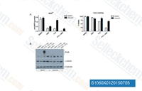
![]() ![]() Role of PARP and BER in the synergy between PTX and GMX in A549 cells. A) Cells were pre-treated +/- 1 uM olaparib (2h) then sequentially +/- 150nM PTX (24h) then +/- GMX 12nM (48h). Cells were harvested for (left) NAD+ quantification by LC-MS/MS (mean +/-SD of quadruplicates) or (right) viability by CellTiter-Glo (mean +/-SD of duplicates) B) PAR modification of proteins and γ-H2AX levels were measured in extracts treated as in A) by western blotting.
|
|
S1267 |
Vemurafenib (PLX4032)Vemurafenib (PLX4032, RG7204) is a novel and potent inhibitor of B-RafV600E with IC50 of 31 nM in cell-free assay. 10-fold selective for B-RafV600E over wild-type B-Raf in enzymatic assays and the cellular selectivity can exceed 100-fold. Vemurafenib (PLX4032, RG7204) induces autophagy. |
![]() ![]() The regressing tumour microenvironment stimulates the outgrowth, infiltration and metastasis of drug-resistant clones. b, Bioluminescent signal of drug-resistant A375RTGL cells in vemurafenib-sensitive, A375 tumours, treated with vehicle or vemurafenib for 5 days (vehicle, n = 36; vemurafenib, n = 15 tumours). D, day. c, EdU incorporation in A375R-TGL cells in A375/A375R-TGL tumours treated with vehicle or vemurafenib for 4 days, as determined by FACS (vehicle, n = 8; vemurafenib, n = 6 tumours). d, Bioluminescent signal of A375R-TGL tumours alone, treated with vehicle or vemurafenib for 5 days (vehicle, n 5 38; vemurafenib, n = 15 tumours). e, Bioluminescent signal of TGLexpressing drug-resistant cancer cells (A375R, M249R4, PC9 and H2030) in drug-sensitive tumours (Colo800, LOX, UACC62, M249, H3122 and HCC827) treated with vehicle or drugs (vemurafenib, crizotinib and erlotinib) for 5 days (n (from left to right on the graph) = 6, 7, 12, 12, 9, 9, 25, 26, 9, 12, 12, 12, 16 and 11 tumours). f, Spontaneous lung metastasis by A375R cells in mice bearing A375/A375R-TGL tumours treated with vehicle or vemurafenib (10 days), visualized by BLI (n = 4).
|
|
S1047 |
Vorinostat (SAHA)Vorinostat (suberoylanilide hydroxamic acid, SAHA, MK0683) is an HDAC inhibitor with IC50 of ~10 nM in a cell-free assay. Vorinostat abrogates productive HPV-18 DNA amplification. |
![]() ![]() Western blot analysis of histone H3 acetylation in the spleen of untreated and vorinostat-treated hNF-E2 tg mice (n = 4 of each genotype). |
|
S6957New |
β-Elemeneβ-Elemene ((-)-β-Elemene, Levo-β-elemene) is a sesquiterpene compound extracted from the herb Curcuma Rhizoma with antitumor activities. β-elemene can inhibit cell proliferation, arrest the cell cycle, and induce cell apoptosis or autophagy. |
||
S6797New |
QX77QX77 is a novel activator of chaperone-mediated autophagy (CMA). QX77 induces the up-regulation of Rab11 expression and up-regulates LAMP2A expression. |
||
S1002 |
ABT-737ABT-737 is a BH3 mimetic inhibitor of Bcl-xL, Bcl-2 and Bcl-w with EC50 of 78.7 nM, 30.3 nM and 197.8 nM in cell-free assays, respectively; no inhibition observed against Mcl-1, Bcl-B or Bfl-1. ABT-737 induces mitochondrial pathway apoptosis and mitophagy. Phase 2. |
![]() ![]() Cardiomyocytes transduced with or without Ad-Mst1 were treated with ABT-737 (0, 0.1, 1, 10 uM) for 12 hours. Representative immunoblots with antibodies to p62/SQSTM1, LC3 and GAPDH are shown. |
|
S1049 |
Y-27632 2HClY-27632 2HCl is a selective ROCK1 (p160ROCK) inhibitor with Ki of 140 nM in a cell-free assay, exhibits >200-fold selectivity over other kinases, including PKC, cAMP-dependent protein kinase, MLCK and PAK. |
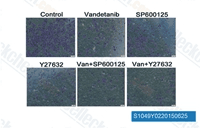
![]() ![]() The Rho GTPase-JNK pathway is required for the inhibitory effects of vandetanib on Calu-6 cells invasion. Calu-6 cells were incubated for 24 h in the presence or absence of vandetanib (1 or 2 uM), SP600125 (50 or 100 uM), and Y27632 (5 or 10 uM). The morphology of the Calu-6 cells was examined under a light microscope. Scale bar: 50 um.
|
|
S2619 |
MG-132MG132 is a potent cell-permeable proteasome and calpain inhibitor with IC50s of 0.1 μM and 1.2 μM for the inhibition of proteasome and calpain, respectively. MG132 activates autophagy and induces apoptosis in tumor cells. |
![]() ![]() MDA-MB-231 cells were treated with 10 uM MG132 and incubated under normoxia or hypoxia for 4 h. Endogenous interaction between LATS2 and SIAH2 was analysed by immunoprecipitation.
|
|
S1009 |
Dactolisib (BEZ235)Dactolisib (BEZ235, NVP-BEZ235) is a dual ATP-competitive PI3K and mTOR inhibitor for p110α/γ/δ/β and mTOR(p70S6K) with IC50 of 4 nM /5 nM /7 nM /75 nM /6 nM in cell-free assays, respectively. Inhibits ATR with IC50 of 21 nM in 3T3TopBP1-ER cell. Dactolisib induces autophagy and suppresses HIV-1 replication. Phase 2. |
![]() ![]() Three-dimensional responses of MCF7/IGF-1R cells to TAM (1 μM), E2 and IGF-1. Compared to parental MCF7 cells (a), MCF7/IGF-1R cells (b) in three-dimensional (3D) culture formed bigger acini in response to IGF-1 stimulation and displayed significant TAM resistance when treated with TAM (1 μM) + E2 + IGF-1, which was removable by kinase inhibitors BMS-536924, U0126 and BEZ235 (c). Cells (10,000/well) were seeded in 96-well plates. Acini were formed on 100% Matrigel and cultured for 14 days in starving medium containing 2% Matrigel and 5% charcoal/dextran-stripped fetal bovine serum with the treatments as indicated. Concentrations used: TAM (1 μM), E2 (1 nM) and IGF-1 (100 ng/mL). Confocal image original magnification, × 20. Red, rhodamine phalloidin (actin). Blue, Hoechst blue stain. Results are representative of two individual experiments. |
|
S1040 |
Sorafenib TosylateSorafenib Tosylate (Bay 43-9006) is a multikinase inhibitor of Raf-1 and B-Raf with IC50 of 6 nM and 22 nM in cell-free assays, respectively. Sorafenib Tosylate inhibits VEGFR-2, VEGFR-3, PDGFR-β, Flt-3 and c-KIT with IC50 of 90 nM, 20 nM, 57 nM, 59 nM and 68 nM, respectively. Sorafenib Tosylate induces autophagy and apoptosis and activates ferroptosis with anti-tumor activity. |
![]() ![]()
Inhibition of breast cancer cell growth using sorafenib. MCF-7 breast cancer cells were treated with increasing concentrations of sorafenib for 5 days. Cell number was measured using a colorimetric growth assay (crystal violet stain) and expressed relative to DMSO treated control cells. |
|
S1021 |
DasatinibDasatinib (BMS-354825) is a novel, potent and multi-targeted inhibitor that targets Abl, Src and c-Kit, with IC50 of <1 nM, 0.8 nM and 79 nM in cell-free assays, respectively. Dasatinib induces autophagy and apoptosis with anti-tumor activity. |
![]() ![]() Combinational treatment of kinase inhibitors induces the similar phenotype produced by PP1. All images are lateral view with dorsal to the top and anterior to the left. The combinational treatment of Dasatinib (D) or U0126 (U) with Sunitinib (SU),PTK787 (PTK), or ZM323881 (Z) resulted in the shrinkage of dorsal aorta. |
|
S1039 |
Rapamycin (Sirolimus)Rapamycin (Sirolimus, AY 22989, NSC-2260804) is a specific mTOR inhibitor with IC50 of ~0.1 nM HEK293 cells. |
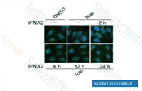
![]() ![]() H4-LC3-GFP cells were treated with 1 nM IFNA2 for the indicated periods in the presence of 200 nM rapamycin. Images of the cells were collected using an ArrayScan HCS 4.0 Reader. Representative cells are shown. The average spot intensity in 500 cells from each indicated sample was determined. Data are displayed as means ?SD of the spot intensity per cell (below). RLU, relative leight unit.
|
|
S1068 |
Crizotinib (PF-02341066)Crizotinib (PF-02341066) is a potent inhibitor of c-Met and ALK with IC50 of 11 nM and 24 nM in cell-based assays, respectively. It is also a potent ROS1 inhibitor with Ki value less than 0.025 nM. Crizotinib induces autophagy through inhibition of the STAT3 pathway in multiple lung cancer cell lines. |
![]() ![]() (c) Western blot analyses of p-Akt (Ser473) and p-S6RP (Ser235 and Ser236) in two RCT-E565 transplanted tumors treated with vehicle or PF02341066. Samples were isolated 4 h after the last dose from mice treated with PF02341066 for 3 d. (d) Responses of RCT-E565 transplanted tumors in athymic mice to PF02341066 or vehicle. Data are means ±s.e.m. (each group, n = 6). *P < 0.005, **P < 0.001 (Student,s t test). |
|
S1023 |
Erlotinib HCl (OSI-744)Erlotinib HCl (OSI-744, CP358774, NSC 718781) is an EGFR inhibitor with IC50 of 2 nM in cell-free assays, >1000-fold more sensitive for EGFR than human c-Src or v-Abl. |
![]() ![]() Effects of combined treatment with erlotinib and NPS-1034 in HCC827/ER cells with AXL activation. Lysates were immunoprecipitated with an anti-AXL antibody and immunoblotted with antibodies for phosphotyrosine (p-Tyr) and AXL. HCC827/ER cells were treated with erlotinib. E, erlotinib; N, NPS-1034. **, P < 0.001 for the combination of erlotinib plus NPS-1034 versus either the control or drug alone. |
|
S1120 |
Everolimus (RAD001)Everolimus (RAD001) is an mTOR inhibitor of FKBP12 with IC50 of 1.6-2.4 nM in a cell-free assay. Everolimus induces cell apoptosis and autophagy and inhibits tumor cells proliferation. |
![]() ![]() Cytoskeleton organisation of 786-O SuR treated with NVP-LDE225 (2.5 uM), everolimus (1 uM), and their combination for 24 h was analysed by confocal microscopy. Actin-based structures were revealed by rhodaminated phalloidin staining (red fluorescence). Localisation of focal adhesion points was obtained by immunofluorescent staining of p-paxillin (green fluorescence). Merged row images show overlapping of p-paxillin and actin signals. Moreover, all captures were shown in transmitted light. Scale bars, 10 um.
|
|
S1025 |
Gefitinib (ZD1839)Gefitinib (ZD-1839, Iressa) is an EGFR inhibitor for Tyr1173, Tyr992, Tyr1173 and Tyr992 in the NR6wtEGFR and NR6W cells with IC50 of 37 nM, 37nM, 26 nM and 57 nM, respectively. Gefitinib promotes autophagy and apoptosis of lung cancer cells via blockade of the PI3K/AKT/mTOR pathway. |
![]() ![]() Perturbation of EGFR by its ligand EGF and gefitinib (ZD-1839 Iressat; inhibits EGFR) produces opposite responses in the predicted EGFR target genes SOCS2 and NR2E1. |
|
S1004 |
Veliparib (ABT-888)Veliparib (ABT-888, NSC 737664) is a potent inhibitor of PARP1 and PARP2 with Ki of 5.2 nM and 2.9 nM in cell-free assays, respectively. It is inactive to SIRT2. Veliparib increases autophagy and apoptosis. Phase 3. |
![]() ![]()
Number of foci detected using laser confocal microscopy and fluorescent Fluor 647 anti-H2A.X-phosphorylated (Ser139) antibody. Double-stranded breaks (red) are clearly augmented in cells incubated with 500 nmol/l of ABT-888 and 500 nmol/l of AZD-2281 compared with PBS and 1% dimethyl sulfoxide controls. Image analysis was performed using ImageJ and the ‘analyze particle’ function. |
|
S1053 |
Entinostat (MS-275)Entinostat (MS-275, SNDX-275) strongly inhibits HDAC1 and HDAC3 with IC50 of 0.51 μM and 1.7 μM in cell-free assays, compared with HDACs 4, 6, 8, and 10. Entinostat induces autophagy and apoptosis. Phase 3. |
![]() ![]() (A) U87 cells were cultured in the presence of DMSO, 1 uM MS-275 alone, 100 ng/ml IFN-λ1 alone, or both for the course of 4 d. Cell numbers were manually determined by hemacytometer counting at the indicated time points. (B, F) Cell proliferation of U87 cells or U87 spheroids in 3D culture with indicated treatment were performed using the WST-1 assay, which measures active cellular metabolism. (C) U87 spheroid formation in 3D culture was photographed at day 14 in culture (representative images are shown; 200x magnification). (D-E) Quantification of the relative sizes and numbers of U87 spheroids in (C). (G) Cell cycle analysis was performed in U87 cells with indicated treatment using propidium iodide staining. Numbers in the histogram show fractions (percent) of sub-G1, N, 2N, and polyploidy from left to right. (H) U87 cells with indicated treatment were stained with Annexin V-FITC and 7-AAD. Numbers indicate the percentage of FITC-positive cells (upper left quadrant). FITC, fluorescein isothiocyanate; 7-AAD, 7-Aminoactinomycin. In all panels, data represent the mean and SEM of at least three experiments.
|
|
S1065 |
Pictilisib (GDC-0941)Pictilisib (GDC-0941, RG7321) is a potent inhibitor of PI3Kα/δ with IC50 of 3 nM in cell-free assays, with modest selectivity against p110β (11-fold) and p110γ (25-fold). Pictilisib (GDC-0941) induces autophagy and apoptosis. Phase 2. |
![]() ![]() TGF-β induces mTORC2 activation. ( A ) NMuMG cells were treated with TGF-β for the indicated times before lysis and immunoblotting. (B ) NMuMG cells were treated or not with TGF-β for the indicated times, in the presence or absence of SB431542 or LY294002. Cell lysates were subjected to anti-Rictor immunoprecipitation, and the immunoprecipitates were subjected to in vitro kinase assays using kinase-inactive Akt1 as a substrate, before immunoblotting of the kinase reactions, immunoprecipitates and cell lysates. The left panels are from the same gel, without differential exposure. ( C ) NMuMG cells were treated or not with TGF-β or insulin for the indicated times, in the presence or absence of SB431542 or GDC-0941. The kinase activity of mTORC2 was then assessed as in B. The top panels are from the same gel, without differential exposure. (D) NMuMG cells were treated or not with TGF-β for the indicated times, in the presence or absence of SB431542 or LY294002. Cell lysates were subjected to immunoprecipitation using Rictor antibody, and/or immunoblotted. |
|
S2673 |
Trametinib (GSK1120212)Trametinib (GSK1120212, JTP-74057, Mekinist) is a highly specific and potent MEK1/2 inhibitor with IC50 of 0.92 nM/1.8 nM in cell-free assays, no inhibition of the kinase activities of c-Raf, B-Raf, ERK1/2. Trametinib activates autophagy and induces apoptosis. |
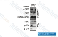
![]() ![]() ERK phosphorylates FBW7 at T205. PANC-1 cells were pretreated with the proteasome inhibitor MG132 and trametinib, as indicated, overnight before harvest. Endogenous FBW7 phosphorylation status was examined by immunoblot analysis after immunoprecipitates (IP).
|
|
S1378 |
Ruxolitinib (INCB018424)Ruxolitinib (INCB018424) is the first potent, selective, JAK1/2 inhibitor to enter the clinic with IC50 of 3.3 nM/2.8 nM in cell-free assays, >130-fold selectivity for JAK1/2 versus JAK3. Ruxolitinib kills tumor cells through toxic mitophagy. Ruxolitinib induces autophagy and enhances apoptosis. |
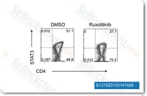
![]() ![]() STAT3 phosphorylation as determined by phospho flow, mixed lymphocyte reactions containing BALB/c spleen-derived CD4+ T cells co-cultured with or without C57BL/6 BM-derived DC preactivated with 20 ng/mL LPS.
|
|
S1030 |
Panobinostat (LBH589)Panobinostat (LBH589, NVP-LBH589) is a novel broad-spectrum HDAC inhibitor with IC50 of 5 nM in a cell-free assay. Panobinostat (LBH589) induces autophagy and apoptosis. Panobinostat effectively disrupts HIV latency in vivo. Phase 3. |
![]() ![]() LSD1 and HDAC inhibitors exhibit synergistic growth inhibition. Cells were simultaneously treated with pargyline or HDAC inhibitors for 48 h.
|
|
S1026 |
Imatinib (STI571) MesylateImatinib (STI571, CGP057148B, Gleevec) Mesylate is an orally bioavailability mesylate salt of Imatinib, which is a multi-target inhibitor of v-Abl, c-Kit and PDGFR with IC50 of 0.6 μM, 0.1 μM and 0.1 μM in cell-free or cell-based assays, respectively. Imatinib Mesylate (STI571) induces autophagy. |
![]() ![]() THOC5 phospho-Y225 levels were assessed in the populations shown by flow cytometry following 24 h treatment with 5 μM imatinib, 150 nM dasatinib or 5 μM nilotinib. Results are displayed as mean fluorescence intensity (n= 4) ±s.e.m. |
|
S1250 |
Enzalutamide (MDV3100)Enzalutamide (MDV3100) is an androgen-receptor (AR) antagonist with IC50 of 36 nM in LNCaP cells. Enzalutamide is shown to increase autophagy. |
![]() ![]() |
|
S1133 |
Alisertib (MLN8237)Alisertib (MLN8237) is a selective Aurora A inhibitor with IC50 of 1.2 nM in a cell-free assay. It has >200-fold higher selectivity for Aurora A than Aurora B. Alisertib induces cell cycle arrest, apoptosis and autophagy. Phase 3. |
![]() ![]() Alisertib inhibits AURKA and AURKB in a concentration-dependent manner. (a) Alisertib induces G 2 /M delay or genome reduplication. HeLa cells were exposed to buffer or the indicated concentrations of Alisertib. After 24 h, the cells were harvested and analyzed with flow cytometry. The positions of 2N, 4N and 8N DNA contents are indicated. (b) Alisertib delays mitotic exit or induces slippage. HeLa cells stably expressing histone H2B-GFP were exposed to buffer or the indicated concentrations of Alisertib. Individual cells were then tracked for 24 h with time-lapse microscopy. Each horizontal bar represents one cell (n ¼ 50). Key: light gray ¼ interphase; black ¼ mitosis (from DNA condensation to anaphase or mitotic slippage); dark gray ¼ interphase after mitotic slippage; truncated bars ¼ cell death. (c) Different concentrations of Alisertib are involved in delaying mitotic exit and inducing slippage. Live-cell imaging of cells treated with Alisertib was described in panel (b). The duration of mitosis (mean±90% confidence interval) and the percentage of cells that underwent mitotic slippage during the imaging period was quantified. (d) Alisertib promotes apoptosis in a concentration-dependent manner. HeLa cells were incubated with the indicated concentrations of Alisertib for 48 h. The cells were then harvested and analyzed with flow cytometry. (e) Concentration-dependent cytotoxicity of Alisertib. HeLa cells were cultured in the presence of the indicated concentrations of Alisertib for 48 h. The number of live and dead cells was analyzed with trypan blue exclusion assay. (f) Concentration-dependent suppression of long-term survival by Alisertib. HeLa cells were seeded on 60-mm culture plates and grown in the presence of 250 n M or 1 m M of Alisertib. After 24 h, the cells were washed gently and propagated in normal medium for another 10–12 days. Colonies were fixed and stained with crystal violet solution (examples of the plates are shown). Average±s.d. from three independent experiments. (g) Both AURKA and AURKB are inhibited by Alisertib.Mitotic HeLa cells were obtained by exposure to nocodazole for 16 h followed by mechanical shake off. The cells were incubated with the indicated concentrations of Alisertib for 2 h. Lysates were then prepared and activated phospho-AURKAThr288 and AURKBThr232were detected with immunoblotting. The asterisk indicates the position of an AURKB-like protein (the same throughout this study). Uniform loading was confirmed by immunoblotting for actin. In this assay, nocodazole and MG132 (a proteasome inhibitor) were added to prevent the cells from exiting mitosis. Accordingly, the total AURKA and AURKB levels remained constant throughout the experiment. (h) Alisertib prevents activation of AURKA and AURKB. HeLa cells were incubated with the indicated concentrations of Alisertib for 8 h. Nocodazole was then added for another 6 h to trap cells that entered mitosis. Lysates were prepared and analyzed with immunoblotting. Actin analysis was included to assess loading and transfer. |
|
S1011 |
Afatinib (BIBW2992)Afatinib (BIBW2992) inhibits EGFR/ErbB irreversibly in vitro with IC50 of 0.5, 0.4, 10, 14, 1 nM for EGFRwt, EGFR L858R , EGFR L858R/T790M ErbB2 (HER2) and ErbB4 (HER4), respectively. Afatinib induces autophagy. |
![]() ![]() Inhibition of signaling pathway activation in lung tumor cell lines by kinase inhibitors. Lung tumor cells were cultured in 10% FBS until reaching ∼80% confluence and then the cells were starved in serum-free medium for overnight, followed by 4-hour treatment with the inhibitors. Cell lysates were then prepared and used for determination of the pathway activation signals by the CEER assay. |
|
S2226 |
Idelalisib (CAL-101, GS-1101)Idelalisib (CAL-101, GS-1101) is a selective p110δ inhibitor with IC50 of 2.5 nM in cell-free assays; shown to have 40- to 300-fold greater selectivity for p110δ than p110α/β/γ, and 400- to 4000-fold more selectivity to p110δ than C2β, hVPS34, DNA-PK and mTOR. Idelalisib also stimulates autophagy. |
![]() ![]() 293T cells were transfected with HA-tagged Fbxo45. At 48 h after transfection, cells were treated with AKT inhibitor (CAL-101; 10 uM, 4 h), cell extracts from the cytoplasm or nuclei were subjected to IP with anti-HA resin followed by western blot analysis with indicated antibodies.
|
|
S1048 |
Tozasertib (VX-680, MK-0457)Tozasertib (VX-680, MK-0457) is a pan-Aurora inhibitor, mostly against Aurora A with Kiapp of 0.6 nM in a cell-free assay, less potent towards Aurora B/Aurora C and 100-fold more selective for Aurora A than 55 other kinases. The only exceptions are Fms-related tyrosine kinase-3 (FLT-3) and BCR-ABL tyrosine kinase, which are inhibited by the Tozasertib with both Ki of 30 nM. Tozasertib induces apoptosis and autophagy. Phase 2. |
![]() ![]() Senescence induction upon PKCι depletion combined with aurora kinase inhibition. ( a) MCF7 cells were transfected as above to deplete PKCι . Two days after transfection, cells were treated for the indicated time period with 400 n M VX-680. Medium with VX-680 was then removed and fresh medium was added. Cells were stained for SA-b -gal activity 5 days after the start of transfection.* indicates a P value <0.05. ( b) MCF7 cells were treated as above. Five days after transfection, cells were fixed and assessed for the presence of gH2AX foci by immunofluorescence microscopy. (c, d) MCF7 cells were treated with dimethyl sulfoxide (DMSO) control or 400 n M VX-680 for the indicated time periods. Total cell lysates were then analyzed by western blotting for levels of p21 and GAPDH (as loading control). A representative blot is shown in panel c. Quantitation of changes in p21 levels (normalized to vehicle-treated controls) is shown in panel d. The data shown are the means ±s.e. of three independent experiments. |
|
S1555 |
AZD8055AZD8055 is a novel ATP-competitive mTOR inhibitor with IC50 of 0.8 nM in MDA-MB-468 cells with excellent selectivity (∼1,000-fold) against PI3K isoforms and ATM/DNA-PK. AZD8055 induces caspase-dependent apoptosis and also induces autophagy. Phase 1. |
![]() ![]() mTOR kinase inhibitor AZD8055 activates PI3K accompanied with induction of expression of EGFR, HER2, HER3 and IRS1. Serum-deprived CHO-EGFP-AKT cells were incubated with 50 nM AZD8055 for 24 hr. The EGFP signal was detected using confocal microscopy. The white arrows indicate EGFP-AKT located on cellular membrane.
|
|
S1006 |
Saracatinib (AZD0530)Saracatinib (AZD0530) is a potent Src inhibitor with IC50 of 2.7 nM in cell-free assays, and potent to c-Yes, Fyn, Lyn, Blk, Fgr and Lck; less active for Abl and EGFR (L858R and L861Q). Saracatinib induces autophagy. Phase 2/3. |
![]() ![]() C and D, in vivo subcutaneous tumor growth curves (C) and tumor weight quantification of intersected subcutaneous tumor tissues (D) of Huh7 cells after stable LHBs expression under saracatinib treatment (25 mg/kg body weight daily for 4 weeks; n =18). *, P < 0.05. E and F,in vivo subcutaneous tumor growth curves (E) and tumor weight quantification of intersected subcutaneous tumor tissues (F) of SK-Hep1 cells after stable LHBs expression under saracatinib treatment (25 mg/kg body weight daily for 4 weeks; n = 18). *, P < 0.05. |
|
S1057 |
Obatoclax Mesylate (GX15-070)Obatoclax Mesylate (GX15-070) is an antagonist of Bcl-2 with Ki of 0.22 μM in a cell-free assay, can assist in overcoming MCL-1 mediated resistance to apoptosis. Phase 3. |
![]() ![]() In vivo antitumor efficacies of AZD2281 and GX15-070 alone or in combination in a BxPC-3 xenograft model. Tumor specimens were fixed in 10% formalin, embedded in paraffin, and cut into 4 lm-thick slides for H&E, PCNA, and CD34 staining.
|
|
S1141 |
Tanespimycin (17-AAG)Tanespimycin (17-AAG, CP127374, NSC-330507, KOS 953) is a potent HSP90 inhibitor with IC50 of 5 nM in a cell-free assay, having a 100-fold higher binding affinity for HSP90 derived from tumour cells than HSP90 from normal cells. Tanespimycin (17-AAG) induces apoptosis, necrosis, autophagy and mitophagy. Phase 3. |
![]() ![]() SKBR3 cells were treated with FW-04-806 at 10, 20, 40 uM for 24 h; 17AAG was used as a positive control at 1 and 2 uM. Hsp70, Hsp90, and Cdc37 protein level were analyzed with western blotting using relevant antibodies.
|
|
S1130 |
YM155 (Sepantronium Bromide)YM155 (Sepantronium Bromide) is a potent survivin suppressant by inhibiting Survivin promoter activity with IC50 of 0.54 nM in HeLa-SURP-luc and CHO-SV40-luc cells; does not significantly inhibit SV40 promoter activity, but is observed to slightly inhibit the interaction of Survivin with XIAP. YM155 down-regulates survivin and XIAP, modulates autophagy and induces autophagy-dependent DNA damage in breast cancer cells. Phase 2. |
![]() ![]() MM cell lines UM9 (left panel) or U266 (right panel) were cultured in presence or absence of accessory cells prior to incubation with CTLs alone, YM155 alone or the combination. Survival of UM9 and U266 MM cells was assessed by CS-BLI. Results show % lysis of MM cells after 24 hour incubation and are depicted for three doses. |
|
S1166 |
CisplatinCisplatin (cisplatinum, cis-diamminedichloroplatinum II, CDDP) is an inorganic platinum complex, which is able to inhibit DNA synthesis by conforming DNA adducts in tumor cells. Cisplatin activates ferroptosis and induces autophagy. Solutions are best fresh-prepared. |
![]() ![]() Cell viabilities with increasing concentrations of cisplatin (CP) and doxorubicin (DOXO) under normoxic and hypoxic condition for 48 hours were determined by MTT assay. IC50 values are presented as the means ?SDs (n=4) and * denotes p<0.05. |
|
S2475 |
Imatinib (STI571)Imatinib (STI571, CGP057148B, Gleevec) is a multi-target inhibitor of tyrosine kinase with inhibition for v-Abl, c-Kit and PDGFR, IC50 values are 0.6 μM, 0.1 μM and 0.1 μM in cell-free or cell-based assays, respectively. Imatinib (STI571) induces autophagy. |
![]() ![]()
Inhibition of thymidine (a and b) and cytarabine (c and d) uptake with imatinib. K562 cells (a and c) and MEG-01 cells (b and d) were incubated at 37 ◦C for 15 min with imatinib transport buffer, and then incubated with 0.5 Ci of [3H] thymidine or [3H] cytarabine for an additional 5 min in presence of imatinib. Cells were then washed 3 times, lysed and radioactivity associated to cell pellets was quantified. DMSO, dimethylsulfoxide; DPD, dipyridamole. |
|
S1033 |
Nilotinib (AMN-107)Nilotinib (AMN-107) is a selective Bcr-Abl inhibitor with IC50 less than 30 nM in Murine myeloid progenitor cells. Nilotinib induces autophagy through AMPK activition. |
![]() ![]() Effect of nilotinib on Bcr-Abl kinase activity in ABCB1- and ABCG2- overexpressing CD34+CD38- cells. K562 parental cells and CD34+CD38- subpopulation isolated from K562 cells were treated with nilotinib at 0.01, 0.1 and 1.0 umol/L for 12 h. Equal amount of protein was loaded for western blot analysis as described in the Experimental section. The experiments were repeated at least three times independently, and a representative experiment is shown. |
|
S1044 |
Temsirolimus (CCI-779)Temsirolimus (CCI-779, NSC 683864) is a specific mTOR inhibitor with IC50 of 1.76 μM in a cell-free assay. Temsirolimus induces autophagy and apoptosis. |
![]() ![]() mTOR inhibitors attenuate ganetespib-driven elevation of HSPs in multiple tumor cell types. A375 melanoma cells were treated with vehicle, ganetespib (25 nmol/L), BEZ235 (500 nmol/L), or temsirolimus (500 nmol/L), either alone or in combination, for 24 hours. The levels of HSP90α, HSP70, HSP27, and GAPDH were determined by immunoblotting.
|
|
S1038 |
PI-103PI-103 is a multi-targeted PI3K inhibitor for p110α/β/δ/γ with IC50 of 2 nM/3 nM/3 nM/15 nM in cell-free assays, less potent to mTOR/DNA-PK with IC50 of 30 nM/23 nM. PI-103 induces apoptosis in murine T-cell Lymphoma. |
![]() ![]() We treated all of drugs in T47D which has a PI3KCA H1044R mutation with the concentration shown below for 1 hour and performed western blot analysis using antibodies to phospho-AKT(SERINE 472), and total AKT.
|
|
S1069 |
Luminespib (NVP-AUY922)Luminespib (AUY-922, NVP-AUY922) is a highly potent HSP90 inhibitor for HSP90α/β with IC50 of 13 nM /21 nM in cell-free assays, weaker potency against the HSP90 family members GRP94 and TRAP-1, exhibits the tightest binding of any small-molecule HSP90 ligand. Luminespib (AUY-922, NVP-AUY922) effectively downregulates and destabilizes the IGF-1Rβ protein and results in growth inhibition, autophagy and apoptosis. Phase 2. |
![]() ![]() |
|
S1046 |
Vandetanib (ZD6474)Vandetanib (ZD6474) is a potent inhibitor of VEGFR2 with IC50 of 40 nM in a cell-free assay. It also inhibits VEGFR3 and EGFR with IC50 of 110 nM and 500 nM, respectively. Not sensitive to PDGFRβ, Flt1, Tie-2 and FGFR1 with IC50 of 1.1-3.6 μM. No activity against MEK, CDK2, c-Kit, erbB2, FAK, PDK1, Akt and IGF-1R with IC50 above 10 μM. Vandetanib (ZD6474) increases apoptosis and induces autophagy by increasing the level of reactive oxygen species (ROS). |
![]() ![]() Vandetanib reduced extracellular nitrite levels in endothelial cells. MS1 endothelial cells (ECs) were incubated with 1 mol/L of vandetanib or matched vehicle (dimethyl sulfoxide [DMSO]), 50 ng/mL of vascular endothelial growth factor (VEGF) or matched vehicle (PBS; 0.5 hours), and L-arginine and soluble N-ethylmaleamide sensitive factor attachment protein (SNAP) added (1.5 hours). Vandetanib lowered nitrite levels in MS1 Ecs (*P0.0003). VEGF was used a positive control and increased nitrite levels (**P0.02). These findings indicate that vandetanib lowered endothelial cell NO levels. |
|
S1149 |
Gemcitabine HClGemcitabine HCl (LY188011) is a DNA synthesis inhibitor with IC50 of 50 nM, 40 nM, 18 nM and 12 nM in PANC1, MIAPaCa2, BxPC3 and Capan2 cells, respectively. |
![]() ![]() RNA incorporating drugs induce SG assembly. HeLa cells were treated with the RNA incorporating agents 5-azacytidine (50 uM) and 6-thioguanine (10 uM), or the DNA incorporating agents trifluorothymidine (10 uM) and gemcitabine (100 nM) for 72 h. Subsequently, the cellular localization of the SG marker protein TIAR (green) and the P-body marker protein DCP1 (red) was analyzed. Nuclei were stained with Hoechst. Scale bars represent 20 um. |
|
S1122 |
Mocetinostat (MGCD0103)Mocetinostat (MGCD0103, MG0103) is a potent HDAC inhibitor with most potency for HDAC1 with IC50 of 0.15 μM in a cell-free assay, 2- to 10- fold selectivity against HDAC2, 3, and 11, and no activity to HDAC4, 5, 6, 7, and 8. Mocetinostat (MGCD0103) induces apoptosis and autophagy. Phase 2. |
![]() ![]()
Comparison of MCAS ovarian cancer cells harboring control and CtBP2 knockdown shRNAs for sensitivity to chemotherapeutic agents. For each cell line, the MTT reading of the untreated cells was assigned as 100%. HDAC inhibitors: (a) Trichostatin A; (b) Vorinostat; (c) Belinostat; (d) MGCD0103; (e) valproic acid; and (f ) carboplatin, a non-HDAC inhibitor. |
|
S1178 |
Regorafenib (BAY 73-4506)Regorafenib (BAY 73-4506, Fluoro-Sorafenib, Resihance, Stivarga) is a multi-target inhibitor for VEGFR1, VEGFR2, VEGFR3, PDGFRβ, Kit (c-Kit), RET (c-RET) and Raf-1 with IC50 of 13 nM/4.2 nM/46 nM, 22 nM, 7 nM, 1.5 nM and 2.5 nM in cell-free assays, respectively. Regorafenib induces autophagy. |
![]() ![]() Hepatoma cells 24 h after plating were treated with vehicle (DMSO), regorafenib (REGO, 0.5 µM), PDE5 inhibitor (sildenafil, 2 µM); or the drugs in combination. 24 hours after treatment cells were isolated and viability determined by trypan blue (n=3, SEM). *P 0.05
|
|
S1129 |
SRT1720 HClSRT1720 HCl is a selective SIRT1 activator with EC50 of 0.16 μM in a cell-free assay, but is >230-fold less potent for SIRT2 and SIRT3. SRT1720 induces autophagy. |
![]() ![]() PAI-1 expression in HUVECs treated with drugs as indicated.(D) senescent HUVECs were treated with SRT1720, culturing for 24, 48 hours. PAI-1 mRNA and protein (E) levels were analyzed using real-time RT–PCR and Western blotting, respectively. The RNA and protein levels were normalized to the internal control β-actin. Data are presented as the mean±SEM of three independent experiments. *P < 0.05 vs. corresponding control. **P < 0.01 vs. corresponding control ***P < 0.001 vs. corresponding control.
|
|
S1035 |
Pazopanib HCl (GW786034 HCl)Pazopanib HCl (GW786034 HCl) is a novel multi-target inhibitor of VEGFR1, VEGFR2, VEGFR3, PDGFR, FGFR, c-Kit and c-Fms with IC50 of 10 nM, 30 nM, 47 nM, 84 nM, 74 nM, 140 nM and 146 nM in cell-free assays, respectively. Pazopanib induces autophagic Type II cell death. |
![]() ![]() Effect of HDIL-2/TKI on apoptosis of RCC cells. Three RCC cell lines treated with different concentrations of Pazopanib and HDIL-2 and incubated for 48 h. Microscopic images show apoptotic materials 48 h following treatment (arrows show the apoptotic materials in the pazopanib-treated cells).
|
|
S1014 |
Bosutinib (SKI-606)Bosutinib (SKI-606) is a novel, dual Src/Abl inhibitor with IC50 of 1.2 nM and 1 nM in cell-free assays, respectively. Bosutinib also effectively decreases the activity of PI3K/AKT/mTOR, MAPK/ERK and JAK/STAT3 signaling pathways by blocking the phosphorylation levels of p-ERK, p-S6, and p-STAT3. Bosutinib promotes autophagy. |
![]() ![]() A,IC50 of Bosutinib that block ANDV-induced EC permeability. Endothelial cells were ANDV infected, and 3 days postinfection the permeability of cells in response to VEGF addition was determined in the presence or absence of increasing amounts of kinase inhibitor. The effect of inhibitors is presented as the percentage of ANDV-induced permeability of inhibitor-treated monolayers 3 days postinfection and 30 min post-VEGF and FITC-dextran addition. B, VEGFR2-Src inhibitors block ANDV-induced permeability. Endothelial cells were plated on vitronectin-coated Transwell inserts and infected at an MOI of 0.5 in triplicate with ANDV. Three days postinfection, the permeability of ANDV- and mock-infected endothelial cell monolayers was determined as described for Fig. 1 at indicated times in the presence or absence of Bosutinib . |
|
S1017 |
Cediranib (AZD2171)Cediranib (AZD2171, NSC-732208) is a highly potent VEGFR(KDR) inhibitor with IC50 of <1 nM, also inhibits Flt1/4 with IC50 of 5 nM/≤3 nM, similar activity against c-Kit and PDGFRβ, 36-, 110-fold and >1000-fold selective more for VEGFR than PDGFR-α, CSF-1R and Flt3 in HUVEC cells. Cediranib (AZD2171) induces autophagic vacuole accumulation. Phase 3. |
![]() ![]() Western blots of EZH2 expression in A549, HCC461, and HCC4006 cells upon treatment with different doses of VEGFR-2-inhibitor AZD2171 (0, 5 and 10 nM). AZD2171 decreased the expression of EZH2 in HCC4006 and HCC461 cells expressing VEGFR-2 in a dose-dependent manner but did not do so in A549 cells lacking expression of VEGFR-2.
|
|
S1085 |
Belinostat (PXD101)Belinostat (PXD101, NSC726630, PX-105684) is a novel HDAC inhibitor with IC50 of 27 nM in a cell-free assay, with activity demonstrated in cisplatin-resistant tumors. Belinostat (PXD101) induces autophagy. |
![]() ![]() Inhibition of LSD1 activity by HDAC inhibitors. a MDA-MB-231 and MDA-MB-468 cells were exposed to indicated HDAC inhibitors for 24 h. |
|
S1077 |
SB202190 (FHPI)SB202190 (FHPI) is a potent p38 MAPK inhibitor targeting p38α/β with IC50 of 50 nM/100 nM in cell-free assays, sometimes used instead of SB 203580 to investigate potential roles for SAPK2a/p38 in vivo. SB202190 inhibits endothelial cell apoptosis via induction of autophagy and heme oxygenase-1. SB202190 significantly suppresses Erastin‐dependent ferroptosis. |
![]() ![]() C, Jurkat cells with SB202190 at 1, 5, and 10 μM were tested, and a decreased SRP72 expression was found when using at 10 μM (lanes 8 and 9). D, results were analyzed and RUA illustrated, finding significant results at 10 μM at 240 versus 0 and 120 versus 0 min (p<0.05).
|
|
S2853 |
Carfilzomib (PR-171)Carfilzomib (PR-171) is an irreversible proteasome inhibitor with IC50 of <5 nM in ANBL-6 cells, displayed preferential in vitro inhibitory potency against the ChT-L activity in the β5 subunit, but little or no effect on the PGPH and T-L activities. Carfilzomib activates prosurvival autophagy and induces cell apoptosis. |
![]() ![]() Validation of activity and specificity of chemical inhibitors of; ATM, ATR, and DNAPK. H460 cells were treated with 1 uM camptothecin (CPT) or 20 ug/ml bleomycin for 1 h in the presence of the indicated inhibitors: DNAPK-i1—NU7026, DNAPK-i2—NU7441. MSH6,
|
|
S1191 |
FulvestrantFulvestrant (ICI-182780, ZD 9238) is an estrogen receptor (ER) antagonist with IC50 of 0.94 nM in a cell-free assay. Fulvestrant also induces autophagy and apoptosis and has antitumor activity. |
![]() ![]() PTPH1 confers breast cancer cell sensitivity to fulvestrant. E and F, PTPH1 overexpression increases the growth inhibition by fulvestrant. PTPH1 was overexpressed by a Tet-on system or a stable transfection, and resultant cells were incubated with fulvestrant as indicated for about 2 weeks. Colony formed was stained and counted. Results shown are normalized to its own solvent control of Vector and PTPH1-overexpressed cells, respectively (means ± SD; n = 3–5) with insets showing PTPH1 overexpression. *, versus vector or no Tet cells for E and F.
|
|
S1075 |
SB216763SB216763 is a potent and selective GSK-3 inhibitor with IC50 of 34.3 nM for GSK-3α and equally effective at inhibiting human GSK-3β. SB216763 activates autophagy. |
![]() ![]() |
|
S1080 |
SU11274SU11274 (PKI-SU11274) is a selective Met (c-Met) inhibitor with IC50 of 10 nM in cell-free assays, no effects on PGDFRβ, EGFR or Tie2. SU11274 induces autophagy, apoptosis and cell cycle arrest. |
![]() ![]() Effect of crizotinib, tivantinib, and SU11274 on levels of c-MET phosphorylation and downstream signaling pathway in SW1736 and TL3 thyroid cancer cells. Cells were prestarved in culture medium containing 0.5% FBS (24 hour) ?either crizotinib or tivantinib (0.1, 1.0, and 10 umol/L) or SU11274 (10 umol/L), and stimulated with 20 ng/mL recombinant human HGF for 10 minutes before lysates were made for Western blotting. A series of c-MET downstream signaling pathway proteins and phosphor proteins were detected using Western blotting. β-Actin was used as a loading balance control.
|
|
S1003 |
Linifanib (ABT-869)Linifanib (ABT-869, AL39324, RG3635) is a novel, potent ATP-competitive VEGFR/PDGFR inhibitor for KDR, CSF-1R, Flt-1/3 and PDGFRβ with IC50 of 4 nM, 3 nM, 3 nM/4 nM and 66 nM respectively, mostly effective in mutant kinase-dependent cancer cells (i.e. FLT3). Linifanib (ABT-869) induces autophagy and apoptosis. Phase 3. |
![]() ![]() (B and C) KMCH-1 cells were plated alone (monoculture) or together with PDGF-BB-secreting LX-2 cells (co-culture) in a transwell insert co-culture system (KMCH-1 cells in the bottom wells and LX-2 cells in the inserts; 1:1 ratio) for 2 days. Cells were treated as indicated with vehicle, rhTRAIL (10 ng/ml for 6 h on day 2), rhTRAIL plus imatinib [rhTRAIL:10 ng/ml for 6 h on day 2; Imatinib: 5 μmol/L for 24 h (day2)], or rhTRAIL plus linifanib [rhTRAIL: 10 ng/ml for 6 h on day 2; Linifanib:0.5 μmol/L for 24 h (day2)]. After rhTRAIL treatment for 6 h,KMCH-1 cells were analysed for apoptotic nuclear morphology by DAPI-staining (B) and for DNA fragmentation by transferasemediated dUTP nick end labelling assay (C) with quantification of apoptotic nuclei by fluorescence microscopy. |
|
S1135 |
PemetrexedPemetrexed (LY-231514) is a novel antifolate and antimetabolite for TS, DHFR and GARFT with Ki of 1.3 nM, 7.2 nM and 65 nM in cell-free assays, respectively. Pemetrexed induces autophagy and apoptosis. |
![]() ![]() Role of PARP and BER in the synergy between PTX and GMX in A549 cells. Cells were pretreated ±1 umol/L olaparib (2 hours) then sequentially ±150 nmol/L PTX (24 hours), then ± GMX 12 nmol/L (48 hours). PAR modification of proteins and γ-H2AX levels were measured in extracts treated as in A by Western blotting.
|
|
S2218 |
Torkinib (PP242)Torkinib (PP242) is a selective mTOR inhibitor with IC50 of 8 nM in cell-free assays; targets both mTOR complexes with >10- and 100-fold selectivity for mTOR than PI3Kδ or PI3Kα/β/γ, respectively. Torkinib (PP242) induces mitophagy and apoptosis. |
![]() ![]() Synergistic effect of BMS-777607 with mTOR inhibitors in reduction of CSCs+24/44/ESA viability. CSCs+24/44/ESA at 5,000 cells per well with stem cell culture media in triplicate in an ultra-low adhesion plate were treated with 5 umol/L BMS-777607, 1 umol/L AZD8055, 1 umol/L RAD001, and 1 umol/L PP242 alone, or in their different combinations. Cells were cultured for 72 hours. Percentages of polyploid cells were determined by counting 300 cells from two different regions. Results shown here were from one of two experiments with similar results.
|
|
S1225 |
EtoposideEtoposide (VP-16, VP-16213) is a semisynthetic derivative of podophyllotoxin, which inhibits DNA synthesis via topoisomerase II inhibition activity. Etoposide induces autophagy, mitophagy and apoptosis. |
![]() ![]() Cellular biomarker responses in HT29 cells exposed to various cytotoxic chemotherapeutic agents in combination with the Chk1 inhibitor V158411. HT29 cells were exposed to the combination GI80 of gemcitabine (0.2 uM), camptothecin (0.44 uM), cisplatin (68 uM), oxaliplatin (131 uM), doxorubicin (1.2 uM) or etoposide (59 uM) for 18 hours followed by DMSO (-) or 400 nM V158411 (+) for a further 24 hours. Protein expression was characterized by immunoblotting.
|
|
S1107 |
Danusertib (PHA-739358)Danusertib (PHA-739358) is an Aurora kinase inhibitor for Aurora A/B/C with IC50 of 13 nM/79 nM/61 nM in cell-free assays, modestly potent to Abl, TrkA, c-RET and FGFR1, and less potent to Lck, VEGFR2/3, c-Kit, CDK2, etc. Danusertib induces apoptosis, cell cycle arrest, and autophagy. Phase 2. |
![]() ![]() Mice bearing subcutaneous allografts of conditional patched mutant tumor cells were treated twice weekly with vehicle (saline) or 30 mg/kg PHA-739358. (B)Images of tumors. (C) Tumor weights. Each point represents a single tumor, and grey lines represent mean tumor weights, which were significantly different between vehicle and PHA-739358 treated mice (p < 0.05, based on paired two-tailed t-test).
|
|
S2248 |
Silmitasertib (CX-4945)Silmitasertib (CX-4945) is a potent and selective inhibitor of CK2 (casein kinase 2) with IC50 of 1 nM in a cell-free assay, less potent to Flt3, Pim1 and CDK1 (inactive in cell-based assay). Silmitasertib induces autophagy and promotes apoptosis. Phase 1/2. |
![]() ![]() |
|
S8048 |
Venetoclax (ABT-199)Venetoclax (ABT-199, GDC-0199) is a Bcl-2-selective inhibitor with Ki of <0.01 nM in cell-free assays, >4800-fold more selective versus Bcl-xL and Bcl-w, and no activity to Mcl-1. Venetoclax is reported to induce cell growth suppression, apoptosis, cell cycle arrest, and autophagy in triple negative breast cancer MDA-MB-231 cells. Phase 3. |
![]() ![]() THP-1 cells were treated with cytarabine alone and in combination with ABT-199 for 8 h. Whole cell lysates were extracted and subjected to Western blotting, and probed with anti-γH2AX or -β-actin antibody. Densitometry for γH2AX expression was measured, normalized to β-actin, and graphed as fold change compared to the no drug control. The data are presented as mean ± standard error from at least 3 independent Western blots. * indicates p < 0.05.
|
|
S1230 |
Flavopiridol (Alvocidib)Flavopiridol (Alvocidib, NSC 649890, HMR-1275) competes with ATP to inhibit CDKs including CDK1, CDK2, CDK4, CDK6, and CDK9 with IC50 values in the 20-100 nM range. It is more selective for CDK1, 2, 4, 6, 9 versus CDK7. Flavopiridol is initially found to inhibit EGFR and PKA. Flavopiridol induces autophagy and ER stress. Flavopiridol blocks HIV-1 replication. Phase 1/2. |
![]() ![]() (C) In vivo treatment of Tg:Pomc-Pttg;Pomc-eGFP embryos with small-molecule CDK inhibitors (50 μM) or 0.2% DMSO as control from 18 to 40 hpf. One hundred to one hundred fifty embryos were treated with each compound. Representative images of live embryos are shown with gross morphology (Right) and pituitary Pomc-GFP-positive cells at higher magnification (Left) at 40 hpf. Embryos exposed to flavopiridol developed early developmental defect before pituitary POMC cell ontogeny occurs. (D) Relative expression of pituitary Pomc-eGFP fluorescence analyzed using Volocity 5.2 software (Improvision; mean ±SE of relative expression, n = 7). (E) R-roscovitine specifically suppresses expansion of pituitary POMC cells overexpressing zPttg from 18 to 48 hpf. Double transgenic Tg:Pomc-Pttg;Prl-RFP embryos were generated by breeding Tg:Pomc-Pttg fish with a previously generated PRL-RFP transgenic line, in which RFP was targeted to pituitary lactotrophs by a zebrafish Prolactin promoter (34). Representative fluorescent microscopy of pituitary POMC-eGFP (a and b) and PRL-RFP (c and d) expression in live Tg:Pomc-Pttg; Pomc-eGFP and Tg:Pomc-Pttg;Prl-RFP embryos treated with 0.2% DMSO (a and c) or 50 μM R-roscovitine (b and d). (F) Relative expression of pituitary POMC-eGFP or PRL-RFP fluorescence were analyzed (mean ±SE of relative expression; n = 10). Results represent one of three similar experiments;*P < 0.02 and **P < 0.000005. (Scale bar, 50 μm.) |
|
S2198 |
SGI-1776 free baseSGI-1776 free base is a novel ATP competitive inhibitor of Pim1 with IC50 of 7 nM in a cell-free assay, 50- and 10-fold selective versus Pim2 and Pim3, also potent to Flt3 and haspin. SGI-1776 induces apoptosis and autophagy. Phase 1. |
![]() ![]() VCaP cells were steroid starved overnight then treated with 10nM R1881 and PIM kinase inhibitor SGI-1776 as indicated for 4 hours. Total protein lysates were analyzed by Western blot with antibodies against P-AR S213, AR (total), P-4EBP1 Thr 37/46, and tubulin.
|
|
S2111 |
LapatinibLapatinib (GSK572016), used in the form of Lapatinib Ditosylate, is a potent EGFR and ErbB2 inhibitor with IC50 of 10.8 and 9.2 nM in cell-free assays, respectively. Lapatinib induces ferroptosis and autophagic cell death. |
![]() ![]() Aberrantly activated PI3K/AKT pathway mediates lapatinib resistance in SK-BR-3-LR cells. (A and B) After drug treatment, phosphorylation of HER2, EGFR, AKT, and ERK1/2 was determined by Western blotting using specific antibodies. |
|
S1237 |
TemozolomideTemozolomide (CCRG81045, NSC 362856, TMZ) is a monofunctional SN-1 alkylating agent that can modify nitrogen atoms in the DNA ring and the extracyclic oxygen group, chemically converted to MTIC and degrades to methyldiazonium cation, which transfers methyl groups to DNA at physiologic pH. A DNA damage inducer in L-1210 and L-1210/BCNU cells. Temozolomide induces apoptosis and exhibits antitumor activity. |
![]() ![]() C57BL/6 mice were implanted in the striatum with citrine-GL26-Cherry-HMGB1, which were stably transfected to express the YFP citrine and HMGB1 fused to red fluorescent protein cherry. Fourteen days later, they were treated with saline, Ad-TK+Ad-Flt3L, or Ad-TK+Ad-Flt3L+TMZ (temozolomide). Five days after treatment, the cellular location of cherry-HMGB1 in these cells was assessed by confocal microscopy. Arrows, tumor cells (green) with cytoplasmic HMGB1 (red).
|
|
S1190 |
BicalutamideBicalutamide (ICI-176334) is an androgen receptor (AR) antagonist with IC50 of 0.16 μM in LNCaP/AR(cs)cell line. Bicalutamide promotes autophagy. |
![]() ![]() Immunohistochemical staining of Ki67 was performed to determine cell proliferation in the tumors. Each tissue section was counted manually in three different areas to assess the Ki67-positive cell index. Data were then presented as number of Ki67-positive cells per x 400 microscope field. Results are presented as the means s.d. **P < 0.001 by Student 's t-test, compared with control. |
|
S5003 |
Tacrolimus (FK506)Tacrolimus (FK506, FR900506, Fujimycin, Prograf) is a 23-membered macrolide lactone, it reduces peptidyl-prolyl isomerase activity in T cells by binding to the immunophilin FKBP12 (FK506 binding protein) creating a new complex. Tacrolimus also inhibits the phosphatase activity of calcineurin. Tacrolimus induces vascular endothelial autophagy. |
![]() ![]() Effect of FK506 on cytosolic calcium homeostasis induced by TG and thrombin in human platelets. (A-B) Human platelets were suspended in a Ca 2+-free medium (100 µM of EGTA was added as indicated by arrowhead), and preincubated for 5 min at 37oC in the absence (solid black traces) or presence of increased concentrations (0.01-100 µM) of FK506 (light-doted and dark solid-grey traces, respectively). Cells were then stimulated with TG (200 nM; A) or Thr (0.1 U/mL; B) and 3 min later 300 μM of CaCl2 was added to extracellular medium to visualize calcium entry. |
|
S1950 |
Metformin HClMetformin HCl decreases hyperglycemia in hepatocytes primarily by suppressing glucose production by the liver (hepatic gluconeogenesis). Metformin promotes mitophagy in mononuclear cells. Metformin induces apoptosis of lung cancer cells through activating JNK/p38 MAPK pathway and GADD153. |
![]() ![]() Cropped immunoblot analyses for downstream effector proteins of the MAPK and PI3K/AKT/mTOR signaling pathways for NRASQ61 mutant lung carcinoma and neuroblastoma cell lines. Dual pathway inhibition can be achieved by combining metformin and trametinib, as evidenced by the abolishment of p-ERK and p-S6.
|
|
S1573 |
Fasudil (HA-1077) HClFasudil(HA-1077), a potent and selective inhibitor of Rho kinase, displays less potent inhibiton over PKA, PKG, PKC and MLCK with Ki of 1.6, 1.6, 3.3, and 36 μM in cell-free assays, respectively. Fasudil is also a calcium channel blocker. |
![]() ![]() The ROCK inhibitors fasudil and Y27632 prevented SCP2 cell bone metastasis in nude mice (n = 10 per group). Shown are BLI images of bone metastases, IHC analyses of SMAD3 C-tail phosphorylation and PTHLH, osteoclast TRAP staining, and BLI quantitation. |
|
S1224 |
OxaliplatinOxaliplatin (L-OHP) is a DNA alkylating agent that activates autophagy. Oxaliplatin inhibits DNA synthesis by conforming DNA adducts in RT4, TCCSUP, A2780, HT-29, U-373MG, U-87MG, SK-MEL-2, and HT-144 cells. DMF is recommended for dissolution. Solutions are best fresh-prepared. |
![]() ![]() Immunocytochemical staining of SW620 (metastatic) cells after treatment with 10 uM oxaliplatin (F) or 10 uM ginsenosides 20(S)-Rg 3 (G) and negative staining (H). Cells demonstrated differential expression of histone H4. |
|
S2626 |
Rabusertib (LY2603618)Rabusertib (LY2603618, IC-83) is a highly selective Chk1 inhibitor with potential anti-tumor activity in a cell-free assay. IC50=7 nM, showing approximately 100-fold more potent against Chk1 than against any of the other protein kinases evaluated. Rabusertib (LY2603618) induces cell cycle arrest, DNA damage response and autophagy in cancer cells. Rabusertib (LY2603618) induces bak-dependent apoptosis in AML cell lines. |
![]() ![]() MK-1775 and LY2603618 synergize to induce apoptosis in AML cell lines and primary patient samples. U937 and CTS cells were treated for 8 h. Whole cell lysates were subjected to Western blotting and probed with anti-γH2AX, -pCHK1, -p-cdc25c, -p-CDK1, -p-CDK2, -CDK1, or -β-actin antibody. Densitometry measurements, as described in the Materials and methods section, are shown below the corresponding Western blot.
|
|
S2679 |
Flavopiridol HClFlavopiridol HCl (NSC 649890 HCl, alvocidib, L86-8275, HMR-1275, DSP-2033) competes with ATP to inhibit CDKs including CDK1, CDK2, CDK4 and CDK6 with IC50 of ~ 40 nM in cell-free assays. It is 7.5-fold more selective for CDK1/2/4/6 than CDK7. Flavopiridol is initially found to inhibit EGFR and PKA. Flavopiridol HCl induces autophagy and ER stress. Flavopiridol HCl blocks HIV-1 replication. Phase 1/2. |
![]() ![]() Comparative efficacy of anticancer therapies (Flavopiridol, vincristine, daunorubicin, et al.) in NMC vs non-NMC cell lines. Mean IC50 (± s.e.m.) of the indicated agents in three NMC (PER-403, PER-624, and PER-704) and two non-NMC cell lines (PER-535 and SAOS2), ***P<0.001, unpaired t-test, corrected for multiple testing. |
|
S7110 |
(+)-JQ1(+)-JQ1 is a BET bromodomain inhibitor, with IC50 of 77 nM/33 nM for BRD4(1/2) in cell-free assays, binding to all bromodomains of the BET family, but not to bromodomains outside the BET family. (+)-JQ1 suppresses cell proliferation via inducing autophagy. (+)-JQ1 inhibits the expression of Nuclear receptor binding SET domain protein 3 (NSD3) target genes. |
![]() ![]() The BET protein inhibitor JQ1 reduces c-Myc expression and attenuates primary MCC cell proliferation. A, decreased c-Myc expression in MCC-3 and MCC-5 treated with JQ1 (800 nmol/L) for 72 hours by qRT-PCR and immunoblotting. The mRNA expression of target genes was normalized to that of MRPS2 and a value of 1.0 was assigned to the mRNA expression of target genes in the control group (means+SEM; **, P < 0.01 vs. control); b-actin was used as a loading control for immunoblotting. |
|
S1314 |
Zoledronic AcidZoledronic acid (ZA, Zoledronate, CGP-4244), a potent osteoclast inhibitor, induces apoptosis in osteoclasts by inhibiting enzymes of the mevalonate pathway and preventing the isoprenylation of small GTP-binding proteins such as Ras and Rho. Zoledronic acid (ZA) also induces autophagy. |
![]() ![]() Isolation of cd T cells. PBMCs were cultured in the presence of 400 nM ZA and 200 IU/ml IL-2 over 10 days. cd T cells were isolated by negative MACS isolation. Flow cytometric analysis revealed co-expression of CD3 and TCRcd on over 99% of cells of the flow through fraction (A). The T cells expressed the TCRVd2 chain (B) and the CD56 protein (C).
|
|
S2219 |
Momelotinib (CYT387)Momelotinib (CYT387, LM-1149 , CYT11387) is an ATP-competitive inhibitor of JAK1/JAK2 with IC50 of 11 nM/18 nM, ~10-fold selectivity versus JAK3. Momelotinib (CYT387) induces apoptosis and autophagy. Phase 3. |
![]() ![]() IL-6- supported INA-6 cells were treated with the JAK inhibitors ruxolitinib (Rux; 10 nM) or CYT387 (CYT; 50 nM) for 1 hour and assessed for inhibition of STAT3 phosphorylation by immunoblotting.
|
|
S2181 |
Ixazomib Citrate (MLN9708)Ixazomib Citrate (MLN9708) immediately hydrolyzed to Ixazomib (MLN2238), the biologically active form, on exposure to aqueous solutions or plasma. Ixazomib (MLN2238) inhibits the chymotrypsin-like proteolytic (β5) site of the 20S proteasome with IC50/Ki of 3.4 nM/0.93 nM in cell-free assays, less potent to β1 and little activity to β2. Ixazomib (MLN2238) induces autophagy. Phase 3. |
![]() ![]() LAT2 is degraded by proteasomes after treatment with alkylphospholipids. (A): 3-h treatment of NB4 cells before exposure to the proteasome inhibitor MG132 (10 M) prevented the reduction of LAT2 induced by 25 M ODPC. (B)(C): a similar effect was observed after exposure (30 min) of NB4 cells to the proteasome inhibitor MLN9708 (5 M) followed by treatment with 25 M ODPC (B) or 25 M perifosine (C).
|
|
S1972 |
Tamoxifen CitrateTamoxifen Citrate (ICI 46474) is a selective estrogen receptor modulator (SERM). Tamoxifen Citrate is also a potent Hsp90 activator and enhances the Hsp90 molecular chaperone ATPase activity. Tamoxifen induces apoptosis and autophagy. |
![]() ![]() Effects of DPN, LY500307, Raloxifene and Tamoxifen on cell viability in BSO-treated FRDA fibroblasts. BSO concentration was 1 mM and all steroid concentrations were 100 nM. Depicted are mean ± SD for n= 8 per group. * indicated p<0.05 versus BSO alone-treated cells. |
|
S1235 |
LetrozoleLetrozole (CGS 20267) is a third generation inhibitor of aromatase with IC50 of 0.07-20 nM in cell-free assays.It has no effect on the plasma levels of 17α-OH progesterone, thyroid-stimulating hormone (TSH), luteinizing hormone (LH), follicle-stimulating hormone (FSH), or androstenedione and does not affect normal urine electrolyte excretion or thyroid function in clinical studies. Letrozole induces autophagy. |
![]() ![]() Uterus from mice treated with Letrozole. (A) 13 weeks old aP2-Cre/ERαflox/flox mice treated with vehicle have swollen abdomen while littermates treated with Letrozole for 17 days looks normal (B). (C) Uterus from vehicle treated aP2-/ERαflox/flox mice with severe hydrometra. (D) Uterus from Letrozole treated aP2-Cre/ERαflox/flox mice looks normal.
|
|
S1231 |
Topotecan HClTopotecan HCl (NSC609699, Nogitecan, SKFS 104864A) is a topoisomerase I inhibitor for MCF-7 Luc cells and DU-145 Luc cells with IC50 of 13 nM and 2 nM in cell-free assays, respectively. Topotecan HCl induces autophagy and apoptosis. |
![]() ![]() Pax3:Foxo1a knockdown increases select chemotherapy sensitivities. MTS assay was performed for Pax3:Foxo1a knockdown mouse aRMS tumor cells treated with DNA damaging agents and microtubule inhibitors. Pax3:Foxo1a knockdown reduced the concentration at which viability was impaired by 50% (IC50) of topotecan by 4.8 fold, respectively, yet did not affect the IC50 of mafosfamide.
|
|
S2827 |
Torin 1Torin 1 is a potent inhibitor of mTORC1/2 with IC50 of 2 nM/10 nM in cell-free assays; exhibits 1000-fold selectivity for mTOR than PI3K. |
![]() ![]() Huh-7.5 cells were treated with increasing concentration of Torin 1, a known autophagy inducer, and the autophagy response was assessed by measuring LC3II and p62 levels.
|
|
S2658 |
Omipalisib (GSK2126458)Omipalisib (GSK2126458, GSK458) is a highly selective and potent inhibitor of p110α/β/δ/γ, mTORC1/2 with Ki of 0.019 nM/0.13 nM/0.024 nM/0.06 nM and 0.18 nM/0.3 nM in cell-free assays, respectively. Omipalisib induces autophagy. Phase 1. |
![]() ![]() |
|
S1233 |
2-Methoxyestradiol (2-MeOE2)2-Methoxyestradiol (2-MeOE2, NSC 659853) depolymerizes microtubules and blocks HIF-1α nuclear accumulation and HIF-transcriptional activity. 2-Methoxyestradiol induces both autophagy and apoptosis in various carcinogenic cell lines. Phase 2. |
![]() ![]() Pancreatic cancer cell lines (CFPAC-1 and BxPC-3) were treated with siHIF-1 and 2-ME and then evaluate the expression HIF-1α, CRT and P-eIF2α by Western blotting experiment.
|
|
S1782 |
Azacitidine (5-Azacytidine)Azacitidine (5-Azacytidine, 5-AzaC, Ladakamycin, AZA, 5-Aza) is a nucleoside analogue of cytidine that specifically inhibits DNA methylation by trapping DNA methyltransferases. Azacitidine induces mitochondrial apoptosis and autophagy. |
![]() ![]() R-2HG treatment or chemotherapeutic treatment, especially their combinations, decrease MYC levels in leukemic cells (using MONOMAC-6 as a representative). AZA:Azacitidine. |
|
S2624 |
OSI-027OSI-027 (ASP4786) is a selective and potent dual inhibitor of mTORC1 and mTORC2 with IC50 of 22 nM and 65 nM in cell-free assays, and more than 100-fold selectivity observed for mTOR than PI3Kα, PI3Kβ, PI3Kγ or DNA-PK. OSI-027 induces autophagy in cancer cells. Phase 1. |
![]() ![]() |
|
S2180 |
Ixazomib (MLN2238)Ixazomib (MLN2238) inhibits the chymotrypsin-like proteolytic (β5) site of the 20S proteasome with IC50 and Ki of 3.4 nM and 0.93 nM in cell-free assays, respectively, also inhibits the caspase-like (β1) and trypsin-like (β2) proteolytic sites, with IC50 of 31 and 3500 nM. Ixazomib (MLN2238) induces autophagy. Phase 3. |
![]() ![]() Primary myoblasts from patient 2 harboring a homozygous Arg555Trp DYSF mutation that were treated with the indicated amounts of MLN2238 for 24 hours.
|
|
S1590 |
TWS119TWS119 is a GSK-3β inhibitor with IC50 of 30 nM in a cell-free assay; capable of inducing neuronal differentiation and may be useful to stem cell biology. GSK-3β inhibition triggers autophagy. |
![]() ![]() Lysates of HCT116p53KO cells were harvested 24 hs after treatment with different GSK3 inhibitors and GSK3A/B activation/inactivation checked by western blot: a mix of pSer21-GSK3A and pSer9-GSK3B antibodies and antibody cross-reacting with both pTyr279-GSK3A and pTyr216-GSK3B were used to assess the specificity of the inhibitor for GSK3A. BIO: 6-bromoindirubin-3'-oxime, TWS: TWS119, SB2: SB216763, SB4: SB415286.
|
|
S2696 |
Apitolisib (GDC-0980)Apitolisib (GDC-0980, RG7422) is a potent, class I PI3K inhibitor for PI3Kα/β/δ/γ with IC50 of 5 nM/27 nM/7 nM/14 nM in cell-free assays, respectively. Also a mTOR inhibitor with Ki of 17 nM in a cell-free assay, and highly selective versus other PIKK family kinases. Apitolisib activates autophagy and apoptosis simultaneously in pancreatic cancer cells. Phase 2. |
![]() ![]() Immunoblots from AR + TNBC cell lines treated with either CDX (25 uM), GDC-0941 (300 nM) or GDC0980 (100 nM) as single agents or CDX in combination with either GDC-0941 or GDC-0980 for 48 h analyzed for AR, p-AKT, AKT, p-S6, S6 and glyceraldehyde-3-phosphate dehydrogenase (GAPDH) protein.
|
|
S2843 |
BI-D1870BI-D1870 is an ATP-competitive inhibitor of S6 ribosome for RSK1/2/3/4 with IC50 of 31 nM/24 nM/18 nM/15 nM in cell-free assays, respectively; 10- to 100-fold selectivity for RSK than MST2, GSK-3β, MARK3, CK1 and Aurora B. BI-D1870 exhibits anticancer attributes including the generation of reactive oxygen species (ROS) and increases in endoplasmic reticulum (ER) stress and autophagy. |
![]() ![]() Expressions of active forms of all p90RSK isoforms were assessed by immunoblotting in Control vs. GR cells following treatment with increasing concentrations of BI-D1870 for 24 hours. Actin was included as a loading control.
|
|
S1396 |
ResveratrolResveratrol has a wide spectrum of targets including cyclooxygenases(i.e. COX, IC50=1.1 μM), lipooxygenases(LOX, IC50=2.7 μM), kinases, sirtuins and other proteins. It has anti-cancer, anti-inflammatory, blood-sugar-lowering and other beneficial cardiovascular effects. Resveratrol induces mitophagy/autophagy and autophagy-dependent apoptosis. |
![]() ![]() Cellular senescence and SIRT1 phosphorylation was monitored in PAECs (P2) incubated in DMEM containing HDL (50 mg/L), LDL (50 mg/L), or resveratrol (100 μM) for 24 hours. |
|
S1322 |
DexamethasoneDexamethasone is a potent synthetic member of the glucocorticoid class of steroid drugs, and an interleukin receptor modulator that has anti-inflammatory and immunosuppressant effects. Dexamethasone induces autophagy and mitophagy. Dexamethasone is tested in hospitalized patients with COVID-19 and is found to have benefits for critically ill patients. |
![]() ![]() Dexamethasone and largazole cooperate to suppress invasion and to restore E-cadherin localization to the cell peripher y. ( a) Phase contrast micrographs showing morphological changes in MDA-MB-231 cells induced by E-cadherin expression combined with 100 nM dexamethasone and 10 nM largazole treatments. Insets show the cells at higher magnification. (b ) Fluorescence (E-Cad-GFP) or immunofluorescence microscopy (g -catenin (g-Cat.)) of 231/E-Cad-GFP cells treated for 72 h with vehicle (Control), 100 n M dexamethasone, 10 nM largazole or 100 nM dexamethasone + 10 nM largazole (Dex. + Larg.). (c ) Invasion assays were per formed with the indicated cell lines treated for 72 h with or without 100 nM dexamethasone + 10 nM largazole using modified Boyden chambers impregnated with matrigel. The results are presented as the average number of cells that invaded through the membrane per field s.d. of five randomly chosen fields, and are representative of three independently per formed experiments. |
|
S1648 |
CytarabineCytarabine (Cytarabin, Ara-C, Arabinofuranosyl Cytidine) is an antimetabolic agent and DNA synthesis inhibitor with IC50 of 16 nM in wild-type CCRF-CEM cells. Cytarabine induces autophagy and apoptosis. |
![]() ![]() Viability and CI vs Fa after 24-h exposure to cytarabine alone or in combination with ABT-199 in Riva, U2932 and VavP-Bcl2/c-MYC murine tumor cells. Viability shown at 500 nM (500 ng/ml for cytarabine; quadruplicates±s.e.m.).
|
|
S1792 |
SimvastatinSimvastatin (MK-0733) is a competitive inhibitor of HMG-CoA reductase with Ki of 0.1-0.2 nM in cell-free assays. Simvastatin induces ferroptosis, mitophagy, autophagy and apoptosis. |
![]() ![]() Statin-Related Inhibition of Dehydroepiandrosterone Sulfate (DHEAS) Uptake by SLCO2B1 in Prostate Cancer (PC) Cells. B, Uptake of DHEAS in PC cells with 2.5 µM DHEAS and different concentrations of statins when incubated for 60 minutes. Statistical analysis was performed by comparing each condition with the DHEAS 2.5 µM and no statin state except when indicated. C, Uptake of DHEAS in PC cells before (scrambled short hairpin RNA) and after (short hairpin RNA 2B1) SLCO2B1 is knocked down when incubated with 2.5 μM DHEAS and 100 μM atorvastatin for 10 and 60 minutes. Statistical analysis was performed by comparing each condition with scrambled short hairpin RNA after 10 minutes with DHEAS except when indicated. P = .02 for the comparison between scrambled short hairpin RNA with 10 vs 60 minutes of DHEAS incubation for LNCaP and .01 for 22RV1. Other P values are indicated in the figure. Bars indicate means and error bars indicate standard deviation. |
|
S2783 |
Vistusertib (AZD2014)Vistusertib (AZD2014) is a novel mTOR inhibitor with IC50 of 2.8 nM in a cell-free assay; highly selective against multiple PI3K isoforms (α/β/γ/δ). AZD2014 showed no or weak binding to the majority of kinases when tested at 1 μM. AZD2014 induces proliferation suppression, apoptosis, cell cycle arrest, and autophagy in HCC cells with antitumor activity. |
![]() ![]() |
|
S2635 |
CCT128930CCT128930 is a potent, ATP-competitive and selective inhibitor of Akt2 with IC50 of 6 nM in a cell-free assay, 28-fold greater selectivity for Akt2 than the closely related PKA kinase. CCT128930 induces cell cycle arrest, DNA damage, and autophagy independent of Akt inhibition. High dose of CCT128930 triggers cell apoptosis in HepG2 cells. |
![]() ![]() PI3K/AKT were involved in the E2 induced decrease of Caov-3 cell anoikis. Caov-3 cells were pretreated by different signaling pathway inhibitors and Bit1 expression was determined by western blotting.
|
|
S1228 |
Idarubicin HClIdarubicin HCl (4-demethoxydaunorubicin, NSC256439, 4-DMDR) is a hydrochloride salt form of Idarubicin which is an anthracycline antibiotic and a DNA topoisomerase II (topo II) inhibitor for MCF-7 cells with IC50 of 3.3 ng/mL in a cell-free assay. Idarubicin induces mTOR-dependent cytotoxic autophagy. |
![]() ![]() Sensitivity of AML cells to conventional induction treatment and small-molecule p53 activators. Cell viability in OCI-AML3 cells treated for 24 hours with cytarabin/idarubicin (CI) and Nultin-3A (Nut; A), CI and Leptomycin-B (LMB; B), or Nut and LMB (D), respectively. CI was tested at 0, 100, 200, and 300 nmol/L CI; Nutlin-3A at 0, 2.5, 5, 7.5; and LMB at 0, 2, 8, 32 ng/mL in dosages 0, 1, 2, and 3, respectively. Cell viability in normal and AML bone marrow cells. |
|
S1714 |
GemcitabineGemcitabine (LY-188011, NSC 613327), a nucleic acid synthesis inhibitor, is a very potent and specific deoxycytidine analogue, used as chemotherapy. Gemcitabine induces a potent p53-dependent apoptosis. |
![]() ![]() RNA incorporating drugs induce SG assembly. HeLa cells were treated with the RNA incorporating agents 5-azacytidine (50 uM) and 6-thioguanine (10 uM), or the DNA incorporating agents trifluorothymidine (10 uM) and gemcitabine (100 nM) for 72 h. Subsequently, the cellular localization of the SG marker protein TIAR (green) and the P-body marker protein DCP1 (red) was analyzed. Nuclei were stained with Hoechst. Scale bars represent 20 um. |
|
S2163 |
PF-4708671PF-4708671 is a cell-permeable inhibitor of p70 ribosomal S6 kinase (S6K1 isoform) with Ki/IC50 of 20 nM/160 nM in cell-free assays, 400-fold greater selectivity for S6K1 than S6K2, and 4- and >20-fold selectivity for S6K1 than MSK1 and RSK1/2, respectively. PF-4708671 induces autophagy. First S6K1-specific inhibitor to be reported. |
![]() ![]() After treated with PF-4708671 (PF, 10 uM) and 4EGI-1 (50 uM) for 48 h, GRP78 and ATF4 in QBC939, RBE and HCCC-9810 cells were analyzed using western blot.
|
|
S2817 |
Torin 2Torin 2 is a potent and selective mTOR inhibitor with IC50 of 0.25 nM in p53−/− MEFs cell line; 800-fold greater selectivity for mTOR than PI3K and improved pharmacokinetic properties. Inhibition of ATM/ATR/DNA-PK with EC50 of 28 nM/35 nM/118 nM,in PC3 cell lines respectively. Torin 2 decreases cell viability and induces autophagy and apoptosis. |
![]() ![]() U2OS cells were plated in six-well plates using complete medium. The next day the cells were washed four times with NaCl/Pi before maintaining them for 6 h in serum- and glucose-free DMEM supplemented as indicated in the absence or presence of 0.1 uM Torin 2 for the last 1 h. The cells were control- treated, treated with 1 ug/mL insulin or treated with 1 mM H2O2 for 15 min. Thereafter, cell lysates were prepared and western blotting was performed using the indicated antibodies.
|
|
S1312 |
Streptozotocin (STZ)Streptozotocin (STZ, NSC-85998, Streptozocin) is a glucosamine-nitrosourea derivative, which is a DNA-methylating, carcinogenic, antibiotic and diabetes inducing agent. Streptozotocin induces autophagy and apoptosis. |
![]() ![]() Effect of telmisartan and other treatments on (a) nitric oxide, (b) serum cortisol level. Data is expressed as mean ± SEM (n = 6). Statistical significances were determined using one way ANOVA followed by dunnetts post hoc test. ###p < 0.001 as compared with normal, *p < 0.05, *p < 0.01, ***p < 0.001 as compared to STZ control. TMS: telmisartan, MET: metformin, FLX: fluoxetine. The figure in parenthesis indicates the dose in mg/kg po. |
|
S1582 |
H 89 2HClH 89 2HCl is a potent PKA inhibitor with Ki of 48 nM in a cell-free assay, 10-fold selective for PKA than PKG,500-fold greater selectivity than PKC, MLCK, calmodulin kinase II and casein kinase I/II. H 89 2HCl induces autophagy. |
![]() ![]() The effects of H89 on bTSH-induced FASN downregulation in 3T3-L1 adipocytes. Differentiated 3T3-L1 adipocytes were pretreated with either 20 µmol/L H89 or vehicle for 1 h, then treated with 0.2 µM bTSH for 8 h. Levels of FASN, pCREB, pERK1/2 and pJNK were determined by Western blotting.
|
|
S2713 |
GeldanamycinGeldanamycin (NSC 122750) is a natural existing HSP90 inhibitor with Kd of 1.2 μM, specifically disrupts glucocorticoid receptor (GR)/HSP association. Geldanamycin attenuates virus infection-induced ALI (acute lung injury)/ARDS (acute respiratory distress syndrome) by reducing the host's inflammatory responses. |
![]() ![]() Phenotypic effect of Genetic or Pharmacologic Compromise of the 477 Hsp70-StiA-Hsp90 Complex. The impact of each genetic modification on radial growth, conidiation, and response to various stress conditions was assessed after inoculation of a suspension of 104 conidia on glucose minimal medium (GMM) agar plates and incubation at 37ºC for 5 days.
|
|
S2770 |
MK-5108 (VX-689)MK-5108 (VX-689) is a highly selective Aurora A inhibitor with IC50 of 0.064 nM in a cell-free assay and is 220- and 190-fold more selective for Aurora A than Aurora B/C, while it inhibits TrkA with less than 100-fold selectivity. MK-5108 (VX-689) induces autophagy. Phase 1. |
![]() ![]() MK-5108 specifically inhibits AURKA and delays mitotic exit. (a) MK-5108 inhibits AURKA but not AURKB. Mitotic HeLa cells were obtained by exposure to nocodazole for 16 h followed by mechanical shake off. The cells were incubated with the indicated concentrations of MK-5108 for 2 h. Nocodazole and MG132 were included to prevent mitotic exit. Lysates were then prepared and activated phospho-AURKAThr288 and AURKBThr232 were detected with immunoblotting. Uniform loading was confirmed by immunoblotting for actin. (b) MK-5108 prevents activation of AURKA but not AURKB. HeLa cells were incubated with the indicated concentrations of MK-5108 for 8 h. Nocodazole was then added for another 6 h to trap any cells that entered mitosis. Mitotic cells were isolated by mechanical shake off. Lysates were prepared and analyzed with immunoblotting. Actin analysis was included to assess loading and transfer. (c) MK-5108 induces a G 2 /M delay. HeLa cells were treated with the indicated concentrations of MK-5108 for 24 h. DNA contents were analyzed with flow cytometry. |
|
S2449 |
Colforsin (Forskolin)Colforsin (Forskolin, Coleonol) is a ubiquitous activator of eukaryotic adenylyl cyclase (AC) in a wide variety of cell types, commonly used to raise levels of cAMP in the study and research of cell physiology. Forskolin also activates PXR and FXR activity. Forskolin stimulates autophagy. |
![]() ![]() |
|
S1168 |
Valproic acid sodium salt (Sodium valproate)Valproic acid sodium salt (Sodium valproate) is a HDAC inhibitor by selectively inducing proteasomal degradation of HDAC2, used in the treatment of epilepsy, bipolar disorder and prevention of migraine headaches. Valproic acid induces Notch1 signaling in small cell lung cancer (SCLC) cells. Valproic acid is under investigation for treatment of HIV and various cancers. Valproic acid (VPA) induces autophagy and mitophagy by upregulation of BNIP3 and mitochondrial biogenesis by upregulating PGC-1α. |
![]() ![]() Western blot analysis of Acetylated Histone and Histone. 0-10μM sodium valproate was added.
|
|
S1759 |
Pitavastatin CalciumPitavastatin Calcium (NK-104, P-872441, itavastatin, nisvastatin), a novel member of the medication class of statins, is a calcium salt formulation of pitavastatin which is a highly effective HMG-CoA reductase inhibitor. Pitavastatin Calcium attenuates AGEs-induced mitophagy via inhibition of ROS generation. Pitavastatin Calcium induces autophagy and apoptosis. |
![]() ![]() Western blotting showed that in U87 cells treated with pitavastatin, the LC3-II isoform dramatically increased after statin treatment and showed at day 2, 3 and 4.
|
|
S7306 |
Dorsomorphin (Compound C) 2HClDorsomorphin 2HCl (BML-275, Compound C) is a potent, reversible, selective AMPK inhibitor with Ki of 109 nM in cell-free assays, exhibiting no significant inhibition of several structurally related kinases including ZAPK, SYK, PKCθ, PKA, and JAK3. Also inhibits type Ⅰ BMP receptor activity. Dorsomorphin induces autophagy in cancer cell line. |
![]() ![]() AMPK inhibition by compound C(Dorsomorphin) inhibited autophagy activation and neuroprotection induced by IPC in PC12 cells. (A) Compound C (Comp C) abolished IPC induced neuroprotection in PC12 cells. Cells were incubated with compound C 5 μM 60 min before the onset of IPC. Twelve hours after IPC, the cells were subjected to OGD for 10 h and cell viability was examined with CCK-8 kit. (B) Compound C reduced LC3II/LC3I ratio. The cells were incubated with compound C 5 μM 60 min before the onset of IPC. Then the cells were harvested 12 h after IPC and subjected to Western blot analysis. Bar represents mean ± SD, n = 3. *P < 0.05, ***P < 0.001 compared with the control group; $$$ P < 0.001 compared with the OGD group; %%% P < 0.001 compared with the IPC + OGD group; # P < 0.05 compared with the IPC group. |
|
S2061 |
LovastatinLovastatin (MK-803) is an inhibitor of HMG-CoA reductase with IC50 of 3.4 nM in a cell-free assay, used for lowering cholesterol (hypolipidemic agent). Lovastatin triggers autophagy. |
![]() ![]() To evaluate the role of cell cycle progression in androgen induced DSB formation, LAPC4 cells were either treated with solvent control or 50 μM lovastatin for 36 h prior to stimulation with 100 nM DHT for 6 h. Note that androgen induced γH2A.X foci formation was not different between control and lovastatin pre-treated cells. |
|
S2505 |
Rosiglitazone maleateRosiglitazone maleate (BRL 49653), a member of the thiazolidinedione class of antihyperglycaemic agents, is a high-affinity selective agonist of the peroxisome proliferator-activated receptor-γ (PPAR-γ) with IC50 of 42 nM. Rosiglitazone maleate also modulates TRP channels and induces autophagy. Rosiglitazone prevents ferroptosis. |
![]() ![]() Total and phosphorylated Akt were detected by western blot in protein lysates of liver, inguinal WAT and gastrocnemius muscle from mice that were treated with an insulin bolus after an overnight fast. |
|
S3012 |
PazopanibPazopanib (GW786034) is a novel multi-target inhibitor of VEGFR1, VEGFR2, VEGFR3, PDGFR, FGFR, c-Kit and c-Fms/CSF1R with IC50 of 10 nM, 30 nM, 47 nM, 84 nM, 74 nM, 140 nM and 146 nM in cell-free assays, respectively. Pazopanib induces cathepsin B activation and autophagy. |
![]() ![]() Three RCC cell lines treated with different concentrations of TKI and HDIL-2 and incubated for 48 h. Microscopic images show apoptotic materials 48 h following treatment (arrows show the apoptotic materials in the pazopanib-treated cells).
|
|
S2799 |
Daporinad (FK866, APO866)Daporinad (FK866, APO866) effectively inhibits nicotinamide phosphoribosyltransferase (NMPRTase; Nampt) with IC50 of 0.09 nM in a cell-free assay. Daporinad (FK866, APO866) triggers autophagy. Phase 1/2. |
![]() ![]() Effect of metabolic inhibitors on SRC-1 protein levels. A549 cells grown in glucose-containing medium were treated with NAM or FK866 at the indicated concentration.
|
|
S1218 |
ClofarabineClofarabine inhibits the enzymatic activities of ribonucleotide reductase (RNR) (IC50 = 65 nM) and DNA polymerase. Clofarabine induces autophagy and apoptosis. |
![]() ![]() Immunoblot analysis of cell lysates of NCI-H929 cells treated with CLO (5 μM, 3-48 h) GAPDH served as the loading control for each membrane, and data are representative of at least two independent experiments |
|
S3022 |
CabazitaxelCabazitaxel (RPR-116258A, XRP6258, TXD 258) is a semi-synthetic derivative of a natural taxoid that kills cancer cells by inhibiting cell division and growth. Cabazitaxel exerts its effects by inhibiting microtubule growth and assembly, processes that are essential for cells to divide. Cabazitaxel induces autophagy via the PI3K/Akt/mTOR pathway. |
![]() ![]() ABCB1 functional inhibition using elacridar re-sensitizes TaxR and DU145-DTXR cells to cabazitaxel treatment. A. TaxR and DU145-DTXR cells were subjected to cell growth assays using either vehicle (DMSO), elacridar (0.5μM), cabazitaxel (1nM), or a combination of elacridar and cabazitaxel. Cells were counted 72 hours post-treatment. B. TaxR and DU145-DTXR cells were treated with either vehicle (DMSO), elacridar (0.5μM), cabazitaxel (1nM), or a combination of both elacridar and cabazitaxel for 72 hours. Whole cell lysates were then prepared and subjected to western blot analysis using indicated antibodies. Tubulin served as a loading control. c-PARP = cleaved-PARP, C = control (DMSO) treatment, Elac = elacridar, CTX = cabazitaxel. All data is presented as percent of control mean ± standard deviation. * = p-value ≤ 0.05. |
|
S2077 |
Atorvastatin CalciumAtorvastatin Calcium is an inhibitor of HMG-CoA reductase used as a cholesterol-lowering medication that blocks the production of cholesterol. Atorvastatin Calcium induces apoptosis and autophagy. |
![]() ![]() Micrographs of hematoxylin and eosin-stained liver sections from untreated and atorvastatin-treated TO mouse. Results of representative experiment of liver specimens incubated in vitro at 37癈 with and without 1.0 礛 atorvastatin for 6 hr is shown. A liver specimen at 0 hr is also shown. Liver structure and cytology are preserved in treated and untreated specimens. Inflammation and cholestasis are absent. (Hematoxylin and eosin, 10?and 40?. |
|
S2812 |
(R)-(-)-Gossypol acetic acid(R)-(-)-Gossypol (AT-101) acetic acid, the R-(-) enantiomer of Gossypol acetic acid, binds with Bcl-2, Bcl-xL and Mcl-1 with Ki of 0.32 μM, 0.48 μM and 0.18 μM in cell-free assays; does not inhibit BIR3 domain and BID. AT-101 simultaneously triggers apoptosis and a cytoprotective type of autophagy. Phase 2. |
![]() ![]() (B and C) assessment of antimigration capacity in each group by transwell migration assay. Abbreviations: CDDP, cisplatin; DMSO, dimethyl sulfoxide. |
|
S2929 |
Pifithrin-α (PFTα) HBrPifithrin-α is an inhibitor of p53, inhibiting p53-dependent transactivation of p53-responsive genes. Pifithrin-α is also a potent agonist of the aryl hydrocarbon receptor (AhR). |
![]() ![]() A, HAECs were per-incubated with PFTα (3 μmol/L) for 60 min followed by transfected with siB-myb or siNC in the presence of PFTα (3 μmol/L) for 7 d. The expression levels of B-myb, p-p53, p53 and p21 proteins were analysed by western blotting. GAPDH was used as a loading control. A typical group of blots is shown from one of three independent experiments. |
|
S4908 |
SN-38SN-38 is an active metabolite of CPT-11, inhibits DNA topoisomerase I, DNA synthesis and causes frequent DNA single-strand breaks. SN-38 induces autophagy. |
![]() ![]() HCT116 cells were pretreated with tested compounds for 1 hour and then cotreated with 1 μM SN-38 for 2 hours. Cell lysates were then subjected to Western blot analysis. Data shown are representative of three independent experiments. Con, concentration. |
|
S7164 |
GSK343GSK343 is a potent and selective EZH2 inhibitor with IC50 of 4 nM in a cell-free assay, showing 60 fold selectivity against EZH1, and >1000 fold selectivity against other histone methyltransferases. GSK343 induces autophagy. |
![]() ![]() Representative confocal microscopy images of oocytes with DZNep or GSK343 treatment. The oocytes presented with a typical barrel-shaped spindle and well-aligned chromosomes on the metaphase plate not only in DZNep or GSK343 treated group but also in the control group. Spindle was recognized by α-tubulin (green) and DNA was recognized by PI (Propidium iodide, red). Scale bar = 4 μm.
|
|
S8006 |
BIX 01294BIX01294 is an inhibitor of G9a histone methyltransferase with IC50 of 2.7 μM in a cell-free assay, reduces H3K9me2 of bulk histones, also weakly inhibits GLP (primarily H3K9me3), no significant activity observed at other histone methyltransferases. BIX01294 induces autophagy. BIX01294 also inhibits H3K36 methylation by oncoproteins NSD1, NSD2 and NSD3. |
![]() ![]() The H3K9me2 inhibitor BIX01294 mimics the effects of BPA and DEHP on the expression of meiotic genes and Plzf in cultured testes. (A) representative double IF staining for Mvh (green) and Stra8 (red) and Scp3 (red) and (B) Plzf (red) in histological sections of control and BIX01294-treated testes. (C) RT-qPCR analysis of Stra8, Scp3, Rec8, Spo11 and plzf in testes from the control and BIX01294-treated groups. Data are represented as the mean ± SEM. ns, P > 0.05, *P < 0.05; **P < 0.01.
|
|
S8059 |
Nutlin-3aNutlin-3a ((-)-Nutlin-3), the active enantiomer of Nutlin-3, inhibits the p53/MDM2 interaction with IC50 of 90 nM in a cell-free assay. Nutlin-3a induces autophagy and apoptosis in a p53-dependent manner. |
![]() ![]() Nutlin-3a preserved p53 expression without influencing high glucose (HG)-induced podocyte impairment. A-D: cultured podocytes were pre-treated by nutlin-3a for 2 hrs before subjected to HG treatment. Western blotting gel documents (A) and summarized data (B) showing the expression of p53 and MDM2 in podocytes under HG exposure for 24 hrs. n = 4. Western blotting gel documents (C) and summarized data (D) showing the expression of Desmin in podocytes under HG exposure for 24 hrs. n = 3. *P < 0.05 vs. Ctrl, #P < 0.05 vs. Vehl + HG. Ctrl: control; Vehl: vehicle; nutlin-3a: nutlin-3a treatment. |
|
S1403 |
TigecyclineTigecycline (GAR-936) is bacteriostatic and is a protein synthesis inhibitor by binding to the 30S ribosomal subunit of bacteria and thereby blocking entry of Aminoacyl-tRNA into the A site of the ribosome during prokaryotic translation. Tigecycline induces autophagy by downregulating the PI3K-AKT-mTOR pathway. |
![]() ![]() CHX blocked Pim-1 AUG isoform translocation from the cytosol to mitochondria and led to reduced pBADS112. Cox-2 was examined to assess the effect of tigecycline on mitochondrial translation. Cy-Cox-2: cytoplasmic Cox-2; Mt-Cox-2: mitochondrial Cox-2.
|
|
S7007 |
Binimetinib (MEK162)Binimetinib (MEK162, ARRY-162, ARRY-438162) is a potent inhibitor of MEK1/2 with IC50 of 12 nM in a cell-free assay. Binimetinib induces G1 cell cycle arrest and apoptosis in human NSCLC cell lines and induces autophagy. Phase 3. |
![]() ![]() Whole-cell lysates from NRAS- or BRAF-mutant melanoma cells treated with encorafenib or/and binimetinib or DMSO as a control for 24 hours were subjected to Western blot analysis to detect pERK, ERK, and
b-actin. The experiment shown is a representative of three independent experiments.
|
|
S2476 |
ItraconazoleItraconazole (R 51211) is a relatively potent inhibitor of CYP3A4 with IC50 of 6.1 nM, used as a triazole antifungal agent. Itraconazole is a potent antagonist of the Hedgehog (Hh) signaling pathway. Itraconazole suppresses the growth of glioblastoma through induction of autophagy. |
![]() ![]() Resistance to itraconazole in MA cells. The parental SUM149-Luc cell line and MA1 cells were treated in parallel with 1 mM itraconazole for 9 days (which killed most of the cells in the parental cell line) and were allowed to recover and grow in a drug-free medium for 5 days before being stained. Since itraconazole was ineffective in killing MA1 cells, the cells grew into a continuous monolayer rather than colonies. |
|
S2751 |
Milciclib (PHA-848125)Milciclib (PHA-848125) is a potent, ATP-competitive CDK inhibitor for CDK2 with IC50 of 45 nM. It is >3-fold more selective for CDK2 than CDK1, 2, 4, 5, and 7. Milciclib (PHA-848125) induces cell death through autophagy. Phase 2. |
![]() ![]() Cell survival curves of A549 cells treated with increasing doses of A-674563, PHA-848125, and H89 2HCl for 72 hours. Cells were incubated with cell proliferation reagent WST-1 for 2 hours and absorbance was read at 450nm. Optical density was then normalized to a 1% DMSO control. The data is presented as the percentage of cell survival relative to the DMSO control ± SEM of three independent trials. Statistical significance was determined with multiple T-tests using the Holm-Sidak method without assuming a consistent SD and is represented by *p<0.05.
|
|
S7397 |
SorafenibSorafenib (BAY 43-9006) is a multikinase inhibitor of Raf-1 and B-Raf with IC50 of 6 nM and 22 nM in cell-free assays, respectively. Sorafenib inhibits VEGFR-2, VEGFR-3, PDGFR-β, Flt-3 and c-KIT with IC50 of 90 nM, 20 nM, 57 nM, 59 nM and 68 nM, respectively. Sorafenib induces autophagy and apoptosis and activates ferroptosis with anti-tumor activity. |
![]() ![]() Involvement of EV linc-VLDLR in tumor cell responses to chemotherapy. Cells were incubated with sorafenib, camptothecin, or doxorubicin. EVs were obtained after 24 hours, and qRT-PCR was performed for linc-VLDLR. The bars represent the mean ?SEM of the increase in cell viability from 3 independent studies. *, P < 0.05.
|
|
S2556 |
RosiglitazoneRosiglitazone (BRL 49653) is a potent antihyperglycemic agent and a potent thiazolidinedione insulin sensitizer with IC50 of 12, 4 and 9 nM for rat, 3T3-L1 and human adipocytes, respectively. Rosiglitazone is a pure ligand of PPAR-gamma, and has no PPAR-alpha-binding action. Rosiglitazone modulates TRP channels and induces autophagy. Rosiglitazone prevents ferroptosis. |
![]() ![]() |
|
S1219 |
YM201636YM201636 is a selective PIKfyve inhibitor with IC50 of 33 nM, less potent to p110α and insensitive to Fabl (yeast orthologue). YM-201636 suppresses the growth of liver cancer via the induction of autophagy. |
![]() ![]() Validation study for vacuolin-1 and YM201636. (A) HeLa cells were treated with 3 lM vacuolin-1 or YM201636 in the presence or absence of the lysosomal protease inhibitor E64d (10 μg/mL) and pepstatin A (10 μg/mL). After 24 h of treatment, cell lysates (10 μg) were separated by 10% polyacrylamide gel electrophoresis, and LC3 was detected via immunoblotting.
|
|
S7152 |
C646C646 is an inhibitor for histone acetyltransferase, and inhibits p300 with a Ki of 400 nM in a cell-free assay. Preferentially selective for p300 versus other acetyltransferases. C646 induces cell cycle arrest, apoptosis and autophagy. |
![]() ![]() Phosphorylation of STAT6 in butyrate-treated M2-BMDMs. Western blotting was performed with anti-phospho-STAT6, STAT6, and β-actin. Data are representative of three independent experiments. M2:M2 macrophage; But:butyrate.
|
|
S1896 |
HydroxyureaHydroxyurea (nci-c04831, nsc32065) is an antineoplastic agent that inhibits DNA synthesis through the inhibition of ribonucleoside diphosphate reductase. Hydroxyurea activates apoptosis and autophagy. Hydroxyurea is used to treat HIV infection. |
![]() ![]() Lethally irradiated C57BL/6 recipient mice were injected with a 1:1 mixture of GFP+JAK2(V671F) and wild-type bone marrow cells. Five weeks later, mice were treated with vehicle (C), hydroxyurea (H; 30 mg/kg twice daily IP), ruxolitinib (R; 30 mg/kg twice daily oral gavage), BMN673 (B; 0.33 mg/kg IV), H+R, H+B, R+B, and H+R+B for 3 weeks. Percentage of GFP+JAK2(V617F) was measured in (panel B) bone marrow cells, (panel C) splenocytes, and (panel D) peripheral blood leukocytes; (panel E) number of GFP+JAK2(V617F) Lin−Sca1+c-Kit+ (LSK) cells per 106 bone marrow cells was calculated, too. *P < .05, **P < .05, and ***P < .05 when compared with control, single treatment, and double treatment, respectively, from 6 to 7 mice using the Student t test.
|
|
S8078 |
Bardoxolone MethylBardoxolone Methyl (RTA 402, TP-155, NSC 713200, CDDO Methyl Ester, CDDO-Me) is an IKK inhibitor, showing potent proapoptotic and anti-inflammatory activities; Also a potent Nrf2 activator and nuclear factor-κB (NF-κB) inhibitor. Bardoxolone Methyl abrogates ferroptosis. Bardoxolone methyl induces apoptosis and autophagy in cancer cells. |
![]() ![]() Renal Nrf2 activity was shown in RTA402-treated acFSGS (RTA402 + acFSGS) mice as early as day 7 (RTA402 + acFSGS) and persisted to day 28, compared to vehicle + acFSGS mice. Kidney in situ ROS production demonstrated by DHE detection.
|
|
S1999 |
Sodium butyrateSodium butyrate (NaB), sodium salt of butyric acid, is a histone deacetylase inhibitor and competitively binds to the zinc sites of class I and II histone deacetylases (HDACs). Sodium butyrate (NaB) inhibits cell cycle progression, promotes differentiation, and induces apoptosis and autophagy in several types of cancer cells. |
![]() ![]() U87 cells were cultured with DMSO or 10 µM 5azadC for 72 h. For the latter, 1 µM Trichostatin A (TSA), 10 mM sodium butyrate (NaBu), 5 mM nicotinamide (NAM), or 0.5 µM apicidin were added in the last 24 h. IFNLR1 expression was determined by RT-qPCR. |
|
S2606 |
MifepristoneMifepristone (RU486, C-1073) is a remarkably active antagonist of progesterone receptor and glucocorticoid receptor with IC50 of 0.2 nM and 2.6 nM, respectively. Mifepristone promotes cell autophagy and apoptosis, decreases Bcl-2 level and increases Beclin1 level, accompanied by weakened interaction between Bcl-2 and Beclin1. |
![]() ![]() Myogenic differentiation assay to determine the GR specificity of DEX by using RU-486 (10 uM). Immunofluorescence detection of MyHC (red) and DAPI counterstaining of nuclei (blue) was used to detect myotubes. The scale bar is 50 um.
|
|
S1847 |
Clemastine FumarateClemastine Fumarate (Clemastine) is a selective histamine H1 receptor antagonist with IC50 of 3 nM. Clemastine stimulates autophagy via mTOR pathway. |
![]() ![]() Detection of P2X7 receptor and histamine receptor H1 (HRH1) in hippocampus and mPFC. (A–F) CUMS treatment upregulated the protein expression of the purinergic receptor P2X, ligand-gated ion channel 7 (P2X7R) in both hippocampus and mPFC subregions. In the meantime, continuous intraperitoneal administration of clemastine protected mice from stress-induced upregulation of P2X7R in the hippocampus rather than mPFC. There is no statistical alteration of HRH1 expression in both subfields of hippocampus and mPFC, even when the mice were subjected to stress stimulation or clemastine injection (n = 4 for (A–F), *p < 0.05, **p < 0.01).
|
|
S7130 |
PR-619PR-619 is a non-selective, reversible inhibitor of the deubiquitinylating enzymes (DUBs) with EC50 of 1-20 μM in a cell-free assay. PR-619 activates autophagy. |
![]() ![]() Ubiquitination inhibitor PYR-41 and deubiquitination inhibitor PR-619 pretreated macrophages with or without Teuvincenone F (25 μM) for 2 h, following stimulated with LPS (100 ng/ml) for 30 min, and then subjected to immunoprecipitation with anti-NEMO antibody followed by immunoblot analysis with specific antibodies. Similar results were obtained from three independent experiments.
|
|
S2782 |
GW4064GW4064 is an agonist of farnesoid X receptor (FXR) with EC50 of 65 nM in CV1 cell line and displays no activity at other nuclear receptors at concentrations up to 1 μM. GW4064 stimulates autophagy in MCF-7 cells. |
![]() ![]() FXR agonist GW4064 reduced the association between β-Catenin/TCF4 complex and the TCF binding sites from Cyclin D1 promoter. ChIP analysis was carried out using antibodies against TCF4, β-Catenin, or normal IgG in lysates from Huh7 cells.
|
|
S3031 |
LinagliptinLinagliptin (BI-1356) is a highly potent, selective DPP-4 inhibitor with IC50 of 1 nM and exhibits a 10,000-fold higher selectivity for DPP-4 than for other dipeptidyl peptidases such as DPP-2, DPP-8, and DPP-9. Linagliptin activates glomerular autophagy in a model of type 2 diabetes. DPP4 mediates ferroptosis in TP53-deficient CRC cells. |
![]() ![]() Effect of linagliptin on oxidative stress in rat hearts suffered from 9 h of hypothermic preservation followed by 60 min of reperfusion. (A) Representative image of NOX2 detected by western blotting. (B). Densitometric analysis showing the expression of NOX2 using β‑actin for normalization. Data are mean ± S.E.M. (n = 3) and expressed as fold increase relative to the value of control group. (C-E) Measurement of MnSOD activity, ROS and MDA content. Data are expressed as mean ± S.E.M., n = 8. *P < 0.05, **P < 0.01 vs control group; #P < 0.05, ##P < 0.01 vs Celsior group. NOX2: NADPH oxidase 2; MnSOD: manganese superoxide dismutase; ROS: reactive oxygen species; MDA: malondialdehyde.
|
|
S3030 |
NiclosamideNiclosamide (Niclocide) can inhibit DNA replication and inhibit STAT3 with IC50 of 0.7 μM in a cell-free assay. Niclosamide selectively inhibited the phosphorylation of STAT3 and had no obvious inhibition against the activation of other homologues (e.g., STAT1 and STAT5). |
![]() ![]() Effects of some confirmed hits on IRF7 transcription level in response to IFN-α2a treatment (1 h) in SH-SY5Y cells. Data represent mean expression fold±SEM relative to GAPDH, measured from three independent experiments, each in triplicates. *P<0.05, **P<0.01, and ***P<0.001 compared to IFN-α2a treated cells.
|
|
S7165 |
UNC1999UNC1999 is a potent, orally bioavailable and selective inhibitor of EZH2 and EZH1 with IC50 of 2 nM and 45 nM in cell-free assays, respectively, showing >1000-fold selectivity over a broad range of epigenetic and non-epigenetic targets. UNC1999 is a potent autophagy inducer. UNC1999 specifically suppresses H3K27me3/2 and induces a range of anti-leukemia effects including anti-proliferation, differentiation, and apoptosis. |
![]() ![]() Ex vivo growth of the SCLC PDX LX92 is significantly inhibited by the EZH2 inhibitors EPZ-5687, GSK343 and UNC1999 as measured by resazurin conversion (two-way analysis of variance, adjusted for multiple comparisons by the method of Dunnet).
|
|
S8047 |
DynasoreDynasore is a cell-permeable, reversible non-competitive dynamin inhibitor of GTPase activity of dynamin 1/2, with IC50 of 15 μM in a cell-free assay, also inhibits the mitochondrial dynamin Drp1, with no effect against other small GTPase. Dynasore suppresses mTORC1 activity and induces autophagy. Dynasore inhibits the entry of several viruses, including herpes simplex virus (HSV). |
![]() ![]() Immunocytochemistry demonstrates NR4A1 nuclear location at control (non-chymase treated) in HL-1 cells (a). Chymase (2.5 ug/ml) treatment for 2 h induces NR4A1 (red) cytoplasmic translocation in HL-1 cells as well as myosin (green) disruption (panel b). Active chymase enters HL-1 cells and is prevented by Dynasore (c and d). There marked entry into HL-1 cell nuclei and cytoplasm after treatment with chymase (5 ug/ml) for 2 h that is prevented by pre-treatment with Dynasore. Lack of co-staining with caveolin 3 (green) demonstrates that chymase is not transported via caveolae. Dynasore prevents transferrin uptake in HL-1 cells. |
|
S1848 |
CurcuminCurcumin (Diferuloylmethane) is the principal curcuminoid of the popular Indian spice turmeric, which is a member of the ginger family (Zingiberaceae). It is an inhibitor of p300 histone acetylatransferase(IC50~25 μM) and Histone deacetylase (HDAC); activates Nrf2 pathway and supresses the activation of transcription factor NF-κB. Curcumin induces mitophagy, autophagy, apoptosis, and cell cycle arrest with antitumor activity. Curcumin reduces renal damage associated with rhabdomyolysis by decreasing ferroptosis-mediated cell death. Curcumin exhibits anti-infective properties against various human pathogens like the influenza virus, hepatitis C virus, HIV and so on. |
![]() ![]() |
|
S1290 |
CelastrolCelastrol (Tripterine) is a potent proteasome inhibitor for the chymotrypsin-like activity of a purified 20S proteasome with IC50 of 2.5 μM. Celastrol induces apoptosis and autophagy via the ROS/JNK signaling pathway. Celastrol inhibits dopaminergic neuronal death of Parkinson's disease through activating mitophagy. |
![]() ![]() SK-BR-3, A549, HCT-116 and BT-474 cells were incubated with or without X66 for 1 h before exposed to GM, celastrol or MG132 for 8 h. Cell lysates were analyzed by Western blot with indicated antibodies. |
|
S2310 |
HonokiolHonokiol is the active principle of magnolia extract that inhibits Akt-phosphorylation and promotes ERK1/2 phosphorylation. Honokiol causes G0/G1 phase arrest, induces apoptosis, and autophagy via the ROS/ERK1/2 signaling pathway. Honokiol inhibits hepatitis C virus (HCV) infection. Phase 3. |
![]() ![]() (B) Cleaved PARP, Bax and Bcl2 protein expression was evaluated by immunoblotting of KRAS mutant cells lysates after 48 h of honokiol (10, 20, 40, and 60 μM) treatment. ∗∗P < 0.01 and ∗∗∗P < 0.001 for comparison between control group and honokiol-treated group. |
|
S1263 |
CHIR-99021 (CT99021)CHIR-99021 (CT99021) is a GSK-3α and GSK-3β inhibitor with IC50 of 10 nM and 6.7 nM, respectively. CHIR99201 does not exhibit cross-reactivity against cyclin-dependent kinases (CDKs) and shows a 350-fold selectivity toward GSK-3β compared to CDKs. CHIR99021 functions as a Wnt/β-catenin activator and induces autophagy. |
![]() ![]() For MTT assays, cells (2,000 ~ 5,000 cells/well) were subcultured into 96-well plates according to their growth properties. Cell proliferation was assayed at 72 hr after treatment of CHIR-99021 by adding 20 μl of 5 mg/ml 3-(4,5-Dimethylthiazol-2-yl)-2,5-diphenyltetrazolium bromide (MTT) solution per 100 μl of growth medium. After incubating for 3-4 h at 37°C, the media were removed and 150 µl/well of MTT solvent (either absolute DMSO or isopropanol containing 4 mM HCl and 0.1% Nonidet-40) was added to dissolve the formazan. The absorbance of each well was measured by ELx808 (BioTek, Winooski, VT) or Wallac Victor2 (Perkin-Elmer Life Sciences, Boston, MA) Microplate Reader. Viable cells are presented as percent of control, vehicle-treated cells. |
|
S1351 |
IvermectinIvermectin (MK-933, IVM) is a glutamate-gated chloride channel (GluCls) activator, used as a broad-spectrum antiparasitic drug. Ivermectin (MK-933, IVM) is a specific positive allosteric effector of P2X4 and α7 nicotinic acetylcholine receptors (nAChRs). Ivermectin has potent antiviral activity towards both HIV-1 and dengue virus. Ivermectin induces autophagy through the AKT/mTOR signaling pathway and mitophagy. |
||
S8075 |
GANT61GANT61 (NSC 136476) is an inhibitor for GLI1 as well as GLI2-induced transcription, inhibits hedgehog with IC50 of 5 μM in GLI1 expressing HEK293T cell, displays selectivity over other pathways, such as TNF and glucocorticoid receptor gene transactivation. GANT61 induces apoptosis and activates protective autophagy in LX-2 cells. |
![]() ![]() Apoptosis evaluation of UACC62R, SK-MEL-28 R and R3 cells treated with Gant61 (10 μM) for 48 h by flow cytometry detection of Annexin V staining. |
|
S1389 |
OmeprazoleOmeprazole is a proton pump inhibitor that blocks H(+)-K(+)-ATPase, used to treat dyspepsia, peptic ulcer disease, gastroesophageal reflux disease, laryngopharyngeal reflux, and Zollinger–Ellison syndrome. |
||
S1979 |
Amiodarone HClAmiodarone HCl is a sodium/potassium-ATPase inhibitor and an autophagy activator, used to treat various types of cardiac dysrhythmias. |
![]() ![]() Drugs across therapeutic indications induce lipid formation in hiPS-CM. Lipid accumulation was detected in cardiac cells using the LipidTox plate-based fluorescent assay on the Thermo Scientific CellInsight High Content platform. A) Ten drugs increased lipid levels in hiPS-CM following 48 h treatment. The lowest drug dose that induced a N1.5-fold increase in lipid formation is shown. B) Representative images (20×) from the assay are shown to the right. All drugs had >55% cell viability at 48 h at these tested concentrations. C) Of these 10 drugs, 8 significantly increased lipid accumulation following only 24 h treatment (images not shown). All drugs had >80% cell viability at 24 h at these drug doses. The graphs represent the mean fold-change of the lowest concentration of drug that significantly induced lipid formation >1.5-fold more than vehicle control. *P<0.05, **P<0.01, and ***P<0.0001.
|
|
S2480 |
Loperamide HClLoperamide HCl (ADL 2-1294) is a selective μ-opioid receptor agonist opioid with Ki of 3.3 nM, 15-fold and 350-fold selective over the δ subtype and the κ subtype of the opioid receptor, used against diarrhea resulting from gastroenteritis or inflammatory bowel disease. |
![]() ![]() Low-micromolar amounts of loperamide inhibit MERS-CoV-induced cytopathology. Huh7 cells in 96-well plates were infected with MERS-CoV isolate EMC/2012 (MOI, 0.005) in the presence of 0 to 8 μM LPM (C). Cells were incubated for 2 days, and cell viability was monitored using an MTS assay. In addition, the potential toxicity of compound treatment only was monitored in parallel mock-infected Huh7 cell cultures. Graphs show the results (averages and standard deviations [SD]) of a representative experiment that was performed in quadruplicate. All experiments were repeated at least twice. For each compound, the calculated EC50, CC50, and SI values are given. |
|
S4028 |
Dexamethasone Sodium PhosphateDexamethasone is a potent synthetic member of the glucocorticoid class of steroid drugs, and an interleukin receptor modulator that has anti-inflammatory and immunosuppressant effects. |
![]() ![]() Dexamethasone and largazole cooperate to suppress invasion and to restore E-cadherin localization to the cell peripher y. ( a) Phase contrast micrographs showing morphological changes in MDA-MB-231 cells induced by E-cadherin expression combined with 100 nM dexamethasone and 10 nM largazole treatments. Insets show the cells at higher magnification. (b ) Fluorescence (E-Cad-GFP) or immunofluorescence microscopy (g -catenin (g-Cat.)) of 231/E-Cad-GFP cells treated for 72 h with vehicle (Control), 100 n M dexamethasone, 10 nM largazole or 100 nM dexamethasone + 10 nM largazole (Dex. + Larg.). (c ) Invasion assays were per formed with the indicated cell lines treated for 72 h with or without 100 nM dexamethasone + 10 nM largazole using modified Boyden chambers impregnated with matrigel. The results are presented as the average number of cells that invaded through the membrane per field s.d. of five randomly chosen fields, and are representative of three independently per formed experiments. |
|
S3017 |
AspirinAspirin (Acetylsalicylic acid) is a salicylate, and irreversible COX1 and COX2 inhibitor, used as an analgesic to relieve minor aches and pains, as an antipyretic to reduce fever, and as an anti-inflammatory medication. Aspirin induces autophagy and stimulates mitophagy. |
||
S7106 |
AZD3463AZD3463 is a novel orally bioavailable ALK inhibitor with Ki of 0.75 nM, which also inhibits IGF1R with equivalent potency. AZD3463 suppresses cell viability by inducing both cell apoptosis and autophagy. |
![]() ![]() (E) Immunoblot analysis of lysates of A4573 and TC32 cells following exposure to media only (Control, C); ST/V and V/ST with (+) or without (-) 20 nM AZD3463 using antibodies against ALK, IGF-1R, STAT3 (Y705), p-STAT3, AKT, p-AKT (S473), MAPK, p-MAPK (p42/44). |
|
S1693 |
CarbamazepineCarbamazepine (Carbatrol, NSC 169864) is a sodium channel blocker with IC50 of 131 μM in rat brain synaptosomes. |
||
S2458 |
Clonidine HClClonidine HCl is a direct-acting α2 adrenergic agonist with an ED50 of 0.02±0.01 mg/kg. |
||
S1837 |
FlubendazoleFlubendazole (Flumoxanal, NSC 313680) is an autophagy inducer by targeting Atg4B, used to treat internal parasite and worm infection. |
![]() ![]() Inhibitory curves and IC50 values for the reference compound flubendazole (B) against VEGFR2.
|
|
S1747 |
NimodipineNimodipine (BAY E 9736) is a dihydropyridine calcium channel blocker and an autophagy inhibitor, used in the treatment of high blood pressure. |
![]() ![]() Assessment of the role of Ca2+ channels during RSV replication. HEp-2 cells were infected with RSV strain long at an MOI of 0.1, and treated with calcium channel blockers nifedipine, nimodipine, and tetrandrine (A), at indicated concentrations. Supernatants were collected at 48 h postinfection and viral titers were determined by immunoplaque assay. Grey bars represent cytotoxicity of the compounds. The data presented were obtained from two independent experiments. Error bars represent the standard deviations from two independent experiments. NS, no differences at a significance level of 0.05. |
|
S7134 |
IU1IU1 is a cell-permeable, reversible and selective proteasome inhibitor of human USP14 with IC50 of 4.7 μ M, 25-fold selective to IsoT. IU1 induces autophagy. |
![]() ![]() IU1 treatment increased levels of peIF2a in both WT and S63del L. Sciatic nerves were treated ex vivo with 200 lM IU1 for 16 hr and the levels of p-eIF2a and eIF2a were analyzed by western blot. A Student’s t-test was performed between treated and untreated conditions of the same genotype
|
|
S1576 |
SulfasalazineSulfasalazine (Azulfidine, Salazopyrin, Sulphasalazine) is a sulfa derivative of mesalazine, used as an anti-inflammatory agent to treat bowel disease and rheumatoid arthritis. Sulfasalazine is a potent and specific inhibitor of nuclear factor kappa B (NF-κB), TGF-β and COX-2. Sulfasalazine induces ferroptosis, apoptosis and autophagy. |
||
S7067 |
Tepotinib (EMD 1214063)Tepotinib (EMD 1214063, MSC2156119) is a potent and selective c-Met inhibitor with IC50 of 4 nM, >200-fold selective for c-Met than IRAK4, TrkA, Axl, IRAK1, and Mer. Tepotinib (EMD 1214063) induces autophagy. Phase 1. |
![]() ![]() Ectopic expression of a RNAi-resistant SMARCE1 cDNA resensitizes SMARCE1-knockdown cells to MET inhibition in MET-amplified NSCLC cells. The above-described cells were grown in the absence or presence of 300 nM Crizotinib, 150 nM EMD1214063, or 150 nM PHA665752. Cells were then fixed, stained and photographed after 12 days (untreated) or 28 days (treated).
|
|
S1794 |
FenofibrateFenofibrate (Tricor, Trilipix) is a compound of the fibrate class and fibric acid derivative. Fenofibrate is a selective agonist of PPARα with EC50 of 30 μM. Fenofibrate binds to and inhibits cytochrome P450 epoxygenase (CYP)2C with IC50 of 0.2 μM, 0.7 μM and 9.7 μM for CYP2C19, CYP2B6 and CYP2C9, respectively. Fenofibrate induces autophagy. |
![]() ![]() E. The mTOR-lipin1-SREBP1 pathway in the liver of the Rab8af/f and Rab8a-mKO female mice gavaged with fenofibrate (25 mg/kg) or vehicle (H2O) for 8 days. F. mRNA expression of key genes in lipid metabolism in the liver of the Rab8af/f and Rab8a-mKO female mice gavaged with fenofibrate or vehicle for 8 days. n = 4-6. Statistical analyses were carried out using two-way ANOVA. * indicates p <0.05. n.s., not significant.
|
|
S7177 |
PF-543PF-543, a novel sphingosine-competitive inhibitor of SphK1, inhibits SphK1 with IC50 and Ki of 2.0 nM and 3.6 nM, exhibits >100-fold selectivity over the SphK2 isoform. PF-543 induces apoptosis, necrosis, and autophagy. |
![]() ![]() (C) Western blot analysis of mature TGF-β1, FN and Col-I protein levels in sham, sham+ PF-543, UUO and UUO +PF-543 groups at 7 days (n =3). (average ± SEM; ANOVA; *P < 0.05, **P < 0.01 versus sham group. #P < 0.05 versus UUO group). (D) Periodic acid-Schiff staning showed that tubular atrophy was evident in the obstructed kidneys at UUO 7 days after PF-543 treatment (n =6). (E) Masson's trichrome staining showed that matrix accumulation increased in the obstructed kidneys at UUO 7 days after PF-543 treatment(n = 6). (original magnification, ×400, scale bar =50 μm).
|
|
S8049 |
Tubastatin ATubastatin A is a potent and selective HDAC6 inhibitor with IC50 of 15 nM in a cell-free assay. It is selective against all the other isozymes (1000-fold) except HDAC8 (57-fold). Tubastatin A promotes autophagy and increases apoptosis. |
![]() ![]() Control and MEC17 KD macrophages (RAW264.7) were treated with TBSA or DMSO for 12 hours followed by LPS treatment for indicated time. p38 phosphorylation were determined by immuno-blotting.
|
|
S2491 |
NitrendipineNitrendipine (Bayotensin) is a dihydropyridine calcium channel blocker with an IC50 of 95 nM. |
||
S4211 |
Montelukast SodiumMontelukast (MK-0476) selectively antagonizes leukotriene D4 (LTD4) by binding to it so that block the action of leukotriene D4 on the cysteinyl leukotriene receptor CysLT1. Montelukast improves macroautophagy but not the chaperone-mediated autophagy pathway. Precipitations may form after reconstitution,solution is best fresh-prepared. |
||
S3984 |
Nordihydroguaiaretic acid (NDGA)Nordihydroguaiaretic acid (NDGA) is a phenolic antioxidant found in the leaves and twigs of the evergreen desert shrub, Larrea tridentata (Sesse and Moc. ex DC) Coville (creosote bush). It is a recognized inhibitor of lipoxygenase (LOX) and has antioxidant and free radical scavenging properties. Nordihydroguaiaretic acid (NDGA) is a cytotoxic insulin-like growth factor-I receptor (IGF-1R)/HER2 inhibitor and induces apoptosis. Nordihydroguaiaretic acid (NDGA) inhibits p300 and activates autophagy. Nordihydroguaiaretic acid (NDGA) protects cells from ferroptosis. |
||
S7684 |
SalirasibSalirasib (Farnesylthiosalicylic acid, FTS) is a potent competitive prenylated protein methyltransferase (PPMTase) inhibitor with Ki of 2.6 μM, which inhibits Ras methylation. Salirasib exerts antitumor effects and induces autophagy. Salirasib exerts antitumor effects and induces autophagy. Phase 2. |
||
S5554 |
Lanatoside CLanatoside C is a cardiac glycoside with antiviral and anti-tumor activity. Lanatoside C induces G2/M cell cycle arrest and induces autophagy and apoptosis via attenuating MAPK, Wnt, JAK-STAT, and PI3K/AKT/mTOR signaling pathways. |
||
S5243 |
Ruxolitinib PhosphateRuxolitinib Phosphate (INCB018424, INC424) is the phosphate salt form of Ruxolitinib. Ruxolitinib is the first potent, selective, JAK1/2 inhibitor to enter the clinic with IC50 of 3.3 nM/2.8 nM in cell-free assays, >130-fold selectivity for JAK1/2 versus JAK3. Ruxolitinib kills tumor cells through toxic mitophagy. Ruxolitinib induces autophagy and enhances apoptosis. |
||
S7215 |
Losmapimod (GW856553X)Losmapimod (GW856553X, GW856553, GSK-AHAB) is a selective, potent, and orally active p38 MAPK inhibitor with pKi of 8.1 and 7.6 for p38α and p38β, respectively. P38 MAPKs are involved in cell differentiation, apoptosis and autophagy. Phase 3. |
![]() ![]() Intracellular pathways of b-adrenoceptor-induced proliferation. Urothelial proliferation (% of basal responses) in T24 in the absence and presence of (d) losmapimod (1-30 μM).
|
|
S6650 |
EN6EN6 is a novel covalent autophagy activator and targets cysteine 277 in the ATP6V1A subunit of the lysosomal v-ATPase with an IC50 of 1.7 μM for recombinant human ATP6V1A protein. |
||
S7998 |
Entrectinib (RXDX-101)Entrectinib (RXDX-101, NMS-E628) is an orally bioavailable pan-TrkA/B/C, ROS1 and ALK inhibitor with IC50 ranging between 0.1 and 1.7 nM. Entrectinib (RXDX-101) induces autophagy. Phase 2. |
![]() ![]() Tumor cells were treated with entrectinib (10 nmol/L) for 4 hours or c-PARP for 48 hours, and harvested lysates were assessed by Western blotting. Data shown are representative of three independent experiments with similar results.
|
|
S7781 |
SunitinibSunitinib (SU11248) is a multi-targeted RTK inhibitor targeting VEGFR2 (Flk-1) and PDGFRβ with IC50 of 80 nM and 2 nM, and also inhibits c-Kit. Sunitinib is also a dose-dependent inhibitor of the autophosphorylation activity of IRE1α. Sunitinib induces autophagy and apoptosis. |
![]() ![]() Sunitinib decreases FLT-3 and RET phosphor ylation but increases ERK phosphorylation in a time-dependent manner. H295R and SW13 cells were treated with sunitinib (10 nM) for various time points as indi-cated. Cell lysates were prepared and phospho-FLT-3, RET, and ERK levels were monitored by Western Blot-ting. Re-probing against FLT-3, RET, and ERK was done to ensure equal protein loading. |
|
S3944 |
Valproic acid (VPA)Valproic acid (VPA, 2-Propylvaleric Acid, Sodium valproate) is a fatty acid with anticonvulsant properties used in the treatment of epilepsy. It is also a histone deacetylase (HDAC) inhibitor and is under investigation for treatment of HIV and various cancers. Valproic acid (VPA) induces autophagy and mitophagy by upregulation of BNIP3 and mitochondrial biogenesis by upregulating PGC-1α. Valproic acid activates Notch-1 signaling. |
||
S7343 |
URMC-099URMC-099 is an orally bioavailable, brain penetrant mixed lineage kinase (MLK) inhibitor with IC50 of 19 nM, 42 nM, 14 nM, and 150 nM, for MLK1, MLK2, MLK3, and DLK, respectively, and also inhibits LRRK2 activity with IC50 of 11 nM. URMC-099 also inhibits ABL1 with IC50 of 6.8 nM. URMC-099 induces autophagy. |
||
S7786 |
ErlotinibErlotinib (CP358774, NSC 718781) is an EGFR inhibitor with IC50 of 2 nM, >1000-fold more sensitive for EGFR than human c-Src or v-Abl. Erlotinib induces autophagy. |
![]() ![]() Effects of combined treatment with erlotinib and NPS-1034 in HCC827/ER cells with AXL activation. Lysates were immunoprecipitated with an anti-AXL antibody and immunoblotted with antibodies for phosphotyrosine (p-Tyr) and AXL. HCC827/ER cells were treated with erlotinib. E, erlotinib; N, NPS-1034. **, P < 0.001 for the combination of erlotinib plus NPS-1034 versus either the control or drug alone. |
|
S7724 |
APR-246 (PRIMA-1MET)APR-246, also known as PRIMA-1MET, is a small organic molecule that has been shown to restore tumour-suppressor function primarily to mutant p53 and also to induce cell death in various cancer types. APR-246 induces apoptosis and autophagy. |
||
S8595 |
Tat-beclin 1 (Tat-BECN1)Tat-beclin 1 (Tat-BECN1), a peptide known to stimulate autophagy through mobilization of endogenous Beclin 1, induces autophagy in vitro and in vivo and improves clinical outcomes. |
||
S7785 |
Pemetrexed Disodium HydratePemetrexed Disodium Hydrate (LY-231514) is a novel antifolate and antimetabolite for TS, DHFR and GARFT with Ki of 1.3 nM, 7.2 nM and 65 nM, respectively. Pemetrexed Disodium Hydrate stimulates autophagy and apoptosis. |
![]() ![]() Role of PARP and BER in the synergy between PTX and GMX in A549 cells. Cells were pretreated ±1 umol/L olaparib (2 hours) then sequentially ±150 nmol/L PTX (24 hours), then ± GMX 12 nmol/L (48 hours). PAR modification of proteins and γ-H2AX levels were measured in extracts treated as in A by Western blotting.
|
|
S5715 |
atorvastatinAtorvastatin is a lipid lowering agent. It is a competitive inhibitor of hydroxymethylglutaryl-coenzyme A (HMG-CoA) reductase, the rate-determining enzyme in cholesterol biosynthesis via the mevalonate pathway. Atorvastatin activates autophagy. |
||
S2391 |
QuercetinQuercetin (Sophoretin), a natural flavonoid present in vegetables, fruit and wine, is a stimulator of recombinant SIRT1 and also a PI3K inhibitor with IC50 of 2.4-5.4 μM. Quercetin induces mitophagy, apoptosis and protective autophagy. Phase 4. |
![]() ![]() After starved in serum-free medium for 24h,A549 cells incubated with the indicated concentrations of Quercetin for 3h,followed by 20-minute stimolation of 100ng/ml EGF. |
|
S7810 |
Afatinib (BIBW2992) DimaleateAfatinib (BIBW2992) Dimaleate irreversibly inhibits EGFR/HER2 including EGFR(wt), EGFR(L858R), EGFR(L858R/T790M) and HER2 with IC50 of 0.5 nM, 0.4 nM, 10 nM and 14 nM, respectively; 100-fold more active against Gefitinib-resistant L858R-T790M EGFR mutant. Afatinib (BIBW2992) Dimaleate induces autophagy. |
![]() ![]() Inhibition of signaling pathway activation in lung tumor cell lines by kinase inhibitors. Lung tumor cells were cultured in 10% FBS until reaching ∼80% confluence and then the cells were starved in serum-free medium for overnight, followed by 4-hour treatment with the inhibitors. Cell lysates were then prepared and used for determination of the pathway activation signals by the CEER assay. |
|
S2285 |
CryptotanshinoneCryptotanshinone is a STAT3 inhibitor with IC50 of 4.6 μM in a cell-free assay, strongly inhibits phosphorylation of STAT3 Tyr705, with a small effect on STAT3 Ser727, but none against STAT1 nor STAT5. Cryptotanshinone induces ROS-dependent autophagy and mitochondria-mediated apoptosis. |
![]() ![]() (a) Effect on STAT3 DNA binding activity of the STAT3 inhibitors cryptotanshinone (CTN) and S31-201 in STAT3 mutant cell lines OCI-Ly12 and OCI-Ly13.2. (b) Effect on cell viability at 48 h of the STAT3 inhibitors cryptotanshinone and S31-201 in OCI-Ly12 and OCI-Ly13.2 cells.
|
|
S2290 |
Dihydroartemisinin (DHA)Dihydroartemisinin (DHA) is a semi-synthetic derivative of artemisinin and isolated from the traditional Chinese herb Artemisia annua. Dihydroartemisinin induces autophagy and apoptosis by suppressing NF-κB activation. |
![]() ![]() (D) Western Blot analysis of TCTP in cell lysates of MDA cells after 24, 48 and 72 h of exposition to DHA. β-actin was used as loading control. |
|
S7655 |
Telaglenastat (CB-839)Telaglenastat (CB-839) is a potent, selective, and orally bioavailable glutaminase inhibitor with IC50 of 24 nM for recombinant human GAC. CB-839(Telaglenastat) inudces autophagy and has antitumor activity. Phase 1. |
![]() ![]() Apoptotic sensitivity of K562 resistant (E) cells exposed to A1331852 (10 nM) for 4 h was restored following pharmacological inhibition of glutamine uptake or metabolism with GPNA (5 mM) for 48 h, CB-839 (10 μM) for 72 h, azaserine (25 μM) for 16 h and AOA (500 μM) for 24 h but not with EGCG (50 μM) for 24 h. Western blots confirmed the knockdown efficiency of the different siRNAs. ***P<0.001, **P<0.01; Error bars = Mean ± SEM (n=3).
|
|
S7793 |
Purvalanol APurvalanol A is a potent, and cell-permeable CDK inhibitor with IC50 of 4 nM, 70 nM, 35 nM, and 850 nM for cdc2-cyclin B, cdk2-cyclin A, cdk2-cyclin E, and cdk4-cyclin D1, respectively. Purvalanol A induces endoplasmic reticulum stress-mediated apoptosis and autophagy. |
![]() ![]() After 12 h PAB treatment, cells were treated with PAB in the absence and presence of RO-3306 or purvalanol A for 12 h and 36 h. (A) Expressions of p-histone h3 were detected by western blot.
|
|
S7409 |
AnisomycinAnisomycin (Flagecidin) is a bacterial antibiotic isolated from Streptomyces griseolus, which inhibits protein synthesis, and also act as a JNK activator. Anisomycin upregulates autophagy and increases apoptosis. |
![]() ![]() Effect of TMZ (100 μmol/l for U87 cells, 50 μmol/l for U251 cells), anisomycin (4 μmol/l), SB203580 (10 μmol/l), TMZ+SB203580 (10 μmol/l) treatment on thephosphorylation of p38 and AQP4 for 24 h in U87 cells and U251 cells, detected by Western blotting. (A) Protein expression of p-p38, p38 and AQP4 in U87 cells with differenttreatments. (B) The ration of p-p38/p38 in U87 cells. (C) The proportion of AQP4 in GAPDH in U87 cells. (D) Protein expression of p-p38, p38 and AQP4 in U251 cells withdifferent treatments. (E) The ration of p-p38/p38 in U251 cells. (F) The proportion of AQP4 in GAPDH in U251 cells. *P< 0.05 versus the control group |
|
S3653 |
Spermidine trihydrochlorideSpermidine (4-Azaoctamethylenediamine), a natural polyamine produced from putrescine and decarboxylated S-adenosylmethionine (dcSAM) by spermidine synthase, is a novel autophagy inducer and negatively modulates N-methyl-d-aspartate (NMDA). |
||
S7648 |
OTS964OTS964 is a potent TOPK inhibitor with high affinity and selectivity and IC50 value is 28 nM. OTS964 is also a potent inhibitor of the cyclin-dependent kinase CDK11 with Kd of 40 nM. OTS964 treatment activates autophagy in glioma cells and induces apoptosis of human lung cancer cells in mouse xenografts. |
||
S8432 |
Troglitazone (CS-045)Troglitazone (Rezulin, Romglizone, Prelay, CS045, Romozin) is a potent agonist for the peroxisome proliferator-activated receptor-(PPAR) that is a ligand activated transcription factor regulating cell differentiation and growth. Troglitazone induces autophagy, apoptosis and necroptosis in bladder cancer cells. Troglitazone prevents RSL3-induced ferroptosis and lipid peroxidation in Pfa1 cells. |
||
S9027 |
CorynoxineCorynoxine, a natural oxindole alkaloid, is a new autophagy enhancer. |
||
S4081 |
Sulfacetamide SodiumSulfacetamide Sodium is an anti-biotic. |
||
S4269 |
Vinorelbine TartrateVinorelbine Tartrate is a semi-synthetic vinca alkaloid, and inhibits mitosis through interaction with tubulin. Vinorelbine Tartrate exhibits anti-tumor activities via inducing the mitotic apoptosis, autophagy and inflammation. |
![]() ![]() Activity of vinorelbine in NSCLC cell lines in cell viability assay. Activity of vinorelbine NCI-H23, NCI-H460, and NCC44 cell lines in a Cell Titer Glo cell viability assay. Cells were treated with increasing drug concentrations from 0-10000 nM. The data are plotted as the mean % of control cells against the corresponding drug concentration.
|
|
S7483 |
DMOGDMOG (Dimethyloxalylglycine) is an antagonist of α-ketoglutarate cofactor and inhibitor for HIF prolylhydroxylase, leading to stabilisation and accumulation of HIF-1α protein in the nucleus. DMOG enhances autophagy. |
![]() ![]() Western blot was performed to analyze p65 signaling and the expression of Bcl2 and cleaved Caspase 3. GAPDH served as the loading control
|
|
S8240 |
SMER28SMER28 is a small-molecule enhancer (SMER) of autophagy, inducing autophagy independently of rapamycin in mammalian cells. |
||
S4505 |
Vinblastine sulfateVinblastine sulfate (NSC49842) inhibits microtubule formation and suppresses nAChR activity with IC50 of 8.9 μM in a cell-free assay, used to treat certain kinds of cancer. Vinblastine sulfate induces autophagy and apoptosis. |
||
S7680 |
SP2509SP2509 (HCI-2509) is a selective histone demethylase LSD1 inhibitor with IC50 of 13 nM, showing no activity against MAO-A, MAO-B, lactate dehydrogenase and glucose oxidase. SP2509 induces apoptosis and promotes autophagy. |
![]() ![]() Cells were exposed to drugs for 48 h or for the indicated times. Cell death and Dwm dissipation were determined by flow cytometric analyses of propidium iodide uptake or DiOC6(3) staining, respectively. Caspase 3/7 activity was determined using the fluorogenic substrate Ac-DEVD-AMC; relative caspase 3/7 activities are the ratio of treated cells to untreated cells.
|
|
S7104 |
AZD1208AZD1208 is a potent, and orally available Pim kinase inhibitor with IC50 of 0.4 nM, 5 nM, and 1.9 nM for Pim1, Pim2, and Pim3 in cell-free assays, respectively. AZD1208 induces autophagy, cell cycle arrest and apoptosis. Phase 1. |
![]() ![]() Loss of proliferation in ATL-derived cell lines by the Pim-kinase inhibitor AZD1208 (D). Cell counts were repeated at least twice. Results represent the percentage of cells alive after 5 days of Pim inhibitor treatment, compared with 5 days treated with DMSO. For AZD1208, cells were treated with 0, 5, or 10 μM AZD1208. Normal PBMCs (n = 2) were used as a control. Western blots indicate loss of Pim1 targets, p-4EBP1 (Thr37/46), p-p70S6K (Thr389), and loss of pBad (Ser20) (negligible for Smi-4a) after 24 hours with 0, 5, or 10 μM AZD1208; or DMSO control. |
|
S7842 |
LY3009120LY03009120 is a potent pan-Raf inhibitor with IC50 of 44 nM, 31-47 nM, and 42 nM for A-raf, B-Raf, and C-Raf in A375 cells, respectively. LY03009120 induces autophagy. Phase 1. |
![]() ![]() B, VE or VELV were transiently transfected in SKBR3 for 24 hours. Then, the cells were treated with 1 μmol/L lapatinib for 1 hour, followed by treatment with DMSO or the RAF inhibitors indicated for 1 hour (the dose for vemurafenib, PLX7904, LY3009120, TAK632, BGB3245, and BGB3290 is 1 μmol/L; the dose for dabrafenib and PLX8394 is 300 nmol/L). Untransfected (UT) SKBR3 cells were treated with DMSO or 1 μmol/L lapatinib (lanes 1 and 2)
|
|
S4546 |
XylitolXylitol (Adonitol, Ribitol, Xylite, D-Xylitol, Adonite) is a sugar alcohol derived from XYLOSE by reduction of the carbonyl group. It is as sweet as sucrose and used as a noncariogenic sweetener. |
||
S7840 |
Dorsomorphin (Compound C)Dorsomorphin (Compound C, BML-275) is a potent, reversible, selective AMPK inhibitor with Ki of 109 nM in cell-free assays, exhibiting no significant inhibition of several structurally related kinases including ZAPK, SYK, PKCθ, PKA, and JAK3. Dorsomorphin selectively inhibits the BMP type I receptors ALK2, ALK3 and ALK6. Dorsomorphin is used in promoting specific cell differentiation and inducing cancer cell line autophagy. |
![]() ![]() FGF21 activates myogenic and aerobic myofiber-associated genes expression via AMPK pathway. The activator (acadesine) or inhibitor (dorsomorphin) of AMPK pathway were used to treat the pcDNA3.1-21 or control transfected C2C12 myoblasts. For this experiment, four groups were set up: FGF21-ACA (pcDNA3.1-21‡acadesine), Control-ACA (control‡acadesine), FGF21-DOR (pcDNA3.1-21‡dorsomorphin), and Control-DOR(control‡dorsomorphin). The qRT-PCR was performed to detect the genes expression of FGF21 (A), AMPK (B), Sirt 1, Myoglobin(C), Desmin (D), MEF2c (E), a-actin (F). (G) The C2C12 myoblasts were transiently transfected with pCDNA3.1-21 or pCDNA3.1, Western blot showed FGF21 activated AMPK signal via FGF21-Sirt1-AMPK. For the phosphorylated AMPK (right), the intensity of band was normalized total AMPK, and then normalized by control. (H and I) FGF21, as well as AMPK activator acadesine (ACA), increased phosphorylation of AMPM, and myogenic genes expression, especially MyHC I, which was activated by ACA (FGF21‡ACA) and suppressed by AMPK inhibitor DOR(FGF21‡DOR). The data are presented as mean±SD (*P<0.05, **P<0.01, and P<0.001), n=ˆ3. |
|
S9310 |
IsorhychophyllineIsorhynchophylline (IsoRhy, 7-Isorhyncophylline) is a major tetracyclic oxindole alkaloid isolated from the Chinese herbal medicine Uncaria rhynchophylla (Miq.)Jacks (Gouteng in Chinese). It acts as a neuronal autophagy inducer with therapeutic potential for cardiovascular and central nervous system diseases. |
||
S1238 |
TamoxifenTamoxifen (ICI 46474) is a selective estrogen receptor modulator (SERM). Tamoxifen enhances the Hsp90 molecular chaperone ATPase activity. Tamoxifen induces apoptosis. |
![]() ![]() E, Analysis of BrCSC viability upon treatment with tamoxifen (tam) and fulvestrant (fulv) at the indicated doses. The experiments were performed in triplicates. ** P<0.01. |
|
S5742 |
Deferoxamine mesylateDeferoxamine mesylate (Desferrioxamine B) is the mesylate salt of Deferoxamine, which forms iron complexes and is used as a chelating agent. Deferoxamine is a ferroptosis inhibitor that stabilizes HIF-1α expression and improves HIF-1α transactivity in hypoxic and hyperglycemic states in vitro. Deferoxamine decreases beta-amyloid (Aβ) deposition and induces autophagy. |
||
S7307 |
GSK2606414GSK2606414 is an orally available, potent, and selective PERK inhibitor with IC50 of 0.4 nM, displaying at least 100-fold selectivity over the other EIF2AKs assayed. GSK2606414 impairs GANT-61 induced autophagy in NB cells with MYCN amplification. GSK2606414 exacerbates ER stress-induced apoptosis in HCT116 cells while reduces the apoptosis in SIL1 KD HeLa cells. |
![]() ![]() GSK2606414 decreased the number of TUNEL-positive neurons at 72 h after SAH. Representative microphotographs showed the colocalization of NeuN (red) with TUNEL (green)-positive cells in the bilateral basal cerebral cortex at 72 h after SAH (a). Quantitative analysis of TUNEL-positive neurons showed that GSK2606414 decreased the number of apoptotic cells after SAH (b). Scale bar=100 μm, *p < 0.05 versus sham; #p < 0.05 versus SAH + vehicle; ξp < 0.05 versus SAH+GSK2606414 (30 μg). |
|
S6889New |
Monomethyl FumarateMonomethyl Fumarate (MMF, Monomethylfumarate, Fumaric acid monomethyl ester, Methyl hydrogen fumarate), the active metabolite of the psoriasis drug Fumaderm, is a potent GPR109A agonist. Monomethyl Fumarate prevents major dysfunctions associated with neurodegenerative diseases: oxidative stress, mitochondrial dysfunction, apoptosis and autophagy. |
||
S8597 |
LYN-1604LYN-1604 is a potential ULK1 agonist with IC50 of 1.66 μM against MDA-MB-231 cells and it binds to wild-type ULK1 with a binding affinity in the nanomole range (Kd=291.4 nM). The ULK1 (Y89A) mutant protein caused a sharp decrease in binding affinity with lower response and Kd than wild-type ULK1, ULK1 (K50A) and ULK1 (L53A) mutants. |
||
S2924 |
CHIR-99021 (CT99021) HClCHIR-99021 (CT99021) HCl is hydrochloride of CHIR-99021, which is a GSK-3α/β inhibitor with IC50 of 10 nM/6.7 nM; CHIR-99021 shows greater than 500-fold selectivity for GSK-3 versus its closest homologs Cdc2 and ERK2. CHIR-99021 is a potent pharmacological activators of the Wnt/beta-catenin signaling pathway. CHIR-99021 significantly rescues light-induced autophagy and augments GR, RORα and autophagy-related proteins. |
![]() ![]() Inhibition of GSK3 activity induces an autophagic response in human pancreatic cancer cells. PANC1 cells were treated for 24 h with DMSO, CHIR99021 (CHIR; 5 uM) or the mTOR inhibitor Torin1 (250 nM). Autophagosome (punctate LC3B) (D) and lysosome (E) labeling was visualized with a Zeiss LSM700 confocal microscope. Images were acquired with a 20x (D) or 63x (E) objective. Nuclei were stained with DAPI. Scale bars, 20 祄.
|
|
S2215 |
DAPT (GSI-IX)DAPT (GSI-IX, LY-374973) is a novel γ-secretase inhibitor, which inhibits Aβ production with IC50 of 20 nM in HEK 293 cells. DAPT enhances the apoptosis of human tongue carcinoma cells and regulates autophagy. |
![]() ![]() Western blotting showing increased unconjugated SUMO1 levels in Notch1 ΔE cells treated with 10 uM DAPT for 3 days. Tubulin was used as a loading control.
|
|
S1241 |
Vincristine sulfateVincristine sulfate (Leurocristine) is an inhibitor of polymerization of microtubules by binding to tubulin with IC50 of 32 μM in a cell-free assay. Vincristine sulfate induces apoptosis. |
![]() ![]() for HXO-RB44 cells, IC50 values for VCR were changed in the different SG600 treatment groups. The results are representative of three independent experiments and of f our replicates in each experiment. |
|
S1274 |
BX-795BX-795 is a potent and specific PDK1 inhibitor with IC50 of 6 nM, 140- and 1600-fold more selective for PDK1 than PKA and PKC in cell-free assays, respectively. Meanwhile, in comparison to GSK3β more than 100-fold selectivity observed for PDK1. BX-795 modulates autophagy via inhibiting ULK1. |
![]() ![]() Induction of basal p62 Ser403 phosphorylation in ULK1/2-deficient MEF was suppressed by BX-795, an inhibitor of TBK1. WT ( Ulk1 +/+/Ulk2 +/+) and Ulk1 -/- /Ulk2 -/-(KO) MEF were treated with NaCl/Pi (Con) or 10 uM BX-795 for 6 h, and subjected to immunoblotting with indicated antibodies.
|
|
S1041 |
STF-62247STF-62247 is a molecule targeting VHL-deficient renal cell carcinoma that induces autophagy. STF-62247 shows selective toxicity and growth inhibition of renal cells lacking VHL; 25-fold greater sensitivity observed for cells with VHL deficiency compared to wild-type (VHL+). |
![]() ![]() Columns depict relative densitometric values that were obtained by comparing with (β-actin. Presenilin 1, Nicastrin, and Pen-2 were increased with the 3-MA treatment (*P<0.001) while the other component of the γ-secretase complex (APH-1 and Presenilin 2) has no difference, compared with the STF-62247 treatment. Moreover, Bace1 and ADAM17 respectively had no difference between 3-MA and STF-62247 treatments. Both autophagy inhibitor and inducer activated α-, β-, and γ-secretases (*P<0.005). Abbreviations: Aβ, beta-amyloid; ADAM17, a disintegrin and metalloprotease 17; APH-1, anterior pharynx-defective 1; APP, amyloid protein precursor; Bace1, beta-site APP cleavage enzyme; 3-MA, 3-methyladenine; Con, control, LC3, microtubule-associated protein 1A/1B-light chain 3. |
|
S7153 |
10058-F410058-F4 is a c-Myc inhibitor that specificallly inhibits the c-Myc-Max interaction and prevents transactivation of c-Myc target gene expression. 10058-F4 promotes a caspase-3-dependent apoptosis and modulates autophagy. |
![]() ![]() Immunoblot analysis of HER2, P-AKT, and HOXB7 in BT474 cells after treatment with 10 µM 10058-F4, 100 µg/ml of trastuzumab, or a combination of 10058-F4 and trastuzumab.
|
|
S1627 |
NitazoxanideNitazoxanide is a synthetic nitrothiazolyl-salicylamide derivative and an antiprotozoal agent(IC50 for canine influenza virus ranges from 0.17 to 0.21 μM). Nitazoxanide modulates autophagy and inhibits mTORC1 signaling. |
||
S1204 |
MelatoninMelatonin is a MT receptor agonist, used as a dietary supplement. Melatonin is a selective ATF-6 inhibitor and downregulates COX-2. Melatonin enhances mitophagy and regulates the homeostasis of apoptosis and autophagy. |
![]() ![]() TUNEL staining of treated adipocytes and flow cytometry analysis of positive TUNEL cells (n=3). |
|
S5144 |
NeferineNeferine ((R)-1,2-Dimethoxyaporphine), a natural component of Nelumbo nucifera, has antitumor efficiency. Neferine induces apoptosis in renal cancer cells. Neferine prevents autophagy through activation of Akt/mTOR pathway and Nrf2 in muscle cells. Neferine strongly inhibits NF-κB activation. Neferine possesses a number of therapeutic effects such as anti-diabetic, anti-aging, anti-microbial, anti-thrombotic, anti-arrhythmic, anti-inflammatory and even anti-HIV. |
||
S2271 |
Berberine chlorideBerberine chloride is a quaternary ammonium salt from the group of isoquinoline alkaloids. Berberine activates caspase 3 and caspase 8, cleavage of poly ADP-ribose polymerase (PARP) and the release of cytochrome c. Berberine chloride decreases the expression of c-IAP1, Bcl-2 and Bcl-XL. Berberine chloride induces apoptosis with sustained phosphorylation of JNK and p38 MAPK, as well as generation of the ROS. Berberine chloride is a dual topoisomerase I and II inhibitor. Berberine chloride is also a potential autophagy modulator. |
||
S7980 |
VPS34-IN1Vps34-IN1 is a potent and highly selective Vps34 inhibitor with IC50 of 25 nM invitro,which does not significantly inhibit the isoforms of class I as well as class II PI3Ks. Vps34-IN1 modulates autophagy. |
![]() ![]() C. Lysosomal and endosomal inhibition selectively targets Tsc2−/− MEFs. Proliferation of Tsc2+/+ and Tsc2−/− MEFs treated with vehicle control (DMSO), CQ (5uM), Vps34 inhibitor (SAR405; 1uM) or the combination for 72 hours (crystal violet staining). D. Confirmation of selective growth inhibition using VPS34-IN1 inhibitor. Proliferation of Tsc2+/+ and Tsc2−/− MEFs treated with vehicle control (DMSO), CQ (5uM), VPS34-IN1 (500nM) or the combination for 72 hours (crystal violet staining). Two-sample t-test was carried out between CQ and SAR405 and each drug alone. Results are representative of three independent experiments. Bar graph represent means ±SD.
|
|
S4750 |
Sulfacetamide sodium salt hydrateSulfacetamide is a sulfonamide antibiotic that blocks the synthesis of dihydrofolic acid by inhibiting the enzyme dihydropteroate synthase(DHPS) with IC50 of 9.5 μM. Sulfacetamide is a competitive inhibitor of bacterial para-aminobenzoic acid (PABA), which is required for bacterial synthesis of folic acid. |
Catalog No. | Information | Product Use Citations | Product Validations |
---|---|---|---|
S1013 |
Bortezomib (PS-341)Bortezomib (PS-341, LDP-341, MLM341) is a potent 20S proteasome inhibitor with Ki of 0.6 nM. It exhibits favorable selectivity towards tumor cells over normal cells. Bortezomib (PS-341) inhibits NF-κB and induces ERK phosphorylation to suppress cathepsin B and inhibit the catalytic process of autophagy in ovarian cancer and other solid tumors. |
![]() ![]() Effect of different proteasome inhibitors on dysferlin expression and on membrane resealing in cultured primary myoblasts. Primary myoblasts from patient 2 harboring a homozygous Arg555Trp DYSF mutation that were treated with the indicated amounts of bortezomib for 24 hours. Western blots of protein extracts were stained with anti-dysferlin antibodies and with anti–a-tubulin antibody as loading control.
|
|
S6999New |
ChloroquineChloroquine (Aralen, CHQ, CQ) is an antimalarial drug and autophagy/lysosome inhibitor. Chloroquine also suppresses Toll-like receptor-9 (TLR9) protein expression. Chloroquine is highly effective agianst SARS-CoV-2 (COVID-19) infection with EC50 of 1.13 μM in Vero E6 cells. Chloroquine has anti-HIV-1 activity. |
||
S5248New |
ApatinibApatinib is a potent inhibitor of the VEGF signaling pathway with IC50 values of 1 nM, 13 nM, 429 nM and 530 nM for VEGFR-2, Ret (c-Ret), c-Kit and c-Src, respectively. Apatinib induces both autophagy and apoptosis. |
||
S3239New |
Emetine DihydrochlorideEmetine Dihydrochloride, a principal alkaloid extracted from the root of ipecac clinically used as an emetic and antiprotozoal drug, reduces HIFs (hypoxia-inducible factors; HIF-1α and HIF-2α), PDK1, RhoA, Rho-kinases (ROCK1 and ROCK2) and BRD4. Emetine Dihydrochloride inhibits cellular autophagy and has anti-malarial, anti-viral, anti-bacterial and anti-amoebic effect. |
||
S6912New |
Vacuolin-1Vacuolin-1 is a potent and cell-permeable inhibitor that blocks the Ca(2+)-dependent exocytosis of lysosomes and prevents the release of lysosomal content without affecting the process of resealing. Vacuolin-1 is also a potent and selective PIKfyve inhibitor, and inhibits autophagy by impairing lysosomal maturation via PIKfyve inhibition. |
||
S6847New |
ML-9 HClML-9 HCl (ML-9 hydrochloride) is a selective and potent inhibitor of Akt kinase, myosin light chain kinase (MLCK) and stromal interaction molecule 1 (STIM1). ML-9 HCl is also a potent inhibitor of Ca2+-permeable channels. ML-9 HCl is a lysosomotropic agent targeting autophagy and cell death. |
||
S6851New |
RA-190RA190, a bis-benzylidine piperidon, is a potent, selective and oral effective inhibitor of proteasome ubiquitin receptor RPN13/ADRM1 with anticancer activity. RA190 triggers ER stress response, p53/p21 signaling axis and autophagy in multiple myeloma cells. |
||
S9611New |
ABTL-0812ABTL0812 (α-Hydroxylinoleic acid, LP-10218, SCLN-0812) inhibits Akt/mTOR axis by inducing the overexpression of TRIB3 and activating autophagy in lung squamous carcinoma cell lines. ABTL0812 also induces AMPK activation and ROS accumulation. |
||
S8943New |
VLX600VLX600 is a novel iron-chelating inhibitor of oxidative phosphorylation (OXPHOS), potentiates the effect of radiation in tumor spheroids in a synergistic manner. VLX600 shows enhanced cytotoxic activity under conditions of nutrient starvation. VLX600 induces autophagy and mitochondrial inhibition with antitumor activity. |
||
S0709New |
Tubastatin A TFATubastatin A TFA (Tubastatin A trifluoroacetate salt) is a potent and selective HDAC6 inhibitor with IC50 of 15 nM in a cell-free assay. It is selective against all the other isozymes (1000-fold) except HDAC8 (57-fold). Tubastatin A promotes autophagy and increases apoptosis. |
||
S1027New |
FL-411FL-411 (BRD4-IN-1) is a potent and selective inhibitor of Bromodomain-containing protein 4 (BRD4) with IC50 of 0.43 μM for BRD4(1). FL-411 induces ATG5-dependent autophagy-associated cell death (ACD) by blocking BRD4-AMPK interaction. |
||
S0012New |
E260E260 (Fer and FerT inhibitor E260) is a Fer kinase and FerT inhibitor that selectively evokes metabolic stress in cancer cells by imposing mitochondrial dysfunction and deformation, and onset of energy-consuming autophagy which decreases the cellular ATP level. |
||
S1109 |
BI 2536BI-2536 is a potent Plk1 inhibitor with IC50 of 0.83 nM in a cell-free assay. BI-2536 inhibits Bromodomain 4 (BRD4) with Kd of 37 nM and potently suppresses c-Myc expression. BI-2536 induces apoptosis and attenuates autophagy. Phase 2. |
![]() ![]() |
|
S1105 |
LY294002LY294002 (SF 1101, NSC 697286) is the first synthetic molecule known to inhibit PI3Kα/δ/β with IC50 of 0.5 μM/0.57 μM/0.97 μM, respectively; more stable in solution than Wortmannin, and also blocks autophagosome formation. It not only binds to class I PI3Ks and other PI3K-related kinases, but also to novel targets seemingly unrelated to the PI3K family. LY294002 also inhibits CK2 with IC50 of 98 nM. LY294002 is a non-specific DNA-PKcs inhibitor and activates autophagy and apoptosis. |
![]() ![]() Inhibition of PI3K, ERK and mTOR prevents the activation of S6K1 and S6 induced by suppression of PKD1 activity. A549 cells were incubated in the absence (-) or presence of either 5 uM Kb or 5 uM Kb and 20 uM LY294002 or 5 uM Kb and 10 uM BKM120 (as indicated) for 1 h prior to stimulation of cells with 50 nM PMA for 30 min and 1 h.
|
|
S1092 |
KU-55933 (ATM Kinase Inhibitor)KU-55933 (ATM Kinase Inhibitor) is a potent and specific ATM inhibitor with IC50/Ki of 12.9 nM/2.2 nM in cell-free assays, and is highly selective for ATM as compared to DNA-PK, PI3K/PI4K, ATR and mTOR. KU‑55933 (ATM Kinase Inhibitor) inhibits the activation of autophagy‑initiating kinase ULK1 and results in a significant decrease of autophagy. |
![]() ![]()
Effects of NVP-BKM120 and KU-55933 and their combination on the DNA damage response. A, HCC1937 cells were treated for 18 hours with NVP-BKM120 at 2.5 μmol/L, KU-55933 at 10 μmol/L, or their combination, subjected to ionizing radiation(IR) with 10 Gy or mock, lysed 6 hours later, and subjected to immunoblotting with antibodies as indicated. |
|
S1102 |
U0126-EtOHU0126-EtOH is a highly selective inhibitor of MEK1/2 with IC50 of 0.07 μM/0.06 μM in cell-free assays, 100-fold higher affinity for ΔN3-S218E/S222D MEK than PD98059. U0126 inhibits autophagy and mitophagy with antiviral activity. |
![]() ![]() Cells were stimulated with TPA (10 nM) for 15 min in the presence of the indicated concentrations of U0126. Samples were collected and analyzed by Western blot to detect phosphorylated p42/p44 MAPK. |
|
S1150 |
PaclitaxelPaclitaxel (NSC 125973, PTX, Taxol, Onxal) is a microtubule polymer stabilizer with IC50 of 0.1 pM in human endothelial cells. |
![]() ![]() Microscope image (406) magnification of U937 cells after exposure to SM-TNs. a) Untreated U937 cells. b) U937 cells treated with 200 nM, and 35 nM ODS empty shells. c) U937 cells treated with microcrystalline (free) Taxol prepared by sonicating Taxol in 20%H3PO4/0.8% SDS solution. d) U937 cells treated with 200 nM SM-TN. A SM-TN is seen in the middle of the picture. e) U937 cells treated with 35 nM SMTN.The nanowires are too small to see at this magnification, but their effect on the cells is clearly visible. Scale bar = 10 mm. |
|
S1460 |
SP600125SP600125 (Nsc75890) is a broad-spectrum JNK inhibitor for JNK1, JNK2 and JNK3 with IC50 of 40 nM, 40 nM and 90 nM in cell-free assays, respectively; 10-fold greater selectivity against MKK4, 25-fold greater selectivity against MKK3, MKK6, PKB, and PKCα, and 100-fold selectivity against ERK2, p38, Chk1, EGFR etc. SP600125 is also a broad‐spectrum inhibitor of serine/threonine kinases including Aurora kinase A,FLT3 and TRKA with of IC50 of 60 nM, 90 nM and 70 nM. SP600125 inhibits autophagy and activates apoptosis. |
![]() ![]() Loss of DUSP4 function upregulates IL-6 and IL-8 and enhances mammosphere growth. Immunoblot analysis of MDA-231 cells after treatment of 24 hours with 1 umol/L selumetinib (MEKi) or 10 umol/L SP600125 (JNKi). I, MDA-231 mammosphere formation quantitated by GelCount software 7 days after siRNA transfection. Where indicated, selumetinib (MEKi) or SP600125 (JNK1) or the combination was added to the mammosphere cultures.
|
|
S1490 |
Ponatinib (AP24534)Ponatinib (AP24534) is a novel, potent multi-target inhibitor of Abl, PDGFRα, VEGFR2, FGFR1 and Src with IC50 of 0.37 nM, 1.1 nM, 1.5 nM, 2.2 nM and 5.4 nM in cell-free assays, respectively. Ponatinib (AP24534) inhibits autophagy. |
![]() ![]()
RCH-ACV cells were treated with ponatinib(50 nM), PCI-32765(50 nM), or BMS-599626(500 nM) over a time course, and whole-cell extracts were subjected to immunoblot analysis for total or phospho-AKT.
|
|
S1113 |
GSK690693GSK690693 is a pan-Akt inhibitor targeting Akt1/2/3 with IC50 of 2 nM/13 nM/9 nM in cell-free assays, also sensitive to the AGC kinase family: PKA, PrkX and PKC isozymes. GSK690693 also potently inhibits AMPK and DAPK3 from the CAMK family with IC50 of 50 nM and 81 nM, respectively. GSK690693 affects Unc-51-like autophagy activating kinase 1 (ULK1) activity, robustly inhibits STING-dependent IRF3 activation. Phase 1. |
![]() ![]() UPN cells were treated with GSK690693 or MK2206 (1 uM) for 1h followed by LPA (10 uM), EGF or IGF-1 (10 ng/ml) for another 1h and Western blot was performed. Band intensities of phospho-AKT (p-AKTS473), phospho-S6 (p-S6S240/S244), phospho-YB-1 (p-YB-1S102) and YB-1 were quantified and normalized to the intensity of ERK2. It directly determined the role of AKT using two potent, AKT inhibitors with distinct actions—a catalytic domain inhibitor, GSK690693, and an allosteric inhibitor, MK2206 -in UPN and SKOV3 cells, which showed appreciable AKT and YB-1 phosphorylation upon growth factor stimulation. GSK690693 increased basal and growth factor-induced AKT phosphorylation due to blocking a negative feedback loop downstream of AKT, whereas MK2206 abolished both basal and growth-factor-induced AKT phosphorylation.
|
|
S2758 |
WortmanninWortmannin (KY 12420) is the first described PI3K inhibitor with IC50 of 3 nM in a cell-free assay, with little selectivity within the PI3K family. Wortmannin blocks autophagosome formation and potently inhibits DNA-PK/ATM with IC50 of 16 nM and 150 nM in cell-free assays. Wortmannin also inhibits PLK1 activity. |
![]() ![]() L3.6pl cells at 6,000 cells per well were incubated in MEM with 5% FBS in triplicate in a 96-well culture plate and then treated alone with 5 umol/L BMS-777607, 10 umol/L wortmannin, or with BMS-777607 in combination with individual inhibitors. Polyploidy was examined under BK71 Olympus microscope and photographed 72 hours after treatment. |
|
S2704 |
LY2109761LY2109761 is a novel selective TGF-β receptor type I/II (TβRI/II) dual inhibitor with Ki of 38 nM and 300 nM in a cell-free assay, respectively; shown to negatively affect the phosphorylation of Smad2. LY2109761 blocks autophagy and induces apoptosis. |
![]() ![]() The inhibition of TGF-β1 signaling pathways suppressed EBV-mediated EMT, and prevented the activation of Syk and Src signaling. The EBV-infected HCECs were treated with 100 nM of the dual TGF-β receptor I and II kinase inhibitor, LY2109761, for 48 hours. The EBV-infected HCECs were cultured with anti-TGF-β1 neutralizing antibody (5 ug/mL) or mouse IgG1 antibody (5 ug/mL) for 48 hours. Photographs were taken at x100 magnification by a digital camera.
|
|
S2767 |
3-Methyladenine (3-MA)3-Methyladenine (3-MA, NSC 66389) is a selective PI3K inhibitor for Vps34 and PI3Kγ with IC50 of 25 μM and 60 μM in HeLa cells; blocks class I PI3K consistently, whereas suppression of class III PI3K is transient, and also blocks autophagosome formation. 3-Methyladenine (3-MA) is successfully used to suppress mitophagy. Solutions of 3-MA are best fresh-prepared by heating. |
![]() ![]() granulosa cells (GCs) with 24 h of melatonin (10 μM) treatment were rinsed in PBS, and then exposed to H2O2 (200 μM) for 2 h. The autophagy inhibitor 3-MA (10 mM), or the apoptosis inhibitor Z-VAD-FMK (50 μM) were added 1 h prior to H2O2 incubation. Cell viability was determined using the CCK-8 assay. Data represent mean ± S.E; n = 3 in each group. *P < 0.05 (**P < 0.01) vs. vehicle group at 0 h. # Represents P < 0.05 (## Represents P < 0.01) vs. H2O2-only-treated cells. & Represents P > 0.05 vs. H2O2-only-treated cells. N, not significant, P > 0.05. δ Represents P < 0.05 (δδ Represents P < 0.01) vs. Z-VAD-FMK-treated cells.
|
|
S2243 |
Degrasyn (WP1130)Degrasyn (WP1130) is a selective deubiquitinase (DUB: USP5, UCH-L1, USP9x, USP14, and UCH37) inhibitor and also suppresses Bcr/Abl, also a JAK2 transducer (without affecting 20S proteasome) and activator of transcription (STAT). Degrasyn (WP1130) induces apoptosis and blocks autophagy. |
![]() ![]() Protein expression by western blot analysis of cell lysates from MCF-7, TAMR-4 and 164R-7 cells treated for 3 days with vehicle (0.1% DMSO), 1 uM or 1.5 uM WP1130.
|
|
S1786 |
VerteporfinVerteporfin (CL 318952, Visudyne) is a small molecule that inhibits TEAD–YAP association and YAP-induced liver overgrowth. It is also a potent second-generation photosensitizing agent derived from porphyrin. Verteporfin is an autophagy inhibitor. Verteporfin inhibits cell proliferation and induces apoptosis. |
![]() ![]() Verteporfin treatment inhibits proliferation and induces apoptosis of Tsc1-null cells in vivo. Mice were administered i.p. with vehicle or verteporfin at a dose of 100 mg/kg every other day for 10 d before sacrifice. Mice were sacrificed at 6 wk of age. Three independent experiments were performed and mice in different treatments were pooled for analysis. Percentage of Ki67 and αSMA double-positive cells in α-SMA+ mesenchymal lesions in the indicated kidneys. Immunofluorescence staining and counting were performed on three sagittal sections from different kidney regions for each mouse.
|
|
S7046 |
Brefeldin ABrefeldin A is a lactone antibiotic and ATPase inhibitor for protein transport with IC50 of 0.2 μM in HCT 116 cells, induces cancer cell differentiation and apoptosis. It could also improve the HDR(homology-directed repair) efficiency and be an enhancer of CRISPR-mediated HDR. Brefeldin A is also an inhibitor of autophagy and mitophagy. |
![]() ![]() Cells were treated with brefeldin A or manumycin A, and the resulting supernatant was collected after 48 h for exosomal preparation (lanes 1 and 2), or exosomes obtained from C81 cells were trypsin-treated or freeze/thawed (F/T) and then trypsin-treated (lanes 3 and 4). Lanes 5 and 6, input exosome controls from C81 or CEM cells, respectively. Resulting exosomes were assayed for the presence of Tax by Western blotting. |
|
S8037 |
Necrostatin-1Necrostatin-1 is a specific RIP1 (RIPK1) inhibitor and inhibits TNF-α-induced necroptosis with EC50 of 490 nM in 293T cells. Necrostatin-1 also blocks IDO and suppresses autophagy and apoptosis. |
![]() ![]() Cytosolic extracts or nuclear extracts were examined by Western blot analysis using Abs against p105/p50, p100/p52 and phospho-p65. Solid arrowhead indicates a non-specific band. A nuclear marker, PARP, and cytosolic marker, b-tubulin, were used to assess the purity of each fraction.
|
|
S2775 |
NocodazoleNocodazole (Oncodazole) is a rapidly-reversible inhibitor of microtubule polymerization, also inhibits Abl, Abl(E255K) and Abl(T315I) with IC50 of 0.21 μM, 0.53 μM and 0.64 μM in cell-free assays, respectively. Nocodazole induces apoptosis. |
![]() ![]() A, HeLa cells were treated with DMSO, Taxol (100 nM for 16 h), or Nocodazole (Noco, 100 ng/ml for 16 h). Total cell lysates were probed with the indicated antibodies against Hippo components on Phos-tag SDS-polyacrylamide gels. O and * mark the non-phosphorylated and phosphorylated proteins, respectively. |
|
S3042 |
PurmorphaminePurmorphamine, which directly binds and activates Smoothened, blocks BODIPY-cyclopamine binding to Smo with IC50 of ~ 1.5 μM in HEK293T cell and also is an inducer of osteoblast differentiation with EC50 of 1 μM. Purmorphamine can reduce both basal and induced autophagy. |
![]() ![]() a-d) LoVo cells were separately or simultaneously treated with 1 μM purmorphamine and 1 μM thiostrepton for the indicated time. a The Gli1, FoxM1, and CCNB1 protein expression levels were examined by immunoblotting after drug treatment for 48 h. b Cell viability was detected after 6 days using an MTT assay. c LoVo cells treated with indicated drugs were cultured for 2 weeks, and outgrowth colonies were stained with crystal violet. d The matched colony count of (c). Error bars represent the mean and S.D. of three independent experiments. **, p < 0.01.
|
|
S1346 |
Heparin sodiumHeparin sodium, a sulfated polysaccharide belonging to the family of glycosaminoglycans, has numerous important biological activities associated with its interaction with diverse proteins. Heparin is used as an anticoagulant primarily through its interaction with AT III by enhancing AT-IIImediated inhibition of blood coagulation factors, including thrombin and factor Xa. Heparin prevents the induction of autophagy. |
||
S4157 |
Chloroquine diphosphateChloroquine diphosphate is a 4-aminoquinoline anti-malarial and anti-rheumatoid agent, also acting as an ATM activator. Chloroquine is also an inhibitor of toll-like receptors (TLRs). |
![]() ![]() NCI-H929 EV and miR-137 OE cells were treated with ATM activator Chloroquine Phosphate (CQ), specific ATM inhibitor KU-55933, and ATR inhibitor AZ20 for 12 hr. Immunoblotting showed the expression of p-ATM, p-Chk2, p-BRCA1, p-ATR and p-Chk1. |
|
S7033 |
GSK2656157GSK2656157 is an ATP-competitive and highly selective inhibitor of PERK with IC50 of 0.9 nM in a cell-free assay, 500-fold greater against a panel of 300 kinases. GSK2656157 decreases apoptosis and inhibits excessive autophagy. |
![]() ![]() GSK2656157 inhibits excessive autophagy in kri1lcas002 HSPCs. Representative confocal images of mCherry-Lc3 puncta (autophagosomes) in kri1lcas002 mutant embryos in Tg (cmyb: egfp) transgenic background are shown. Scale bars, 5 μm.
|
|
S1733 |
MethylprednisoloneMethylprednisolone (NSC-19987) is a synthetic glucocorticoid receptor agonist, used to achieve prompt suppression of inflammation. Methylprednisolone activates ACE2 and reduces IL-6 levels, thus improves severe or critical COVID-19. Methylprednisolone markedly reduces autophagy and apoptosis. |
![]() ![]() Motor disturbances of the rat hindlimbs after SCI, using an inclined plane test. Key: ◇ = PBS group; ■ = Sal B group; ▲ = methylprednisolone group.
|
|
S1413 |
Bafilomycin A1 (Baf-A1)Bafilomycin A1(Baf-A1) is a vacuolar H+-ATPase inhibitor with IC50 of 0.44 nM. Bafilomycin A1 is found to inhibit autophagy while induces apoptosis. |
![]() ![]() Mitochondrial depolarization mediated relocation of mitochondrial RC-TPP into lysosomes and the ensuing lysosomal acidity triggered rhodamine fluorescence. RC-TPP-loaded Tom20-GFP+ HeLa cells were treated without or with CCCP (20 μM) in the presence or absence of BFA (200 nM). Colocalization of Tom20-GFP (in green) and mitochondrial RC-TPP (in blue) is shown in cyan. Scale bar: 10 μm.
|
|
S1835 |
AzithromycinAzithromycin (CP-62993, XZ-450) is an antibiotic by inhibiting protein synthesis, used for the treatment of bacterial infections. |
||
S2930 |
Pifithrin-μPifithrin-μ (NSC 303580, PFTμ) is a specific p53 inhibitor by reducing its affinity to Bcl-xL and Bcl-2, and also inhibits HSP70 function and autophagy. |
![]() ![]() Effect of pifithrin-μ (the inhibitor of mitochondrial translocation of p53) on MEG3-induced apoptosis in TGF-β1-treated LX-2 cells. The inhibitor pifithrin-μ attenuated cleavage of caspase 3 which blocked apoptosis. The results are expressed as relative expression against control expression without treatment.
|
|
S8808 |
DC661DC661 is capable of deacidifying the lysosome and inhibiting autophagy significantly better than hydroxychloroquine (HCQ). DC661 induces apoptosis. |
||
S7289 |
PFK15PFK15 (PFK-015) is a potent and selective 6-phosphofructo-2-kinase (PFKFB3) inhibitor with IC50 of 207 nM. |
![]() ![]() b EdU incorporation assays indicated PFK15 inhibited the cell proliferation of Cal27 cells. c The quantitative data of the EdU incorporation assays. |
|
S7885 |
SBI-0206965SBI-0206965 is a highly selective autophagy kinase ULK1 inhibitor with IC50 of 108 nM, about 7-fold selectivity over ULK2. SBI-0206965 inhibits autophagy and enhances apoptosis in human glioblastoma and lung cancer cells. |
![]() ![]() Inhibition of autophagy advances LPS-induced accumulation of G-MDSCs in vivo and in vitro. (A-E) C57BL/6 mice were treated with SBI-0206965 (5 μg/g) or vehicle for 2 h followed by LPS challenge (10 μg/g). Percentages of CD11b+Gr-1+ MDSCs (A, C), CD11b+Ly6G+Ly6Clow G-MDSCs (B, D) and CD11b+Ly6G-Ly6Chigh M-MDSCs (B, E) in spleens of these mice were analyzed with FACS at 12, 24 and 36 hours. The data are shown as the means ± SEM (n=5 replicates/group) and are representative of three independent experiments. (F, G) Bone marrow cells were pretreated with SBI-0206965 (1 μg/ml) or vehicle for 2 h and then cultured with GM-CSF (40 ng/ml) and IL-6 (40 ng/ml). Four days later, the percentages of CD11b+Gr-1+ MDSCs (F), CD11b+Ly6G+Ly6Clow G-MDSCs (G) and CD11b+Ly6G- Ly6Chigh M-MDSCs (G) were analyzed with FACS. Data are representative from one out of three biological replicates, each with three technical replicates. Error bars represent S.E.M. * p < 0.05, ** p < 0.01, *** p < 0.001, as determined by ANOVA tests; ns denotes p > 0.05. |
|
S8576 |
EAD1EAD1 is a potent autophagy inhibitor with an IC50 of 5.8 μM in the BxPC3 cells. It has antiproliferative activities in lung and pancreatic cancer cells. |
||
S8274 |
STO-609STO-609 is a specific inhibitor of the Ca2+/Calmodulin-dependent protein kinase kinase(CaM-KK) that inhibits the activities of recombinant CaM-KKα and CaM-KKβ isoforms, with Ki values of 80 and 15 ng/ml, respectively, and also inhibits their autophosphorylation activities. STO-609 inhibits AMPKK activity and inhibits autophagy. |
||
S8764 |
IITZ-01IITZ-01 is a potent autophagy inhibitor, enhancing autophagosome accumulation but inhibiting autophagosomal degradation by impairing lysosomal function. |
||
S7888 |
Spautin-1Spautin-1 is a potent and specific autophagy inhibitor, and inhibits the deubiquitinating activity of USP10 and USP13 with IC50 of ∼0.6-0.7 μM. Spautin-1 enhances apoptosis. |
![]() ![]() Western blot analyses show significant inhibition of autophagy-related gene expression in Spautin-1 treated OCI-AML2 cells after Ara-C treatment.
|
|
S8317 |
3BDO3BDO, a butyrolactone derivative, could target FKBP1A and activate the mTOR signaling pathway. It inhibits autophagy in HUVECs. 3BDO inhibits oxLDL-induced apoptosis. |
||
S7811 |
MHY1485MHY1485 is a potent, and cell-permeable mTOR activator, and also potently inhibits autophagy. |
![]() ![]() Exosomal miR-124-3p inhibited neuronal inflammation by suppressing the activity of mTOR signaling. A, B) Immunoblot (A) and quantitative (B) data of p-4E-BP1 and p-P70S6K in neurons after scratch injury and exosomal treatment. Their expression was increased after scratch injury and was suppressed by miR-124-3p–up-regulated exosomes, suggesting that miR-124-3p suppresses the activity of mTOR signaling (n = 6/group). ##P < 0.01 vs. control group; **P < 0.01 vs. injury group. C) Expression levels of inflammatory mediators in the culture medium were detected in the 3 groups of neurons: injured neurons (injury group), injured neurons treated with miR-124-3p–up-regulated exosomes (I+Exo-124), and injured neurons treated with miR-124-3p-up-regulated exosomes and the mTOR activator (MHY1485). Compared with the I+Exo-124 group, the MHY1485 group represented an increased expression on proinflammatory cytokines (TNF-α, IL-1β, and IL-6) and a decreased expression of anti-inflammatory cytokines (IL-10), suggesting that MHY1485 blocks the anti-inflammatory effect of miR-124-3p in injured neurons. Thus, the inhibitory effect of exosomal miR-124-3p on neuronal inflammation was exerted by suppressing the activity of mTOR signaling (n = 6/group). #P < 0.05 vs. injury group; *P < 0.05 vs. I+Exo-124 group.
|
|
S8596 |
AutophinibAutophinib is a potent autophagy inhibitor with a novel chemotype with IC50 values of 90 and 40 nM for autophagy in starvation induced autophagy assay and rapamycin induced autophagy assay. The IC50 value for Vps34 is 19 nM in vitro. |
||
S7423 |
KN-93 PhosphateKN-93 Phosphate is a potent and specific inhibitor of Ca2+/calmodulin-dependent protein kinase II (CaMKII) with Ki of 0.37 μM, no remarkable inhibitory effects on APK, PKC, MLCK or Ca2+-PDE activities. KN-93 attenuates CaMKII-induced autophagy. |
![]() ![]() Paeoniflorin (PF) protects the heart against MI-induced injury in DM mice. Heart function was analyzed by measuring fractional shortening {FS = [LV end diastolic diameter (LVEDD)-LVend systolic diameter (LVESD)]×100/LVEDD} and LV ejection fraction [LVEF = (LVEDD2−LVESD2)/LVEDD2].
|
|
S8807 |
PFK158PFK158 is a potent and selective inhibitor of PFKFB3. It has improved PK properties and causes ~80% growth inhibition in several mouse models of human-derived tumors. |
||
S3758 |
Sinomenine hydrochlorideSinomenine (SN, Cucoline, Kukoline, Sabianine A), extracted from the Chinese medicinal plant, sinomenium acutum, is a potent anti-inflammatory and neuroprotective agent. |
||
S7948 |
MRT67307 HClMRT67307 is a potent and dual IKKϵ and TBK1 inhibitor with IC50 of 160 and 19 nM, respectively. MRT67307 potently inhibits ULK1 and ULK2 and blocks autophagy. |
![]() ![]() |
|
S8369 |
Lys05Lys05 is a new lysosomal autophagy inhibitor which potently accumulates within and deacidifies the lysosome of both cells and tumors, resulting in sustained inhibition of autophagy and tumor growth. |
||
S7455 |
Resatorvid (TAK-242)Resatorvid (TAK-242) is a small-molecule-specific inhibitor of Toll-like receptor (TLR) 4 signaling, blocking LPS-induced production of NO, TNF-α and IL-6 in macrophages with IC50 of 1.8 nM, 1.9 nM and 1.3 nM, respectively. Resatorvid downregulates expression of TLR4 downstream signaling molecules MyD88 and TRIF. Resatorvid inhibits autophagy. |
||
S7660 |
Obeticholic AcidObeticholic Acid (INT-747, 6-ECDCA) is a potent and selective farnesoid X receptor (FXR) agonist with EC50 of 99 nM. Obeticholic Acid inhibits autophagy. Phase 3. |
![]() ![]() (c) Functional bile canaliculi or lack thereof in PHHs within MPTCs (12 d of drug treatment) as visualized by transport of fluorescent (green) dye into the canaliculi between PHHs. DMSO-treated MPCC control image is shown to the far right. (d) Neutral lipid (Nile red, green) staining of PHHs within MPTCs (12 d of drug treatment). DMSO-treated MPCC control image is shown to the far right. (e) NR1I2 (PXR) gene expression in drug-treated MPTCs relative to DMSOtreated MPTC controls (12 d of treatment). (f) ABCC2 (MRP2) gene expression in drug-treated MPTCs relative to DMSO-treated MPTC controls (12 d of treatment). (g) IL-6 levels in drug-treated MPTC supernatants (6 d of treatment). In all panels, statistical significance is displayed relative to DMSO-treated MPTCs. *p r 0.05, **p r 0.01, ***p r 0.001, and ****p r 0.0001. Scale bars on images represent 80 mm. |
|
S7146 |
DMH1DMH1 is a selective BMP receptor inhibitor with IC50 of 107.9 nM for ALK2, exhibiting no inhibition on AMPK, ALK5, KDR (VEGFR-2) or PDGFR. DMH1 inhibits autophagy. |
![]() ![]() The combined treatment with miR-140-5p mimics and some common chemotherapeutics did not have any effect on the SK-MES1 cell line, whereas miR-140-5p and DMH1 and cisplatin decreased the proliferation of
the A549 cells. Data are presented as mean±SEM of three individual experiments undertaken in triplicate. t-Test was used to assess significance with *p<0.05.
|
|
S4430 |
Hydroxychloroquine SulfateHydroxychloroquine Sulfate (HCQ) is an antimalarial agent used for the treatment of systemic lupus erythematosus, rheumatoid arthritis and other autoimmune, inflammatory and dermatologic conditions. Also acts as an inhibitor of autophagy and toll-like receptor (TLR) 7/9. |
![]() ![]() C, SA-beta gal staining results of A549-LKB1 cells treated by trametinib (30 nmol/L), radiotherapy (2 Gy), and HCQ (50 μmol/L). Cells were treated by HCQ and/or trametinib 4 hours prior to radiotherapy. Drugs were washed out 24 hours after radiotherapy. Cells were incubated for additional 48 hours before staining. D, Clonogenic survival assay of A549-LKB1 cells treated with trametinib (30 nmol/L) and HCQ (50 μmol/L).
|
|
S7949 |
MRT68921 HClMRT68921 is a potent and dual autophagy kinase ULK1/2 inhibitor with IC50 of 2.9 nM and 1.1 nM, respectively. |
![]() ![]() |
|
S8744 |
PHY34PHY34 is a late-stage autophagy inhibitor with nanomolar potency and significant antitumor efficacy as a single agent against HGSOC in vivo. |
||
S5920 |
CA-5fCA-5f is a potent late-stage macroautophagy/autophagy inhibitor via inhibiting autophagosome-lysosome fusion. |
||
S8222 |
AS1842856AS1842856 is a cell-permeable inhibitor that blocks the transcription activity of Foxo1 with IC50 of 33 nM. It could directly bind to the active Foxo1, but not the Ser256-phosphorylated form. AS1842856 suppresses autophagy. |
![]() ![]() (E-F) Images and results from microfluidic devices containing human kidney capillaries from young healthy kidneys (n = 6 per group, 2 donors) (E) or aged hypertensive kidneys (n = 5 per group, 1 donor) (F), stimulated to undergo sprouting angiogenesis with VEGF. In the presence of the FOXO1 inhibitor AS1842856 both healthy capillaries and aged capillaries successfully form new capillaries with tip-stalk structures. FOXO1 inhibition enables angiogenic capacity in kidney MVECs like that seen in HUVECs. Capillaries engineered from aged hypertensive kidneys had a higher tendency to form new vascular structures, although these did not show tip cell morphology (F, lower left images). FOXO1 inhibition enhanced new vessel formation with obvious tip cell morphology. Note bpV was less effective in aged capillary.
|
|
S7682 |
SAR405SAR405 is a low-molecular-mass kinase inhibitor of PIK3C3/Vps34 (KD 1.5 nM) showing high selectivity and not be active up to 10 μM on class I and class II PI3Ks as well as on mTOR. SAR405 prevents autophagy and synergizes with MTOR (mechanistic target of rapamycin) inhibition in tumor cells. |
![]() ![]() RT-112 and RT-112 (CisPt-R) cells were treated with cisplatin, SAR405, and a combination of both in absence and presence of QVD (10 µM). Again, the concentrations used were the IC50 or half IC50 values for RT-112 (CisPt-R) and RT-112, respectively. After 48 hours, the cells were lysed, and cleared cellular lysates were subjected to SDS-PAGE and immunoblotting for PARP, caspase-3 (Casp3), and Actin. One representative immunoblot is shown.
|
|
S7683 |
PIK-IIIPIK-III (VPS34-IN2), which is a selective inhibitor of VPS34 enzymatic activity, inhibits autophagy and de novo lipidation of LC3 and leads to the stabilization of autophagy substrates. The IC50 values for VPS34 and PI(3)Kδ are 0.018 μM and 1.2 μM respectively. |
![]() ![]() LC3 and DAPI immunofluorescence staining were performed to detect autophagy in VSC4.1 cells treated with 25 mM HD alone, 25 mM HD + 5 μM PIK-III, 5 μM PIK-III and 0.1% DMSO alone (control group). Scale bar: 20 μm |
|
S8793 |
ULK-101ULK-101 is a potent and selective ULK1 inhibitor with IC50 values of 8.3 nM and 30 nM for ULK1 and ULK2, respectively. |
||
S8527 |
ROC-325ROC-325 is an orally available novel inhibitor of lysosomal-mediated autophagy which diminishes AML cell viability with the IC50 range of 0.7-2.2 μM. |
||
S9424 |
Liensinine diperchlorateLiensinine, a major isoquinoline alkaloid, inhibits late-stage autophagy/mitophagy through blocking autophagosome-lysosome fusion. It is a novel autophagy/mitophagy inhibitor. |
||
S6471 |
LucanthoneLucanthone is a novel autophagic inhibitor and also an orally available thioxanthone-based DNA intercalator and inhibitor of the DNA repair enzyme apurinic-apyrimidinic endonuclease 1. |
||
S7751 |
VER155008VER155008 (C07) is a potent Hsp70 family inhibitor with IC50 of 0.5 μM, 2.6 μM, and 2.6 μM in cell-free assays for HSP70, HSC70, and GRP78 (HSPA5, Bip), respectively, >100-fold selectivity over HSP90. VER155008 inhibits autophagy and causes reduced levels of HSP90 client proteins. |
![]() ![]() (D) Microglial cultures treated with 300 μM H2O2 alone or 100 μM SNAP plus 300 μM H2O2 for 24 h in the absence or presence of a chemical inhibitor (20 μM VER-155008 or 20 μM SnPP) were subjected to the determination of cell viability (MTT reduction assay). ∗p < 0.05 vs. H2O2 (300 μM) alone. +p < 0.05 vs. SNAP (100 μM) plus H2O2 (300 μM). |
|
S6716 |
NSC 185058NSC185058 inhibits ATG4B, the lipidation of LC3B, and autophagy without affecting the MTOR or PtdIns3K pathways. |
||
S9150 |
DaurisolineDaurisoline, a bis-benzylisoquinoline alkaloid isolated from the rhizomes of Menispermum dauricum, is a potent autophagy blockers with antiarrhythmic effects. |
||
S7381 |
Pepstatin APepstatin A (Pepstatin) is a potent aspartic protease inhibitor, and also inhibits HIV replication. Pepstatin A is also an inhibitor of cathepsins D and cathepsins E. Pepstatin A inhibits autophagy by suppressing lysosomal proteases. |
Catalog No. | Information | Product Use Citations | Product Validations |
---|---|---|---|
S1078 |
MK-2206 2HClMK-2206 2HCl is a highly selective inhibitor of Akt1/2/3 with IC50 of 8 nM/12 nM/65 nM in cell-free assays, respectively; no inhibitory activities against 250 other protein kinases observed. MK-2206 2HCl induces autophagy and apoptosis in cancer cells. Phase 2. |
![]() ![]() VE-cadherin-induced Akt activation mediates YAP phosphorylation and translocation in ECs. HUVECs were starved for 1h and treated with thrombin (1U) for 1h. Total cell lysates were probed with anti-pAkt, Akt or b-actin antibody. The representative blots of three independent experiments are depicted, and the normalized values for p-Akt are shown. HUVECs were cultured and starved as described as in d and incubated for 8h in complete medium with the Akt inhibitor, MK-2206 (1 uM). pAkt, Akt, pYAP and YAP were detected by western blotting using specific antibodies.
|
|
S1060 |
Olaparib (AZD2281)Olaparib (AZD2281, KU0059436) is a selective inhibitor of PARP1/2 with IC50 of 5 nM/1 nM in cell-free assays, 300-times less effective against tankyrase-1. Olaparib induces significant autophagy that is associated with mitophagy in cells with BRCA mutations. |
![]() ![]() Role of PARP and BER in the synergy between PTX and GMX in A549 cells. A) Cells were pre-treated +/- 1 uM olaparib (2h) then sequentially +/- 150nM PTX (24h) then +/- GMX 12nM (48h). Cells were harvested for (left) NAD+ quantification by LC-MS/MS (mean +/-SD of quadruplicates) or (right) viability by CellTiter-Glo (mean +/-SD of duplicates) B) PAR modification of proteins and γ-H2AX levels were measured in extracts treated as in A) by western blotting.
|
|
S1267 |
Vemurafenib (PLX4032)Vemurafenib (PLX4032, RG7204) is a novel and potent inhibitor of B-RafV600E with IC50 of 31 nM in cell-free assay. 10-fold selective for B-RafV600E over wild-type B-Raf in enzymatic assays and the cellular selectivity can exceed 100-fold. Vemurafenib (PLX4032, RG7204) induces autophagy. |
![]() ![]() The regressing tumour microenvironment stimulates the outgrowth, infiltration and metastasis of drug-resistant clones. b, Bioluminescent signal of drug-resistant A375RTGL cells in vemurafenib-sensitive, A375 tumours, treated with vehicle or vemurafenib for 5 days (vehicle, n = 36; vemurafenib, n = 15 tumours). D, day. c, EdU incorporation in A375R-TGL cells in A375/A375R-TGL tumours treated with vehicle or vemurafenib for 4 days, as determined by FACS (vehicle, n = 8; vemurafenib, n = 6 tumours). d, Bioluminescent signal of A375R-TGL tumours alone, treated with vehicle or vemurafenib for 5 days (vehicle, n 5 38; vemurafenib, n = 15 tumours). e, Bioluminescent signal of TGLexpressing drug-resistant cancer cells (A375R, M249R4, PC9 and H2030) in drug-sensitive tumours (Colo800, LOX, UACC62, M249, H3122 and HCC827) treated with vehicle or drugs (vemurafenib, crizotinib and erlotinib) for 5 days (n (from left to right on the graph) = 6, 7, 12, 12, 9, 9, 25, 26, 9, 12, 12, 12, 16 and 11 tumours). f, Spontaneous lung metastasis by A375R cells in mice bearing A375/A375R-TGL tumours treated with vehicle or vemurafenib (10 days), visualized by BLI (n = 4).
|
|
S1047 |
Vorinostat (SAHA)Vorinostat (suberoylanilide hydroxamic acid, SAHA, MK0683) is an HDAC inhibitor with IC50 of ~10 nM in a cell-free assay. Vorinostat abrogates productive HPV-18 DNA amplification. |
![]() ![]() Western blot analysis of histone H3 acetylation in the spleen of untreated and vorinostat-treated hNF-E2 tg mice (n = 4 of each genotype). |
|
S6957New |
β-Elemeneβ-Elemene ((-)-β-Elemene, Levo-β-elemene) is a sesquiterpene compound extracted from the herb Curcuma Rhizoma with antitumor activities. β-elemene can inhibit cell proliferation, arrest the cell cycle, and induce cell apoptosis or autophagy. |
||
S6797New |
QX77QX77 is a novel activator of chaperone-mediated autophagy (CMA). QX77 induces the up-regulation of Rab11 expression and up-regulates LAMP2A expression. |
||
S1002 |
ABT-737ABT-737 is a BH3 mimetic inhibitor of Bcl-xL, Bcl-2 and Bcl-w with EC50 of 78.7 nM, 30.3 nM and 197.8 nM in cell-free assays, respectively; no inhibition observed against Mcl-1, Bcl-B or Bfl-1. ABT-737 induces mitochondrial pathway apoptosis and mitophagy. Phase 2. |
![]() ![]() Cardiomyocytes transduced with or without Ad-Mst1 were treated with ABT-737 (0, 0.1, 1, 10 uM) for 12 hours. Representative immunoblots with antibodies to p62/SQSTM1, LC3 and GAPDH are shown. |
|
S1049 |
Y-27632 2HClY-27632 2HCl is a selective ROCK1 (p160ROCK) inhibitor with Ki of 140 nM in a cell-free assay, exhibits >200-fold selectivity over other kinases, including PKC, cAMP-dependent protein kinase, MLCK and PAK. |
![]() ![]() The Rho GTPase-JNK pathway is required for the inhibitory effects of vandetanib on Calu-6 cells invasion. Calu-6 cells were incubated for 24 h in the presence or absence of vandetanib (1 or 2 uM), SP600125 (50 or 100 uM), and Y27632 (5 or 10 uM). The morphology of the Calu-6 cells was examined under a light microscope. Scale bar: 50 um.
|
|
S2619 |
MG-132MG132 is a potent cell-permeable proteasome and calpain inhibitor with IC50s of 0.1 μM and 1.2 μM for the inhibition of proteasome and calpain, respectively. MG132 activates autophagy and induces apoptosis in tumor cells. |
![]() ![]() MDA-MB-231 cells were treated with 10 uM MG132 and incubated under normoxia or hypoxia for 4 h. Endogenous interaction between LATS2 and SIAH2 was analysed by immunoprecipitation.
|
|
S1009 |
Dactolisib (BEZ235)Dactolisib (BEZ235, NVP-BEZ235) is a dual ATP-competitive PI3K and mTOR inhibitor for p110α/γ/δ/β and mTOR(p70S6K) with IC50 of 4 nM /5 nM /7 nM /75 nM /6 nM in cell-free assays, respectively. Inhibits ATR with IC50 of 21 nM in 3T3TopBP1-ER cell. Dactolisib induces autophagy and suppresses HIV-1 replication. Phase 2. |
![]() ![]() Three-dimensional responses of MCF7/IGF-1R cells to TAM (1 μM), E2 and IGF-1. Compared to parental MCF7 cells (a), MCF7/IGF-1R cells (b) in three-dimensional (3D) culture formed bigger acini in response to IGF-1 stimulation and displayed significant TAM resistance when treated with TAM (1 μM) + E2 + IGF-1, which was removable by kinase inhibitors BMS-536924, U0126 and BEZ235 (c). Cells (10,000/well) were seeded in 96-well plates. Acini were formed on 100% Matrigel and cultured for 14 days in starving medium containing 2% Matrigel and 5% charcoal/dextran-stripped fetal bovine serum with the treatments as indicated. Concentrations used: TAM (1 μM), E2 (1 nM) and IGF-1 (100 ng/mL). Confocal image original magnification, × 20. Red, rhodamine phalloidin (actin). Blue, Hoechst blue stain. Results are representative of two individual experiments. |
|
S1040 |
Sorafenib TosylateSorafenib Tosylate (Bay 43-9006) is a multikinase inhibitor of Raf-1 and B-Raf with IC50 of 6 nM and 22 nM in cell-free assays, respectively. Sorafenib Tosylate inhibits VEGFR-2, VEGFR-3, PDGFR-β, Flt-3 and c-KIT with IC50 of 90 nM, 20 nM, 57 nM, 59 nM and 68 nM, respectively. Sorafenib Tosylate induces autophagy and apoptosis and activates ferroptosis with anti-tumor activity. |
![]() ![]()
Inhibition of breast cancer cell growth using sorafenib. MCF-7 breast cancer cells were treated with increasing concentrations of sorafenib for 5 days. Cell number was measured using a colorimetric growth assay (crystal violet stain) and expressed relative to DMSO treated control cells. |
|
S1021 |
DasatinibDasatinib (BMS-354825) is a novel, potent and multi-targeted inhibitor that targets Abl, Src and c-Kit, with IC50 of <1 nM, 0.8 nM and 79 nM in cell-free assays, respectively. Dasatinib induces autophagy and apoptosis with anti-tumor activity. |
![]() ![]() Combinational treatment of kinase inhibitors induces the similar phenotype produced by PP1. All images are lateral view with dorsal to the top and anterior to the left. The combinational treatment of Dasatinib (D) or U0126 (U) with Sunitinib (SU),PTK787 (PTK), or ZM323881 (Z) resulted in the shrinkage of dorsal aorta. |
|
S1039 |
Rapamycin (Sirolimus)Rapamycin (Sirolimus, AY 22989, NSC-2260804) is a specific mTOR inhibitor with IC50 of ~0.1 nM HEK293 cells. |
![]() ![]() H4-LC3-GFP cells were treated with 1 nM IFNA2 for the indicated periods in the presence of 200 nM rapamycin. Images of the cells were collected using an ArrayScan HCS 4.0 Reader. Representative cells are shown. The average spot intensity in 500 cells from each indicated sample was determined. Data are displayed as means ?SD of the spot intensity per cell (below). RLU, relative leight unit.
|
|
S1068 |
Crizotinib (PF-02341066)Crizotinib (PF-02341066) is a potent inhibitor of c-Met and ALK with IC50 of 11 nM and 24 nM in cell-based assays, respectively. It is also a potent ROS1 inhibitor with Ki value less than 0.025 nM. Crizotinib induces autophagy through inhibition of the STAT3 pathway in multiple lung cancer cell lines. |
![]() ![]() (c) Western blot analyses of p-Akt (Ser473) and p-S6RP (Ser235 and Ser236) in two RCT-E565 transplanted tumors treated with vehicle or PF02341066. Samples were isolated 4 h after the last dose from mice treated with PF02341066 for 3 d. (d) Responses of RCT-E565 transplanted tumors in athymic mice to PF02341066 or vehicle. Data are means ±s.e.m. (each group, n = 6). *P < 0.005, **P < 0.001 (Student,s t test). |
|
S1023 |
Erlotinib HCl (OSI-744)Erlotinib HCl (OSI-744, CP358774, NSC 718781) is an EGFR inhibitor with IC50 of 2 nM in cell-free assays, >1000-fold more sensitive for EGFR than human c-Src or v-Abl. |
![]() ![]() Effects of combined treatment with erlotinib and NPS-1034 in HCC827/ER cells with AXL activation. Lysates were immunoprecipitated with an anti-AXL antibody and immunoblotted with antibodies for phosphotyrosine (p-Tyr) and AXL. HCC827/ER cells were treated with erlotinib. E, erlotinib; N, NPS-1034. **, P < 0.001 for the combination of erlotinib plus NPS-1034 versus either the control or drug alone. |
|
S1120 |
Everolimus (RAD001)Everolimus (RAD001) is an mTOR inhibitor of FKBP12 with IC50 of 1.6-2.4 nM in a cell-free assay. Everolimus induces cell apoptosis and autophagy and inhibits tumor cells proliferation. |
![]() ![]() Cytoskeleton organisation of 786-O SuR treated with NVP-LDE225 (2.5 uM), everolimus (1 uM), and their combination for 24 h was analysed by confocal microscopy. Actin-based structures were revealed by rhodaminated phalloidin staining (red fluorescence). Localisation of focal adhesion points was obtained by immunofluorescent staining of p-paxillin (green fluorescence). Merged row images show overlapping of p-paxillin and actin signals. Moreover, all captures were shown in transmitted light. Scale bars, 10 um.
|
|
S1025 |
Gefitinib (ZD1839)Gefitinib (ZD-1839, Iressa) is an EGFR inhibitor for Tyr1173, Tyr992, Tyr1173 and Tyr992 in the NR6wtEGFR and NR6W cells with IC50 of 37 nM, 37nM, 26 nM and 57 nM, respectively. Gefitinib promotes autophagy and apoptosis of lung cancer cells via blockade of the PI3K/AKT/mTOR pathway. |
![]() ![]() Perturbation of EGFR by its ligand EGF and gefitinib (ZD-1839 Iressat; inhibits EGFR) produces opposite responses in the predicted EGFR target genes SOCS2 and NR2E1. |
|
S1004 |
Veliparib (ABT-888)Veliparib (ABT-888, NSC 737664) is a potent inhibitor of PARP1 and PARP2 with Ki of 5.2 nM and 2.9 nM in cell-free assays, respectively. It is inactive to SIRT2. Veliparib increases autophagy and apoptosis. Phase 3. |
![]() ![]()
Number of foci detected using laser confocal microscopy and fluorescent Fluor 647 anti-H2A.X-phosphorylated (Ser139) antibody. Double-stranded breaks (red) are clearly augmented in cells incubated with 500 nmol/l of ABT-888 and 500 nmol/l of AZD-2281 compared with PBS and 1% dimethyl sulfoxide controls. Image analysis was performed using ImageJ and the ‘analyze particle’ function. |
|
S1053 |
Entinostat (MS-275)Entinostat (MS-275, SNDX-275) strongly inhibits HDAC1 and HDAC3 with IC50 of 0.51 μM and 1.7 μM in cell-free assays, compared with HDACs 4, 6, 8, and 10. Entinostat induces autophagy and apoptosis. Phase 3. |
![]() ![]() (A) U87 cells were cultured in the presence of DMSO, 1 uM MS-275 alone, 100 ng/ml IFN-λ1 alone, or both for the course of 4 d. Cell numbers were manually determined by hemacytometer counting at the indicated time points. (B, F) Cell proliferation of U87 cells or U87 spheroids in 3D culture with indicated treatment were performed using the WST-1 assay, which measures active cellular metabolism. (C) U87 spheroid formation in 3D culture was photographed at day 14 in culture (representative images are shown; 200x magnification). (D-E) Quantification of the relative sizes and numbers of U87 spheroids in (C). (G) Cell cycle analysis was performed in U87 cells with indicated treatment using propidium iodide staining. Numbers in the histogram show fractions (percent) of sub-G1, N, 2N, and polyploidy from left to right. (H) U87 cells with indicated treatment were stained with Annexin V-FITC and 7-AAD. Numbers indicate the percentage of FITC-positive cells (upper left quadrant). FITC, fluorescein isothiocyanate; 7-AAD, 7-Aminoactinomycin. In all panels, data represent the mean and SEM of at least three experiments.
|
|
S1065 |
Pictilisib (GDC-0941)Pictilisib (GDC-0941, RG7321) is a potent inhibitor of PI3Kα/δ with IC50 of 3 nM in cell-free assays, with modest selectivity against p110β (11-fold) and p110γ (25-fold). Pictilisib (GDC-0941) induces autophagy and apoptosis. Phase 2. |
![]() ![]() TGF-β induces mTORC2 activation. ( A ) NMuMG cells were treated with TGF-β for the indicated times before lysis and immunoblotting. (B ) NMuMG cells were treated or not with TGF-β for the indicated times, in the presence or absence of SB431542 or LY294002. Cell lysates were subjected to anti-Rictor immunoprecipitation, and the immunoprecipitates were subjected to in vitro kinase assays using kinase-inactive Akt1 as a substrate, before immunoblotting of the kinase reactions, immunoprecipitates and cell lysates. The left panels are from the same gel, without differential exposure. ( C ) NMuMG cells were treated or not with TGF-β or insulin for the indicated times, in the presence or absence of SB431542 or GDC-0941. The kinase activity of mTORC2 was then assessed as in B. The top panels are from the same gel, without differential exposure. (D) NMuMG cells were treated or not with TGF-β for the indicated times, in the presence or absence of SB431542 or LY294002. Cell lysates were subjected to immunoprecipitation using Rictor antibody, and/or immunoblotted. |
|
S2673 |
Trametinib (GSK1120212)Trametinib (GSK1120212, JTP-74057, Mekinist) is a highly specific and potent MEK1/2 inhibitor with IC50 of 0.92 nM/1.8 nM in cell-free assays, no inhibition of the kinase activities of c-Raf, B-Raf, ERK1/2. Trametinib activates autophagy and induces apoptosis. |
![]() ![]() ERK phosphorylates FBW7 at T205. PANC-1 cells were pretreated with the proteasome inhibitor MG132 and trametinib, as indicated, overnight before harvest. Endogenous FBW7 phosphorylation status was examined by immunoblot analysis after immunoprecipitates (IP).
|
|
S1378 |
Ruxolitinib (INCB018424)Ruxolitinib (INCB018424) is the first potent, selective, JAK1/2 inhibitor to enter the clinic with IC50 of 3.3 nM/2.8 nM in cell-free assays, >130-fold selectivity for JAK1/2 versus JAK3. Ruxolitinib kills tumor cells through toxic mitophagy. Ruxolitinib induces autophagy and enhances apoptosis. |
![]() ![]() STAT3 phosphorylation as determined by phospho flow, mixed lymphocyte reactions containing BALB/c spleen-derived CD4+ T cells co-cultured with or without C57BL/6 BM-derived DC preactivated with 20 ng/mL LPS.
|
|
S1030 |
Panobinostat (LBH589)Panobinostat (LBH589, NVP-LBH589) is a novel broad-spectrum HDAC inhibitor with IC50 of 5 nM in a cell-free assay. Panobinostat (LBH589) induces autophagy and apoptosis. Panobinostat effectively disrupts HIV latency in vivo. Phase 3. |
![]() ![]() LSD1 and HDAC inhibitors exhibit synergistic growth inhibition. Cells were simultaneously treated with pargyline or HDAC inhibitors for 48 h.
|
|
S1026 |
Imatinib (STI571) MesylateImatinib (STI571, CGP057148B, Gleevec) Mesylate is an orally bioavailability mesylate salt of Imatinib, which is a multi-target inhibitor of v-Abl, c-Kit and PDGFR with IC50 of 0.6 μM, 0.1 μM and 0.1 μM in cell-free or cell-based assays, respectively. Imatinib Mesylate (STI571) induces autophagy. |
![]() ![]() THOC5 phospho-Y225 levels were assessed in the populations shown by flow cytometry following 24 h treatment with 5 μM imatinib, 150 nM dasatinib or 5 μM nilotinib. Results are displayed as mean fluorescence intensity (n= 4) ±s.e.m. |
|
S1250 |
Enzalutamide (MDV3100)Enzalutamide (MDV3100) is an androgen-receptor (AR) antagonist with IC50 of 36 nM in LNCaP cells. Enzalutamide is shown to increase autophagy. |
![]() ![]() |
|
S1133 |
Alisertib (MLN8237)Alisertib (MLN8237) is a selective Aurora A inhibitor with IC50 of 1.2 nM in a cell-free assay. It has >200-fold higher selectivity for Aurora A than Aurora B. Alisertib induces cell cycle arrest, apoptosis and autophagy. Phase 3. |
![]() ![]() Alisertib inhibits AURKA and AURKB in a concentration-dependent manner. (a) Alisertib induces G 2 /M delay or genome reduplication. HeLa cells were exposed to buffer or the indicated concentrations of Alisertib. After 24 h, the cells were harvested and analyzed with flow cytometry. The positions of 2N, 4N and 8N DNA contents are indicated. (b) Alisertib delays mitotic exit or induces slippage. HeLa cells stably expressing histone H2B-GFP were exposed to buffer or the indicated concentrations of Alisertib. Individual cells were then tracked for 24 h with time-lapse microscopy. Each horizontal bar represents one cell (n ¼ 50). Key: light gray ¼ interphase; black ¼ mitosis (from DNA condensation to anaphase or mitotic slippage); dark gray ¼ interphase after mitotic slippage; truncated bars ¼ cell death. (c) Different concentrations of Alisertib are involved in delaying mitotic exit and inducing slippage. Live-cell imaging of cells treated with Alisertib was described in panel (b). The duration of mitosis (mean±90% confidence interval) and the percentage of cells that underwent mitotic slippage during the imaging period was quantified. (d) Alisertib promotes apoptosis in a concentration-dependent manner. HeLa cells were incubated with the indicated concentrations of Alisertib for 48 h. The cells were then harvested and analyzed with flow cytometry. (e) Concentration-dependent cytotoxicity of Alisertib. HeLa cells were cultured in the presence of the indicated concentrations of Alisertib for 48 h. The number of live and dead cells was analyzed with trypan blue exclusion assay. (f) Concentration-dependent suppression of long-term survival by Alisertib. HeLa cells were seeded on 60-mm culture plates and grown in the presence of 250 n M or 1 m M of Alisertib. After 24 h, the cells were washed gently and propagated in normal medium for another 10–12 days. Colonies were fixed and stained with crystal violet solution (examples of the plates are shown). Average±s.d. from three independent experiments. (g) Both AURKA and AURKB are inhibited by Alisertib.Mitotic HeLa cells were obtained by exposure to nocodazole for 16 h followed by mechanical shake off. The cells were incubated with the indicated concentrations of Alisertib for 2 h. Lysates were then prepared and activated phospho-AURKAThr288 and AURKBThr232were detected with immunoblotting. The asterisk indicates the position of an AURKB-like protein (the same throughout this study). Uniform loading was confirmed by immunoblotting for actin. In this assay, nocodazole and MG132 (a proteasome inhibitor) were added to prevent the cells from exiting mitosis. Accordingly, the total AURKA and AURKB levels remained constant throughout the experiment. (h) Alisertib prevents activation of AURKA and AURKB. HeLa cells were incubated with the indicated concentrations of Alisertib for 8 h. Nocodazole was then added for another 6 h to trap cells that entered mitosis. Lysates were prepared and analyzed with immunoblotting. Actin analysis was included to assess loading and transfer. |
|
S1011 |
Afatinib (BIBW2992)Afatinib (BIBW2992) inhibits EGFR/ErbB irreversibly in vitro with IC50 of 0.5, 0.4, 10, 14, 1 nM for EGFRwt, EGFR L858R , EGFR L858R/T790M ErbB2 (HER2) and ErbB4 (HER4), respectively. Afatinib induces autophagy. |
![]() ![]() Inhibition of signaling pathway activation in lung tumor cell lines by kinase inhibitors. Lung tumor cells were cultured in 10% FBS until reaching ∼80% confluence and then the cells were starved in serum-free medium for overnight, followed by 4-hour treatment with the inhibitors. Cell lysates were then prepared and used for determination of the pathway activation signals by the CEER assay. |
|
S2226 |
Idelalisib (CAL-101, GS-1101)Idelalisib (CAL-101, GS-1101) is a selective p110δ inhibitor with IC50 of 2.5 nM in cell-free assays; shown to have 40- to 300-fold greater selectivity for p110δ than p110α/β/γ, and 400- to 4000-fold more selectivity to p110δ than C2β, hVPS34, DNA-PK and mTOR. Idelalisib also stimulates autophagy. |
![]() ![]() 293T cells were transfected with HA-tagged Fbxo45. At 48 h after transfection, cells were treated with AKT inhibitor (CAL-101; 10 uM, 4 h), cell extracts from the cytoplasm or nuclei were subjected to IP with anti-HA resin followed by western blot analysis with indicated antibodies.
|
|
S1048 |
Tozasertib (VX-680, MK-0457)Tozasertib (VX-680, MK-0457) is a pan-Aurora inhibitor, mostly against Aurora A with Kiapp of 0.6 nM in a cell-free assay, less potent towards Aurora B/Aurora C and 100-fold more selective for Aurora A than 55 other kinases. The only exceptions are Fms-related tyrosine kinase-3 (FLT-3) and BCR-ABL tyrosine kinase, which are inhibited by the Tozasertib with both Ki of 30 nM. Tozasertib induces apoptosis and autophagy. Phase 2. |
![]() ![]() Senescence induction upon PKCι depletion combined with aurora kinase inhibition. ( a) MCF7 cells were transfected as above to deplete PKCι . Two days after transfection, cells were treated for the indicated time period with 400 n M VX-680. Medium with VX-680 was then removed and fresh medium was added. Cells were stained for SA-b -gal activity 5 days after the start of transfection.* indicates a P value <0.05. ( b) MCF7 cells were treated as above. Five days after transfection, cells were fixed and assessed for the presence of gH2AX foci by immunofluorescence microscopy. (c, d) MCF7 cells were treated with dimethyl sulfoxide (DMSO) control or 400 n M VX-680 for the indicated time periods. Total cell lysates were then analyzed by western blotting for levels of p21 and GAPDH (as loading control). A representative blot is shown in panel c. Quantitation of changes in p21 levels (normalized to vehicle-treated controls) is shown in panel d. The data shown are the means ±s.e. of three independent experiments. |
|
S1555 |
AZD8055AZD8055 is a novel ATP-competitive mTOR inhibitor with IC50 of 0.8 nM in MDA-MB-468 cells with excellent selectivity (∼1,000-fold) against PI3K isoforms and ATM/DNA-PK. AZD8055 induces caspase-dependent apoptosis and also induces autophagy. Phase 1. |
![]() ![]() mTOR kinase inhibitor AZD8055 activates PI3K accompanied with induction of expression of EGFR, HER2, HER3 and IRS1. Serum-deprived CHO-EGFP-AKT cells were incubated with 50 nM AZD8055 for 24 hr. The EGFP signal was detected using confocal microscopy. The white arrows indicate EGFP-AKT located on cellular membrane.
|
|
S1006 |
Saracatinib (AZD0530)Saracatinib (AZD0530) is a potent Src inhibitor with IC50 of 2.7 nM in cell-free assays, and potent to c-Yes, Fyn, Lyn, Blk, Fgr and Lck; less active for Abl and EGFR (L858R and L861Q). Saracatinib induces autophagy. Phase 2/3. |
![]() ![]() C and D, in vivo subcutaneous tumor growth curves (C) and tumor weight quantification of intersected subcutaneous tumor tissues (D) of Huh7 cells after stable LHBs expression under saracatinib treatment (25 mg/kg body weight daily for 4 weeks; n =18). *, P < 0.05. E and F,in vivo subcutaneous tumor growth curves (E) and tumor weight quantification of intersected subcutaneous tumor tissues (F) of SK-Hep1 cells after stable LHBs expression under saracatinib treatment (25 mg/kg body weight daily for 4 weeks; n = 18). *, P < 0.05. |
|
S1057 |
Obatoclax Mesylate (GX15-070)Obatoclax Mesylate (GX15-070) is an antagonist of Bcl-2 with Ki of 0.22 μM in a cell-free assay, can assist in overcoming MCL-1 mediated resistance to apoptosis. Phase 3. |
![]() ![]() In vivo antitumor efficacies of AZD2281 and GX15-070 alone or in combination in a BxPC-3 xenograft model. Tumor specimens were fixed in 10% formalin, embedded in paraffin, and cut into 4 lm-thick slides for H&E, PCNA, and CD34 staining.
|
|
S1141 |
Tanespimycin (17-AAG)Tanespimycin (17-AAG, CP127374, NSC-330507, KOS 953) is a potent HSP90 inhibitor with IC50 of 5 nM in a cell-free assay, having a 100-fold higher binding affinity for HSP90 derived from tumour cells than HSP90 from normal cells. Tanespimycin (17-AAG) induces apoptosis, necrosis, autophagy and mitophagy. Phase 3. |
![]() ![]() SKBR3 cells were treated with FW-04-806 at 10, 20, 40 uM for 24 h; 17AAG was used as a positive control at 1 and 2 uM. Hsp70, Hsp90, and Cdc37 protein level were analyzed with western blotting using relevant antibodies.
|
|
S1130 |
YM155 (Sepantronium Bromide)YM155 (Sepantronium Bromide) is a potent survivin suppressant by inhibiting Survivin promoter activity with IC50 of 0.54 nM in HeLa-SURP-luc and CHO-SV40-luc cells; does not significantly inhibit SV40 promoter activity, but is observed to slightly inhibit the interaction of Survivin with XIAP. YM155 down-regulates survivin and XIAP, modulates autophagy and induces autophagy-dependent DNA damage in breast cancer cells. Phase 2. |
![]() ![]() MM cell lines UM9 (left panel) or U266 (right panel) were cultured in presence or absence of accessory cells prior to incubation with CTLs alone, YM155 alone or the combination. Survival of UM9 and U266 MM cells was assessed by CS-BLI. Results show % lysis of MM cells after 24 hour incubation and are depicted for three doses. |
|
S1166 |
CisplatinCisplatin (cisplatinum, cis-diamminedichloroplatinum II, CDDP) is an inorganic platinum complex, which is able to inhibit DNA synthesis by conforming DNA adducts in tumor cells. Cisplatin activates ferroptosis and induces autophagy. Solutions are best fresh-prepared. |
![]() ![]() Cell viabilities with increasing concentrations of cisplatin (CP) and doxorubicin (DOXO) under normoxic and hypoxic condition for 48 hours were determined by MTT assay. IC50 values are presented as the means ?SDs (n=4) and * denotes p<0.05. |
|
S2475 |
Imatinib (STI571)Imatinib (STI571, CGP057148B, Gleevec) is a multi-target inhibitor of tyrosine kinase with inhibition for v-Abl, c-Kit and PDGFR, IC50 values are 0.6 μM, 0.1 μM and 0.1 μM in cell-free or cell-based assays, respectively. Imatinib (STI571) induces autophagy. |
![]() ![]()
Inhibition of thymidine (a and b) and cytarabine (c and d) uptake with imatinib. K562 cells (a and c) and MEG-01 cells (b and d) were incubated at 37 ◦C for 15 min with imatinib transport buffer, and then incubated with 0.5 Ci of [3H] thymidine or [3H] cytarabine for an additional 5 min in presence of imatinib. Cells were then washed 3 times, lysed and radioactivity associated to cell pellets was quantified. DMSO, dimethylsulfoxide; DPD, dipyridamole. |
|
S1033 |
Nilotinib (AMN-107)Nilotinib (AMN-107) is a selective Bcr-Abl inhibitor with IC50 less than 30 nM in Murine myeloid progenitor cells. Nilotinib induces autophagy through AMPK activition. |
![]() ![]() Effect of nilotinib on Bcr-Abl kinase activity in ABCB1- and ABCG2- overexpressing CD34+CD38- cells. K562 parental cells and CD34+CD38- subpopulation isolated from K562 cells were treated with nilotinib at 0.01, 0.1 and 1.0 umol/L for 12 h. Equal amount of protein was loaded for western blot analysis as described in the Experimental section. The experiments were repeated at least three times independently, and a representative experiment is shown. |
|
S1044 |
Temsirolimus (CCI-779)Temsirolimus (CCI-779, NSC 683864) is a specific mTOR inhibitor with IC50 of 1.76 μM in a cell-free assay. Temsirolimus induces autophagy and apoptosis. |
![]() ![]() mTOR inhibitors attenuate ganetespib-driven elevation of HSPs in multiple tumor cell types. A375 melanoma cells were treated with vehicle, ganetespib (25 nmol/L), BEZ235 (500 nmol/L), or temsirolimus (500 nmol/L), either alone or in combination, for 24 hours. The levels of HSP90α, HSP70, HSP27, and GAPDH were determined by immunoblotting.
|
|
S1038 |
PI-103PI-103 is a multi-targeted PI3K inhibitor for p110α/β/δ/γ with IC50 of 2 nM/3 nM/3 nM/15 nM in cell-free assays, less potent to mTOR/DNA-PK with IC50 of 30 nM/23 nM. PI-103 induces apoptosis in murine T-cell Lymphoma. |
![]() ![]() We treated all of drugs in T47D which has a PI3KCA H1044R mutation with the concentration shown below for 1 hour and performed western blot analysis using antibodies to phospho-AKT(SERINE 472), and total AKT.
|
|
S1069 |
Luminespib (NVP-AUY922)Luminespib (AUY-922, NVP-AUY922) is a highly potent HSP90 inhibitor for HSP90α/β with IC50 of 13 nM /21 nM in cell-free assays, weaker potency against the HSP90 family members GRP94 and TRAP-1, exhibits the tightest binding of any small-molecule HSP90 ligand. Luminespib (AUY-922, NVP-AUY922) effectively downregulates and destabilizes the IGF-1Rβ protein and results in growth inhibition, autophagy and apoptosis. Phase 2. |
![]() ![]() |
|
S1046 |
Vandetanib (ZD6474)Vandetanib (ZD6474) is a potent inhibitor of VEGFR2 with IC50 of 40 nM in a cell-free assay. It also inhibits VEGFR3 and EGFR with IC50 of 110 nM and 500 nM, respectively. Not sensitive to PDGFRβ, Flt1, Tie-2 and FGFR1 with IC50 of 1.1-3.6 μM. No activity against MEK, CDK2, c-Kit, erbB2, FAK, PDK1, Akt and IGF-1R with IC50 above 10 μM. Vandetanib (ZD6474) increases apoptosis and induces autophagy by increasing the level of reactive oxygen species (ROS). |
![]() ![]() Vandetanib reduced extracellular nitrite levels in endothelial cells. MS1 endothelial cells (ECs) were incubated with 1 mol/L of vandetanib or matched vehicle (dimethyl sulfoxide [DMSO]), 50 ng/mL of vascular endothelial growth factor (VEGF) or matched vehicle (PBS; 0.5 hours), and L-arginine and soluble N-ethylmaleamide sensitive factor attachment protein (SNAP) added (1.5 hours). Vandetanib lowered nitrite levels in MS1 Ecs (*P0.0003). VEGF was used a positive control and increased nitrite levels (**P0.02). These findings indicate that vandetanib lowered endothelial cell NO levels. |
|
S1149 |
Gemcitabine HClGemcitabine HCl (LY188011) is a DNA synthesis inhibitor with IC50 of 50 nM, 40 nM, 18 nM and 12 nM in PANC1, MIAPaCa2, BxPC3 and Capan2 cells, respectively. |
![]() ![]() RNA incorporating drugs induce SG assembly. HeLa cells were treated with the RNA incorporating agents 5-azacytidine (50 uM) and 6-thioguanine (10 uM), or the DNA incorporating agents trifluorothymidine (10 uM) and gemcitabine (100 nM) for 72 h. Subsequently, the cellular localization of the SG marker protein TIAR (green) and the P-body marker protein DCP1 (red) was analyzed. Nuclei were stained with Hoechst. Scale bars represent 20 um. |
|
S1122 |
Mocetinostat (MGCD0103)Mocetinostat (MGCD0103, MG0103) is a potent HDAC inhibitor with most potency for HDAC1 with IC50 of 0.15 μM in a cell-free assay, 2- to 10- fold selectivity against HDAC2, 3, and 11, and no activity to HDAC4, 5, 6, 7, and 8. Mocetinostat (MGCD0103) induces apoptosis and autophagy. Phase 2. |
![]() ![]()
Comparison of MCAS ovarian cancer cells harboring control and CtBP2 knockdown shRNAs for sensitivity to chemotherapeutic agents. For each cell line, the MTT reading of the untreated cells was assigned as 100%. HDAC inhibitors: (a) Trichostatin A; (b) Vorinostat; (c) Belinostat; (d) MGCD0103; (e) valproic acid; and (f ) carboplatin, a non-HDAC inhibitor. |
|
S1178 |
Regorafenib (BAY 73-4506)Regorafenib (BAY 73-4506, Fluoro-Sorafenib, Resihance, Stivarga) is a multi-target inhibitor for VEGFR1, VEGFR2, VEGFR3, PDGFRβ, Kit (c-Kit), RET (c-RET) and Raf-1 with IC50 of 13 nM/4.2 nM/46 nM, 22 nM, 7 nM, 1.5 nM and 2.5 nM in cell-free assays, respectively. Regorafenib induces autophagy. |
![]() ![]() Hepatoma cells 24 h after plating were treated with vehicle (DMSO), regorafenib (REGO, 0.5 µM), PDE5 inhibitor (sildenafil, 2 µM); or the drugs in combination. 24 hours after treatment cells were isolated and viability determined by trypan blue (n=3, SEM). *P 0.05
|
|
S1129 |
SRT1720 HClSRT1720 HCl is a selective SIRT1 activator with EC50 of 0.16 μM in a cell-free assay, but is >230-fold less potent for SIRT2 and SIRT3. SRT1720 induces autophagy. |
![]() ![]() PAI-1 expression in HUVECs treated with drugs as indicated.(D) senescent HUVECs were treated with SRT1720, culturing for 24, 48 hours. PAI-1 mRNA and protein (E) levels were analyzed using real-time RT–PCR and Western blotting, respectively. The RNA and protein levels were normalized to the internal control β-actin. Data are presented as the mean±SEM of three independent experiments. *P < 0.05 vs. corresponding control. **P < 0.01 vs. corresponding control ***P < 0.001 vs. corresponding control.
|
|
S1035 |
Pazopanib HCl (GW786034 HCl)Pazopanib HCl (GW786034 HCl) is a novel multi-target inhibitor of VEGFR1, VEGFR2, VEGFR3, PDGFR, FGFR, c-Kit and c-Fms with IC50 of 10 nM, 30 nM, 47 nM, 84 nM, 74 nM, 140 nM and 146 nM in cell-free assays, respectively. Pazopanib induces autophagic Type II cell death. |
![]() ![]() Effect of HDIL-2/TKI on apoptosis of RCC cells. Three RCC cell lines treated with different concentrations of Pazopanib and HDIL-2 and incubated for 48 h. Microscopic images show apoptotic materials 48 h following treatment (arrows show the apoptotic materials in the pazopanib-treated cells).
|
|
S1014 |
Bosutinib (SKI-606)Bosutinib (SKI-606) is a novel, dual Src/Abl inhibitor with IC50 of 1.2 nM and 1 nM in cell-free assays, respectively. Bosutinib also effectively decreases the activity of PI3K/AKT/mTOR, MAPK/ERK and JAK/STAT3 signaling pathways by blocking the phosphorylation levels of p-ERK, p-S6, and p-STAT3. Bosutinib promotes autophagy. |
![]() ![]() A,IC50 of Bosutinib that block ANDV-induced EC permeability. Endothelial cells were ANDV infected, and 3 days postinfection the permeability of cells in response to VEGF addition was determined in the presence or absence of increasing amounts of kinase inhibitor. The effect of inhibitors is presented as the percentage of ANDV-induced permeability of inhibitor-treated monolayers 3 days postinfection and 30 min post-VEGF and FITC-dextran addition. B, VEGFR2-Src inhibitors block ANDV-induced permeability. Endothelial cells were plated on vitronectin-coated Transwell inserts and infected at an MOI of 0.5 in triplicate with ANDV. Three days postinfection, the permeability of ANDV- and mock-infected endothelial cell monolayers was determined as described for Fig. 1 at indicated times in the presence or absence of Bosutinib . |
|
S1017 |
Cediranib (AZD2171)Cediranib (AZD2171, NSC-732208) is a highly potent VEGFR(KDR) inhibitor with IC50 of <1 nM, also inhibits Flt1/4 with IC50 of 5 nM/≤3 nM, similar activity against c-Kit and PDGFRβ, 36-, 110-fold and >1000-fold selective more for VEGFR than PDGFR-α, CSF-1R and Flt3 in HUVEC cells. Cediranib (AZD2171) induces autophagic vacuole accumulation. Phase 3. |
![]() ![]() Western blots of EZH2 expression in A549, HCC461, and HCC4006 cells upon treatment with different doses of VEGFR-2-inhibitor AZD2171 (0, 5 and 10 nM). AZD2171 decreased the expression of EZH2 in HCC4006 and HCC461 cells expressing VEGFR-2 in a dose-dependent manner but did not do so in A549 cells lacking expression of VEGFR-2.
|
|
S1085 |
Belinostat (PXD101)Belinostat (PXD101, NSC726630, PX-105684) is a novel HDAC inhibitor with IC50 of 27 nM in a cell-free assay, with activity demonstrated in cisplatin-resistant tumors. Belinostat (PXD101) induces autophagy. |
![]() ![]() Inhibition of LSD1 activity by HDAC inhibitors. a MDA-MB-231 and MDA-MB-468 cells were exposed to indicated HDAC inhibitors for 24 h. |
|
S1077 |
SB202190 (FHPI)SB202190 (FHPI) is a potent p38 MAPK inhibitor targeting p38α/β with IC50 of 50 nM/100 nM in cell-free assays, sometimes used instead of SB 203580 to investigate potential roles for SAPK2a/p38 in vivo. SB202190 inhibits endothelial cell apoptosis via induction of autophagy and heme oxygenase-1. SB202190 significantly suppresses Erastin‐dependent ferroptosis. |
![]() ![]() C, Jurkat cells with SB202190 at 1, 5, and 10 μM were tested, and a decreased SRP72 expression was found when using at 10 μM (lanes 8 and 9). D, results were analyzed and RUA illustrated, finding significant results at 10 μM at 240 versus 0 and 120 versus 0 min (p<0.05).
|
|
S2853 |
Carfilzomib (PR-171)Carfilzomib (PR-171) is an irreversible proteasome inhibitor with IC50 of <5 nM in ANBL-6 cells, displayed preferential in vitro inhibitory potency against the ChT-L activity in the β5 subunit, but little or no effect on the PGPH and T-L activities. Carfilzomib activates prosurvival autophagy and induces cell apoptosis. |
![]() ![]() Validation of activity and specificity of chemical inhibitors of; ATM, ATR, and DNAPK. H460 cells were treated with 1 uM camptothecin (CPT) or 20 ug/ml bleomycin for 1 h in the presence of the indicated inhibitors: DNAPK-i1—NU7026, DNAPK-i2—NU7441. MSH6,
|
|
S1191 |
FulvestrantFulvestrant (ICI-182780, ZD 9238) is an estrogen receptor (ER) antagonist with IC50 of 0.94 nM in a cell-free assay. Fulvestrant also induces autophagy and apoptosis and has antitumor activity. |
![]() ![]() PTPH1 confers breast cancer cell sensitivity to fulvestrant. E and F, PTPH1 overexpression increases the growth inhibition by fulvestrant. PTPH1 was overexpressed by a Tet-on system or a stable transfection, and resultant cells were incubated with fulvestrant as indicated for about 2 weeks. Colony formed was stained and counted. Results shown are normalized to its own solvent control of Vector and PTPH1-overexpressed cells, respectively (means ± SD; n = 3–5) with insets showing PTPH1 overexpression. *, versus vector or no Tet cells for E and F.
|
|
S1075 |
SB216763SB216763 is a potent and selective GSK-3 inhibitor with IC50 of 34.3 nM for GSK-3α and equally effective at inhibiting human GSK-3β. SB216763 activates autophagy. |
![]() ![]() |
|
S1080 |
SU11274SU11274 (PKI-SU11274) is a selective Met (c-Met) inhibitor with IC50 of 10 nM in cell-free assays, no effects on PGDFRβ, EGFR or Tie2. SU11274 induces autophagy, apoptosis and cell cycle arrest. |
![]() ![]() Effect of crizotinib, tivantinib, and SU11274 on levels of c-MET phosphorylation and downstream signaling pathway in SW1736 and TL3 thyroid cancer cells. Cells were prestarved in culture medium containing 0.5% FBS (24 hour) ?either crizotinib or tivantinib (0.1, 1.0, and 10 umol/L) or SU11274 (10 umol/L), and stimulated with 20 ng/mL recombinant human HGF for 10 minutes before lysates were made for Western blotting. A series of c-MET downstream signaling pathway proteins and phosphor proteins were detected using Western blotting. β-Actin was used as a loading balance control.
|
|
S1003 |
Linifanib (ABT-869)Linifanib (ABT-869, AL39324, RG3635) is a novel, potent ATP-competitive VEGFR/PDGFR inhibitor for KDR, CSF-1R, Flt-1/3 and PDGFRβ with IC50 of 4 nM, 3 nM, 3 nM/4 nM and 66 nM respectively, mostly effective in mutant kinase-dependent cancer cells (i.e. FLT3). Linifanib (ABT-869) induces autophagy and apoptosis. Phase 3. |
![]() ![]() (B and C) KMCH-1 cells were plated alone (monoculture) or together with PDGF-BB-secreting LX-2 cells (co-culture) in a transwell insert co-culture system (KMCH-1 cells in the bottom wells and LX-2 cells in the inserts; 1:1 ratio) for 2 days. Cells were treated as indicated with vehicle, rhTRAIL (10 ng/ml for 6 h on day 2), rhTRAIL plus imatinib [rhTRAIL:10 ng/ml for 6 h on day 2; Imatinib: 5 μmol/L for 24 h (day2)], or rhTRAIL plus linifanib [rhTRAIL: 10 ng/ml for 6 h on day 2; Linifanib:0.5 μmol/L for 24 h (day2)]. After rhTRAIL treatment for 6 h,KMCH-1 cells were analysed for apoptotic nuclear morphology by DAPI-staining (B) and for DNA fragmentation by transferasemediated dUTP nick end labelling assay (C) with quantification of apoptotic nuclei by fluorescence microscopy. |
|
S1135 |
PemetrexedPemetrexed (LY-231514) is a novel antifolate and antimetabolite for TS, DHFR and GARFT with Ki of 1.3 nM, 7.2 nM and 65 nM in cell-free assays, respectively. Pemetrexed induces autophagy and apoptosis. |
![]() ![]() Role of PARP and BER in the synergy between PTX and GMX in A549 cells. Cells were pretreated ±1 umol/L olaparib (2 hours) then sequentially ±150 nmol/L PTX (24 hours), then ± GMX 12 nmol/L (48 hours). PAR modification of proteins and γ-H2AX levels were measured in extracts treated as in A by Western blotting.
|
|
S2218 |
Torkinib (PP242)Torkinib (PP242) is a selective mTOR inhibitor with IC50 of 8 nM in cell-free assays; targets both mTOR complexes with >10- and 100-fold selectivity for mTOR than PI3Kδ or PI3Kα/β/γ, respectively. Torkinib (PP242) induces mitophagy and apoptosis. |
![]() ![]() Synergistic effect of BMS-777607 with mTOR inhibitors in reduction of CSCs+24/44/ESA viability. CSCs+24/44/ESA at 5,000 cells per well with stem cell culture media in triplicate in an ultra-low adhesion plate were treated with 5 umol/L BMS-777607, 1 umol/L AZD8055, 1 umol/L RAD001, and 1 umol/L PP242 alone, or in their different combinations. Cells were cultured for 72 hours. Percentages of polyploid cells were determined by counting 300 cells from two different regions. Results shown here were from one of two experiments with similar results.
|
|
S1225 |
EtoposideEtoposide (VP-16, VP-16213) is a semisynthetic derivative of podophyllotoxin, which inhibits DNA synthesis via topoisomerase II inhibition activity. Etoposide induces autophagy, mitophagy and apoptosis. |
![]() ![]() Cellular biomarker responses in HT29 cells exposed to various cytotoxic chemotherapeutic agents in combination with the Chk1 inhibitor V158411. HT29 cells were exposed to the combination GI80 of gemcitabine (0.2 uM), camptothecin (0.44 uM), cisplatin (68 uM), oxaliplatin (131 uM), doxorubicin (1.2 uM) or etoposide (59 uM) for 18 hours followed by DMSO (-) or 400 nM V158411 (+) for a further 24 hours. Protein expression was characterized by immunoblotting.
|
|
S1107 |
Danusertib (PHA-739358)Danusertib (PHA-739358) is an Aurora kinase inhibitor for Aurora A/B/C with IC50 of 13 nM/79 nM/61 nM in cell-free assays, modestly potent to Abl, TrkA, c-RET and FGFR1, and less potent to Lck, VEGFR2/3, c-Kit, CDK2, etc. Danusertib induces apoptosis, cell cycle arrest, and autophagy. Phase 2. |
![]() ![]() Mice bearing subcutaneous allografts of conditional patched mutant tumor cells were treated twice weekly with vehicle (saline) or 30 mg/kg PHA-739358. (B)Images of tumors. (C) Tumor weights. Each point represents a single tumor, and grey lines represent mean tumor weights, which were significantly different between vehicle and PHA-739358 treated mice (p < 0.05, based on paired two-tailed t-test).
|
|
S2248 |
Silmitasertib (CX-4945)Silmitasertib (CX-4945) is a potent and selective inhibitor of CK2 (casein kinase 2) with IC50 of 1 nM in a cell-free assay, less potent to Flt3, Pim1 and CDK1 (inactive in cell-based assay). Silmitasertib induces autophagy and promotes apoptosis. Phase 1/2. |
![]() ![]() |
|
S8048 |
Venetoclax (ABT-199)Venetoclax (ABT-199, GDC-0199) is a Bcl-2-selective inhibitor with Ki of <0.01 nM in cell-free assays, >4800-fold more selective versus Bcl-xL and Bcl-w, and no activity to Mcl-1. Venetoclax is reported to induce cell growth suppression, apoptosis, cell cycle arrest, and autophagy in triple negative breast cancer MDA-MB-231 cells. Phase 3. |
![]() ![]() THP-1 cells were treated with cytarabine alone and in combination with ABT-199 for 8 h. Whole cell lysates were extracted and subjected to Western blotting, and probed with anti-γH2AX or -β-actin antibody. Densitometry for γH2AX expression was measured, normalized to β-actin, and graphed as fold change compared to the no drug control. The data are presented as mean ± standard error from at least 3 independent Western blots. * indicates p < 0.05.
|
|
S1230 |
Flavopiridol (Alvocidib)Flavopiridol (Alvocidib, NSC 649890, HMR-1275) competes with ATP to inhibit CDKs including CDK1, CDK2, CDK4, CDK6, and CDK9 with IC50 values in the 20-100 nM range. It is more selective for CDK1, 2, 4, 6, 9 versus CDK7. Flavopiridol is initially found to inhibit EGFR and PKA. Flavopiridol induces autophagy and ER stress. Flavopiridol blocks HIV-1 replication. Phase 1/2. |
![]() ![]() (C) In vivo treatment of Tg:Pomc-Pttg;Pomc-eGFP embryos with small-molecule CDK inhibitors (50 μM) or 0.2% DMSO as control from 18 to 40 hpf. One hundred to one hundred fifty embryos were treated with each compound. Representative images of live embryos are shown with gross morphology (Right) and pituitary Pomc-GFP-positive cells at higher magnification (Left) at 40 hpf. Embryos exposed to flavopiridol developed early developmental defect before pituitary POMC cell ontogeny occurs. (D) Relative expression of pituitary Pomc-eGFP fluorescence analyzed using Volocity 5.2 software (Improvision; mean ±SE of relative expression, n = 7). (E) R-roscovitine specifically suppresses expansion of pituitary POMC cells overexpressing zPttg from 18 to 48 hpf. Double transgenic Tg:Pomc-Pttg;Prl-RFP embryos were generated by breeding Tg:Pomc-Pttg fish with a previously generated PRL-RFP transgenic line, in which RFP was targeted to pituitary lactotrophs by a zebrafish Prolactin promoter (34). Representative fluorescent microscopy of pituitary POMC-eGFP (a and b) and PRL-RFP (c and d) expression in live Tg:Pomc-Pttg; Pomc-eGFP and Tg:Pomc-Pttg;Prl-RFP embryos treated with 0.2% DMSO (a and c) or 50 μM R-roscovitine (b and d). (F) Relative expression of pituitary POMC-eGFP or PRL-RFP fluorescence were analyzed (mean ±SE of relative expression; n = 10). Results represent one of three similar experiments;*P < 0.02 and **P < 0.000005. (Scale bar, 50 μm.) |
|
S2198 |
SGI-1776 free baseSGI-1776 free base is a novel ATP competitive inhibitor of Pim1 with IC50 of 7 nM in a cell-free assay, 50- and 10-fold selective versus Pim2 and Pim3, also potent to Flt3 and haspin. SGI-1776 induces apoptosis and autophagy. Phase 1. |
![]() ![]() VCaP cells were steroid starved overnight then treated with 10nM R1881 and PIM kinase inhibitor SGI-1776 as indicated for 4 hours. Total protein lysates were analyzed by Western blot with antibodies against P-AR S213, AR (total), P-4EBP1 Thr 37/46, and tubulin.
|
|
S2111 |
LapatinibLapatinib (GSK572016), used in the form of Lapatinib Ditosylate, is a potent EGFR and ErbB2 inhibitor with IC50 of 10.8 and 9.2 nM in cell-free assays, respectively. Lapatinib induces ferroptosis and autophagic cell death. |
![]() ![]() Aberrantly activated PI3K/AKT pathway mediates lapatinib resistance in SK-BR-3-LR cells. (A and B) After drug treatment, phosphorylation of HER2, EGFR, AKT, and ERK1/2 was determined by Western blotting using specific antibodies. |
|
S1237 |
TemozolomideTemozolomide (CCRG81045, NSC 362856, TMZ) is a monofunctional SN-1 alkylating agent that can modify nitrogen atoms in the DNA ring and the extracyclic oxygen group, chemically converted to MTIC and degrades to methyldiazonium cation, which transfers methyl groups to DNA at physiologic pH. A DNA damage inducer in L-1210 and L-1210/BCNU cells. Temozolomide induces apoptosis and exhibits antitumor activity. |
![]() ![]() C57BL/6 mice were implanted in the striatum with citrine-GL26-Cherry-HMGB1, which were stably transfected to express the YFP citrine and HMGB1 fused to red fluorescent protein cherry. Fourteen days later, they were treated with saline, Ad-TK+Ad-Flt3L, or Ad-TK+Ad-Flt3L+TMZ (temozolomide). Five days after treatment, the cellular location of cherry-HMGB1 in these cells was assessed by confocal microscopy. Arrows, tumor cells (green) with cytoplasmic HMGB1 (red).
|
|
S1190 |
BicalutamideBicalutamide (ICI-176334) is an androgen receptor (AR) antagonist with IC50 of 0.16 μM in LNCaP/AR(cs)cell line. Bicalutamide promotes autophagy. |
![]() ![]() Immunohistochemical staining of Ki67 was performed to determine cell proliferation in the tumors. Each tissue section was counted manually in three different areas to assess the Ki67-positive cell index. Data were then presented as number of Ki67-positive cells per x 400 microscope field. Results are presented as the means s.d. **P < 0.001 by Student 's t-test, compared with control. |
|
S5003 |
Tacrolimus (FK506)Tacrolimus (FK506, FR900506, Fujimycin, Prograf) is a 23-membered macrolide lactone, it reduces peptidyl-prolyl isomerase activity in T cells by binding to the immunophilin FKBP12 (FK506 binding protein) creating a new complex. Tacrolimus also inhibits the phosphatase activity of calcineurin. Tacrolimus induces vascular endothelial autophagy. |
![]() ![]() Effect of FK506 on cytosolic calcium homeostasis induced by TG and thrombin in human platelets. (A-B) Human platelets were suspended in a Ca 2+-free medium (100 µM of EGTA was added as indicated by arrowhead), and preincubated for 5 min at 37oC in the absence (solid black traces) or presence of increased concentrations (0.01-100 µM) of FK506 (light-doted and dark solid-grey traces, respectively). Cells were then stimulated with TG (200 nM; A) or Thr (0.1 U/mL; B) and 3 min later 300 μM of CaCl2 was added to extracellular medium to visualize calcium entry. |
|
S1950 |
Metformin HClMetformin HCl decreases hyperglycemia in hepatocytes primarily by suppressing glucose production by the liver (hepatic gluconeogenesis). Metformin promotes mitophagy in mononuclear cells. Metformin induces apoptosis of lung cancer cells through activating JNK/p38 MAPK pathway and GADD153. |
![]() ![]() Cropped immunoblot analyses for downstream effector proteins of the MAPK and PI3K/AKT/mTOR signaling pathways for NRASQ61 mutant lung carcinoma and neuroblastoma cell lines. Dual pathway inhibition can be achieved by combining metformin and trametinib, as evidenced by the abolishment of p-ERK and p-S6.
|
|
S1573 |
Fasudil (HA-1077) HClFasudil(HA-1077), a potent and selective inhibitor of Rho kinase, displays less potent inhibiton over PKA, PKG, PKC and MLCK with Ki of 1.6, 1.6, 3.3, and 36 μM in cell-free assays, respectively. Fasudil is also a calcium channel blocker. |
![]() ![]() The ROCK inhibitors fasudil and Y27632 prevented SCP2 cell bone metastasis in nude mice (n = 10 per group). Shown are BLI images of bone metastases, IHC analyses of SMAD3 C-tail phosphorylation and PTHLH, osteoclast TRAP staining, and BLI quantitation. |
|
S1224 |
OxaliplatinOxaliplatin (L-OHP) is a DNA alkylating agent that activates autophagy. Oxaliplatin inhibits DNA synthesis by conforming DNA adducts in RT4, TCCSUP, A2780, HT-29, U-373MG, U-87MG, SK-MEL-2, and HT-144 cells. DMF is recommended for dissolution. Solutions are best fresh-prepared. |
![]() ![]() Immunocytochemical staining of SW620 (metastatic) cells after treatment with 10 uM oxaliplatin (F) or 10 uM ginsenosides 20(S)-Rg 3 (G) and negative staining (H). Cells demonstrated differential expression of histone H4. |
|
S2626 |
Rabusertib (LY2603618)Rabusertib (LY2603618, IC-83) is a highly selective Chk1 inhibitor with potential anti-tumor activity in a cell-free assay. IC50=7 nM, showing approximately 100-fold more potent against Chk1 than against any of the other protein kinases evaluated. Rabusertib (LY2603618) induces cell cycle arrest, DNA damage response and autophagy in cancer cells. Rabusertib (LY2603618) induces bak-dependent apoptosis in AML cell lines. |
![]() ![]() MK-1775 and LY2603618 synergize to induce apoptosis in AML cell lines and primary patient samples. U937 and CTS cells were treated for 8 h. Whole cell lysates were subjected to Western blotting and probed with anti-γH2AX, -pCHK1, -p-cdc25c, -p-CDK1, -p-CDK2, -CDK1, or -β-actin antibody. Densitometry measurements, as described in the Materials and methods section, are shown below the corresponding Western blot.
|
|
S2679 |
Flavopiridol HClFlavopiridol HCl (NSC 649890 HCl, alvocidib, L86-8275, HMR-1275, DSP-2033) competes with ATP to inhibit CDKs including CDK1, CDK2, CDK4 and CDK6 with IC50 of ~ 40 nM in cell-free assays. It is 7.5-fold more selective for CDK1/2/4/6 than CDK7. Flavopiridol is initially found to inhibit EGFR and PKA. Flavopiridol HCl induces autophagy and ER stress. Flavopiridol HCl blocks HIV-1 replication. Phase 1/2. |
![]() ![]() Comparative efficacy of anticancer therapies (Flavopiridol, vincristine, daunorubicin, et al.) in NMC vs non-NMC cell lines. Mean IC50 (± s.e.m.) of the indicated agents in three NMC (PER-403, PER-624, and PER-704) and two non-NMC cell lines (PER-535 and SAOS2), ***P<0.001, unpaired t-test, corrected for multiple testing. |
|
S7110 |
(+)-JQ1(+)-JQ1 is a BET bromodomain inhibitor, with IC50 of 77 nM/33 nM for BRD4(1/2) in cell-free assays, binding to all bromodomains of the BET family, but not to bromodomains outside the BET family. (+)-JQ1 suppresses cell proliferation via inducing autophagy. (+)-JQ1 inhibits the expression of Nuclear receptor binding SET domain protein 3 (NSD3) target genes. |
![]() ![]() The BET protein inhibitor JQ1 reduces c-Myc expression and attenuates primary MCC cell proliferation. A, decreased c-Myc expression in MCC-3 and MCC-5 treated with JQ1 (800 nmol/L) for 72 hours by qRT-PCR and immunoblotting. The mRNA expression of target genes was normalized to that of MRPS2 and a value of 1.0 was assigned to the mRNA expression of target genes in the control group (means+SEM; **, P < 0.01 vs. control); b-actin was used as a loading control for immunoblotting. |
|
S1314 |
Zoledronic AcidZoledronic acid (ZA, Zoledronate, CGP-4244), a potent osteoclast inhibitor, induces apoptosis in osteoclasts by inhibiting enzymes of the mevalonate pathway and preventing the isoprenylation of small GTP-binding proteins such as Ras and Rho. Zoledronic acid (ZA) also induces autophagy. |
![]() ![]() Isolation of cd T cells. PBMCs were cultured in the presence of 400 nM ZA and 200 IU/ml IL-2 over 10 days. cd T cells were isolated by negative MACS isolation. Flow cytometric analysis revealed co-expression of CD3 and TCRcd on over 99% of cells of the flow through fraction (A). The T cells expressed the TCRVd2 chain (B) and the CD56 protein (C).
|
|
S2219 |
Momelotinib (CYT387)Momelotinib (CYT387, LM-1149 , CYT11387) is an ATP-competitive inhibitor of JAK1/JAK2 with IC50 of 11 nM/18 nM, ~10-fold selectivity versus JAK3. Momelotinib (CYT387) induces apoptosis and autophagy. Phase 3. |
![]() ![]() IL-6- supported INA-6 cells were treated with the JAK inhibitors ruxolitinib (Rux; 10 nM) or CYT387 (CYT; 50 nM) for 1 hour and assessed for inhibition of STAT3 phosphorylation by immunoblotting.
|
|
S2181 |
Ixazomib Citrate (MLN9708)Ixazomib Citrate (MLN9708) immediately hydrolyzed to Ixazomib (MLN2238), the biologically active form, on exposure to aqueous solutions or plasma. Ixazomib (MLN2238) inhibits the chymotrypsin-like proteolytic (β5) site of the 20S proteasome with IC50/Ki of 3.4 nM/0.93 nM in cell-free assays, less potent to β1 and little activity to β2. Ixazomib (MLN2238) induces autophagy. Phase 3. |
![]() ![]() LAT2 is degraded by proteasomes after treatment with alkylphospholipids. (A): 3-h treatment of NB4 cells before exposure to the proteasome inhibitor MG132 (10 M) prevented the reduction of LAT2 induced by 25 M ODPC. (B)(C): a similar effect was observed after exposure (30 min) of NB4 cells to the proteasome inhibitor MLN9708 (5 M) followed by treatment with 25 M ODPC (B) or 25 M perifosine (C).
|
|
S1972 |
Tamoxifen CitrateTamoxifen Citrate (ICI 46474) is a selective estrogen receptor modulator (SERM). Tamoxifen Citrate is also a potent Hsp90 activator and enhances the Hsp90 molecular chaperone ATPase activity. Tamoxifen induces apoptosis and autophagy. |
![]() ![]() Effects of DPN, LY500307, Raloxifene and Tamoxifen on cell viability in BSO-treated FRDA fibroblasts. BSO concentration was 1 mM and all steroid concentrations were 100 nM. Depicted are mean ± SD for n= 8 per group. * indicated p<0.05 versus BSO alone-treated cells. |
|
S1235 |
LetrozoleLetrozole (CGS 20267) is a third generation inhibitor of aromatase with IC50 of 0.07-20 nM in cell-free assays.It has no effect on the plasma levels of 17α-OH progesterone, thyroid-stimulating hormone (TSH), luteinizing hormone (LH), follicle-stimulating hormone (FSH), or androstenedione and does not affect normal urine electrolyte excretion or thyroid function in clinical studies. Letrozole induces autophagy. |
![]() ![]() Uterus from mice treated with Letrozole. (A) 13 weeks old aP2-Cre/ERαflox/flox mice treated with vehicle have swollen abdomen while littermates treated with Letrozole for 17 days looks normal (B). (C) Uterus from vehicle treated aP2-/ERαflox/flox mice with severe hydrometra. (D) Uterus from Letrozole treated aP2-Cre/ERαflox/flox mice looks normal.
|
|
S1231 |
Topotecan HClTopotecan HCl (NSC609699, Nogitecan, SKFS 104864A) is a topoisomerase I inhibitor for MCF-7 Luc cells and DU-145 Luc cells with IC50 of 13 nM and 2 nM in cell-free assays, respectively. Topotecan HCl induces autophagy and apoptosis. |
![]() ![]() Pax3:Foxo1a knockdown increases select chemotherapy sensitivities. MTS assay was performed for Pax3:Foxo1a knockdown mouse aRMS tumor cells treated with DNA damaging agents and microtubule inhibitors. Pax3:Foxo1a knockdown reduced the concentration at which viability was impaired by 50% (IC50) of topotecan by 4.8 fold, respectively, yet did not affect the IC50 of mafosfamide.
|
|
S2827 |
Torin 1Torin 1 is a potent inhibitor of mTORC1/2 with IC50 of 2 nM/10 nM in cell-free assays; exhibits 1000-fold selectivity for mTOR than PI3K. |
![]() ![]() Huh-7.5 cells were treated with increasing concentration of Torin 1, a known autophagy inducer, and the autophagy response was assessed by measuring LC3II and p62 levels.
|
|
S2658 |
Omipalisib (GSK2126458)Omipalisib (GSK2126458, GSK458) is a highly selective and potent inhibitor of p110α/β/δ/γ, mTORC1/2 with Ki of 0.019 nM/0.13 nM/0.024 nM/0.06 nM and 0.18 nM/0.3 nM in cell-free assays, respectively. Omipalisib induces autophagy. Phase 1. |
![]() ![]() |
|
S1233 |
2-Methoxyestradiol (2-MeOE2)2-Methoxyestradiol (2-MeOE2, NSC 659853) depolymerizes microtubules and blocks HIF-1α nuclear accumulation and HIF-transcriptional activity. 2-Methoxyestradiol induces both autophagy and apoptosis in various carcinogenic cell lines. Phase 2. |
![]() ![]() Pancreatic cancer cell lines (CFPAC-1 and BxPC-3) were treated with siHIF-1 and 2-ME and then evaluate the expression HIF-1α, CRT and P-eIF2α by Western blotting experiment.
|
|
S1782 |
Azacitidine (5-Azacytidine)Azacitidine (5-Azacytidine, 5-AzaC, Ladakamycin, AZA, 5-Aza) is a nucleoside analogue of cytidine that specifically inhibits DNA methylation by trapping DNA methyltransferases. Azacitidine induces mitochondrial apoptosis and autophagy. |
![]() ![]() R-2HG treatment or chemotherapeutic treatment, especially their combinations, decrease MYC levels in leukemic cells (using MONOMAC-6 as a representative). AZA:Azacitidine. |
|
S2624 |
OSI-027OSI-027 (ASP4786) is a selective and potent dual inhibitor of mTORC1 and mTORC2 with IC50 of 22 nM and 65 nM in cell-free assays, and more than 100-fold selectivity observed for mTOR than PI3Kα, PI3Kβ, PI3Kγ or DNA-PK. OSI-027 induces autophagy in cancer cells. Phase 1. |
![]() ![]() |
|
S2180 |
Ixazomib (MLN2238)Ixazomib (MLN2238) inhibits the chymotrypsin-like proteolytic (β5) site of the 20S proteasome with IC50 and Ki of 3.4 nM and 0.93 nM in cell-free assays, respectively, also inhibits the caspase-like (β1) and trypsin-like (β2) proteolytic sites, with IC50 of 31 and 3500 nM. Ixazomib (MLN2238) induces autophagy. Phase 3. |
![]() ![]() Primary myoblasts from patient 2 harboring a homozygous Arg555Trp DYSF mutation that were treated with the indicated amounts of MLN2238 for 24 hours.
|
|
S1590 |
TWS119TWS119 is a GSK-3β inhibitor with IC50 of 30 nM in a cell-free assay; capable of inducing neuronal differentiation and may be useful to stem cell biology. GSK-3β inhibition triggers autophagy. |
![]() ![]() Lysates of HCT116p53KO cells were harvested 24 hs after treatment with different GSK3 inhibitors and GSK3A/B activation/inactivation checked by western blot: a mix of pSer21-GSK3A and pSer9-GSK3B antibodies and antibody cross-reacting with both pTyr279-GSK3A and pTyr216-GSK3B were used to assess the specificity of the inhibitor for GSK3A. BIO: 6-bromoindirubin-3'-oxime, TWS: TWS119, SB2: SB216763, SB4: SB415286.
|
|
S2696 |
Apitolisib (GDC-0980)Apitolisib (GDC-0980, RG7422) is a potent, class I PI3K inhibitor for PI3Kα/β/δ/γ with IC50 of 5 nM/27 nM/7 nM/14 nM in cell-free assays, respectively. Also a mTOR inhibitor with Ki of 17 nM in a cell-free assay, and highly selective versus other PIKK family kinases. Apitolisib activates autophagy and apoptosis simultaneously in pancreatic cancer cells. Phase 2. |
![]() ![]() Immunoblots from AR + TNBC cell lines treated with either CDX (25 uM), GDC-0941 (300 nM) or GDC0980 (100 nM) as single agents or CDX in combination with either GDC-0941 or GDC-0980 for 48 h analyzed for AR, p-AKT, AKT, p-S6, S6 and glyceraldehyde-3-phosphate dehydrogenase (GAPDH) protein.
|
|
S2843 |
BI-D1870BI-D1870 is an ATP-competitive inhibitor of S6 ribosome for RSK1/2/3/4 with IC50 of 31 nM/24 nM/18 nM/15 nM in cell-free assays, respectively; 10- to 100-fold selectivity for RSK than MST2, GSK-3β, MARK3, CK1 and Aurora B. BI-D1870 exhibits anticancer attributes including the generation of reactive oxygen species (ROS) and increases in endoplasmic reticulum (ER) stress and autophagy. |
![]() ![]() Expressions of active forms of all p90RSK isoforms were assessed by immunoblotting in Control vs. GR cells following treatment with increasing concentrations of BI-D1870 for 24 hours. Actin was included as a loading control.
|
|
S1396 |
ResveratrolResveratrol has a wide spectrum of targets including cyclooxygenases(i.e. COX, IC50=1.1 μM), lipooxygenases(LOX, IC50=2.7 μM), kinases, sirtuins and other proteins. It has anti-cancer, anti-inflammatory, blood-sugar-lowering and other beneficial cardiovascular effects. Resveratrol induces mitophagy/autophagy and autophagy-dependent apoptosis. |
![]() ![]() Cellular senescence and SIRT1 phosphorylation was monitored in PAECs (P2) incubated in DMEM containing HDL (50 mg/L), LDL (50 mg/L), or resveratrol (100 μM) for 24 hours. |
|
S1322 |
DexamethasoneDexamethasone is a potent synthetic member of the glucocorticoid class of steroid drugs, and an interleukin receptor modulator that has anti-inflammatory and immunosuppressant effects. Dexamethasone induces autophagy and mitophagy. Dexamethasone is tested in hospitalized patients with COVID-19 and is found to have benefits for critically ill patients. |
![]() ![]() Dexamethasone and largazole cooperate to suppress invasion and to restore E-cadherin localization to the cell peripher y. ( a) Phase contrast micrographs showing morphological changes in MDA-MB-231 cells induced by E-cadherin expression combined with 100 nM dexamethasone and 10 nM largazole treatments. Insets show the cells at higher magnification. (b ) Fluorescence (E-Cad-GFP) or immunofluorescence microscopy (g -catenin (g-Cat.)) of 231/E-Cad-GFP cells treated for 72 h with vehicle (Control), 100 n M dexamethasone, 10 nM largazole or 100 nM dexamethasone + 10 nM largazole (Dex. + Larg.). (c ) Invasion assays were per formed with the indicated cell lines treated for 72 h with or without 100 nM dexamethasone + 10 nM largazole using modified Boyden chambers impregnated with matrigel. The results are presented as the average number of cells that invaded through the membrane per field s.d. of five randomly chosen fields, and are representative of three independently per formed experiments. |
|
S1648 |
CytarabineCytarabine (Cytarabin, Ara-C, Arabinofuranosyl Cytidine) is an antimetabolic agent and DNA synthesis inhibitor with IC50 of 16 nM in wild-type CCRF-CEM cells. Cytarabine induces autophagy and apoptosis. |
![]() ![]() Viability and CI vs Fa after 24-h exposure to cytarabine alone or in combination with ABT-199 in Riva, U2932 and VavP-Bcl2/c-MYC murine tumor cells. Viability shown at 500 nM (500 ng/ml for cytarabine; quadruplicates±s.e.m.).
|
|
S1792 |
SimvastatinSimvastatin (MK-0733) is a competitive inhibitor of HMG-CoA reductase with Ki of 0.1-0.2 nM in cell-free assays. Simvastatin induces ferroptosis, mitophagy, autophagy and apoptosis. |
![]() ![]() Statin-Related Inhibition of Dehydroepiandrosterone Sulfate (DHEAS) Uptake by SLCO2B1 in Prostate Cancer (PC) Cells. B, Uptake of DHEAS in PC cells with 2.5 µM DHEAS and different concentrations of statins when incubated for 60 minutes. Statistical analysis was performed by comparing each condition with the DHEAS 2.5 µM and no statin state except when indicated. C, Uptake of DHEAS in PC cells before (scrambled short hairpin RNA) and after (short hairpin RNA 2B1) SLCO2B1 is knocked down when incubated with 2.5 μM DHEAS and 100 μM atorvastatin for 10 and 60 minutes. Statistical analysis was performed by comparing each condition with scrambled short hairpin RNA after 10 minutes with DHEAS except when indicated. P = .02 for the comparison between scrambled short hairpin RNA with 10 vs 60 minutes of DHEAS incubation for LNCaP and .01 for 22RV1. Other P values are indicated in the figure. Bars indicate means and error bars indicate standard deviation. |
|
S2783 |
Vistusertib (AZD2014)Vistusertib (AZD2014) is a novel mTOR inhibitor with IC50 of 2.8 nM in a cell-free assay; highly selective against multiple PI3K isoforms (α/β/γ/δ). AZD2014 showed no or weak binding to the majority of kinases when tested at 1 μM. AZD2014 induces proliferation suppression, apoptosis, cell cycle arrest, and autophagy in HCC cells with antitumor activity. |
![]() ![]() |
|
S2635 |
CCT128930CCT128930 is a potent, ATP-competitive and selective inhibitor of Akt2 with IC50 of 6 nM in a cell-free assay, 28-fold greater selectivity for Akt2 than the closely related PKA kinase. CCT128930 induces cell cycle arrest, DNA damage, and autophagy independent of Akt inhibition. High dose of CCT128930 triggers cell apoptosis in HepG2 cells. |
![]() ![]() PI3K/AKT were involved in the E2 induced decrease of Caov-3 cell anoikis. Caov-3 cells were pretreated by different signaling pathway inhibitors and Bit1 expression was determined by western blotting.
|
|
S1228 |
Idarubicin HClIdarubicin HCl (4-demethoxydaunorubicin, NSC256439, 4-DMDR) is a hydrochloride salt form of Idarubicin which is an anthracycline antibiotic and a DNA topoisomerase II (topo II) inhibitor for MCF-7 cells with IC50 of 3.3 ng/mL in a cell-free assay. Idarubicin induces mTOR-dependent cytotoxic autophagy. |
![]() ![]() Sensitivity of AML cells to conventional induction treatment and small-molecule p53 activators. Cell viability in OCI-AML3 cells treated for 24 hours with cytarabin/idarubicin (CI) and Nultin-3A (Nut; A), CI and Leptomycin-B (LMB; B), or Nut and LMB (D), respectively. CI was tested at 0, 100, 200, and 300 nmol/L CI; Nutlin-3A at 0, 2.5, 5, 7.5; and LMB at 0, 2, 8, 32 ng/mL in dosages 0, 1, 2, and 3, respectively. Cell viability in normal and AML bone marrow cells. |
|
S1714 |
GemcitabineGemcitabine (LY-188011, NSC 613327), a nucleic acid synthesis inhibitor, is a very potent and specific deoxycytidine analogue, used as chemotherapy. Gemcitabine induces a potent p53-dependent apoptosis. |
![]() ![]() RNA incorporating drugs induce SG assembly. HeLa cells were treated with the RNA incorporating agents 5-azacytidine (50 uM) and 6-thioguanine (10 uM), or the DNA incorporating agents trifluorothymidine (10 uM) and gemcitabine (100 nM) for 72 h. Subsequently, the cellular localization of the SG marker protein TIAR (green) and the P-body marker protein DCP1 (red) was analyzed. Nuclei were stained with Hoechst. Scale bars represent 20 um. |
|
S2163 |
PF-4708671PF-4708671 is a cell-permeable inhibitor of p70 ribosomal S6 kinase (S6K1 isoform) with Ki/IC50 of 20 nM/160 nM in cell-free assays, 400-fold greater selectivity for S6K1 than S6K2, and 4- and >20-fold selectivity for S6K1 than MSK1 and RSK1/2, respectively. PF-4708671 induces autophagy. First S6K1-specific inhibitor to be reported. |
![]() ![]() After treated with PF-4708671 (PF, 10 uM) and 4EGI-1 (50 uM) for 48 h, GRP78 and ATF4 in QBC939, RBE and HCCC-9810 cells were analyzed using western blot.
|
|
S2817 |
Torin 2Torin 2 is a potent and selective mTOR inhibitor with IC50 of 0.25 nM in p53−/− MEFs cell line; 800-fold greater selectivity for mTOR than PI3K and improved pharmacokinetic properties. Inhibition of ATM/ATR/DNA-PK with EC50 of 28 nM/35 nM/118 nM,in PC3 cell lines respectively. Torin 2 decreases cell viability and induces autophagy and apoptosis. |
![]() ![]() U2OS cells were plated in six-well plates using complete medium. The next day the cells were washed four times with NaCl/Pi before maintaining them for 6 h in serum- and glucose-free DMEM supplemented as indicated in the absence or presence of 0.1 uM Torin 2 for the last 1 h. The cells were control- treated, treated with 1 ug/mL insulin or treated with 1 mM H2O2 for 15 min. Thereafter, cell lysates were prepared and western blotting was performed using the indicated antibodies.
|
|
S1312 |
Streptozotocin (STZ)Streptozotocin (STZ, NSC-85998, Streptozocin) is a glucosamine-nitrosourea derivative, which is a DNA-methylating, carcinogenic, antibiotic and diabetes inducing agent. Streptozotocin induces autophagy and apoptosis. |
![]() ![]() Effect of telmisartan and other treatments on (a) nitric oxide, (b) serum cortisol level. Data is expressed as mean ± SEM (n = 6). Statistical significances were determined using one way ANOVA followed by dunnetts post hoc test. ###p < 0.001 as compared with normal, *p < 0.05, *p < 0.01, ***p < 0.001 as compared to STZ control. TMS: telmisartan, MET: metformin, FLX: fluoxetine. The figure in parenthesis indicates the dose in mg/kg po. |
|
S1582 |
H 89 2HClH 89 2HCl is a potent PKA inhibitor with Ki of 48 nM in a cell-free assay, 10-fold selective for PKA than PKG,500-fold greater selectivity than PKC, MLCK, calmodulin kinase II and casein kinase I/II. H 89 2HCl induces autophagy. |
![]() ![]() The effects of H89 on bTSH-induced FASN downregulation in 3T3-L1 adipocytes. Differentiated 3T3-L1 adipocytes were pretreated with either 20 µmol/L H89 or vehicle for 1 h, then treated with 0.2 µM bTSH for 8 h. Levels of FASN, pCREB, pERK1/2 and pJNK were determined by Western blotting.
|
|
S2713 |
GeldanamycinGeldanamycin (NSC 122750) is a natural existing HSP90 inhibitor with Kd of 1.2 μM, specifically disrupts glucocorticoid receptor (GR)/HSP association. Geldanamycin attenuates virus infection-induced ALI (acute lung injury)/ARDS (acute respiratory distress syndrome) by reducing the host's inflammatory responses. |
![]() ![]() Phenotypic effect of Genetic or Pharmacologic Compromise of the 477 Hsp70-StiA-Hsp90 Complex. The impact of each genetic modification on radial growth, conidiation, and response to various stress conditions was assessed after inoculation of a suspension of 104 conidia on glucose minimal medium (GMM) agar plates and incubation at 37ºC for 5 days.
|
|
S2770 |
MK-5108 (VX-689)MK-5108 (VX-689) is a highly selective Aurora A inhibitor with IC50 of 0.064 nM in a cell-free assay and is 220- and 190-fold more selective for Aurora A than Aurora B/C, while it inhibits TrkA with less than 100-fold selectivity. MK-5108 (VX-689) induces autophagy. Phase 1. |
![]() ![]() MK-5108 specifically inhibits AURKA and delays mitotic exit. (a) MK-5108 inhibits AURKA but not AURKB. Mitotic HeLa cells were obtained by exposure to nocodazole for 16 h followed by mechanical shake off. The cells were incubated with the indicated concentrations of MK-5108 for 2 h. Nocodazole and MG132 were included to prevent mitotic exit. Lysates were then prepared and activated phospho-AURKAThr288 and AURKBThr232 were detected with immunoblotting. Uniform loading was confirmed by immunoblotting for actin. (b) MK-5108 prevents activation of AURKA but not AURKB. HeLa cells were incubated with the indicated concentrations of MK-5108 for 8 h. Nocodazole was then added for another 6 h to trap any cells that entered mitosis. Mitotic cells were isolated by mechanical shake off. Lysates were prepared and analyzed with immunoblotting. Actin analysis was included to assess loading and transfer. (c) MK-5108 induces a G 2 /M delay. HeLa cells were treated with the indicated concentrations of MK-5108 for 24 h. DNA contents were analyzed with flow cytometry. |
|
S2449 |
Colforsin (Forskolin)Colforsin (Forskolin, Coleonol) is a ubiquitous activator of eukaryotic adenylyl cyclase (AC) in a wide variety of cell types, commonly used to raise levels of cAMP in the study and research of cell physiology. Forskolin also activates PXR and FXR activity. Forskolin stimulates autophagy. |
![]() ![]() |
|
S1168 |
Valproic acid sodium salt (Sodium valproate)Valproic acid sodium salt (Sodium valproate) is a HDAC inhibitor by selectively inducing proteasomal degradation of HDAC2, used in the treatment of epilepsy, bipolar disorder and prevention of migraine headaches. Valproic acid induces Notch1 signaling in small cell lung cancer (SCLC) cells. Valproic acid is under investigation for treatment of HIV and various cancers. Valproic acid (VPA) induces autophagy and mitophagy by upregulation of BNIP3 and mitochondrial biogenesis by upregulating PGC-1α. |
![]() ![]() Western blot analysis of Acetylated Histone and Histone. 0-10μM sodium valproate was added.
|
|
S1759 |
Pitavastatin CalciumPitavastatin Calcium (NK-104, P-872441, itavastatin, nisvastatin), a novel member of the medication class of statins, is a calcium salt formulation of pitavastatin which is a highly effective HMG-CoA reductase inhibitor. Pitavastatin Calcium attenuates AGEs-induced mitophagy via inhibition of ROS generation. Pitavastatin Calcium induces autophagy and apoptosis. |
![]() ![]() Western blotting showed that in U87 cells treated with pitavastatin, the LC3-II isoform dramatically increased after statin treatment and showed at day 2, 3 and 4.
|
|
S7306 |
Dorsomorphin (Compound C) 2HClDorsomorphin 2HCl (BML-275, Compound C) is a potent, reversible, selective AMPK inhibitor with Ki of 109 nM in cell-free assays, exhibiting no significant inhibition of several structurally related kinases including ZAPK, SYK, PKCθ, PKA, and JAK3. Also inhibits type Ⅰ BMP receptor activity. Dorsomorphin induces autophagy in cancer cell line. |
![]() ![]() AMPK inhibition by compound C(Dorsomorphin) inhibited autophagy activation and neuroprotection induced by IPC in PC12 cells. (A) Compound C (Comp C) abolished IPC induced neuroprotection in PC12 cells. Cells were incubated with compound C 5 μM 60 min before the onset of IPC. Twelve hours after IPC, the cells were subjected to OGD for 10 h and cell viability was examined with CCK-8 kit. (B) Compound C reduced LC3II/LC3I ratio. The cells were incubated with compound C 5 μM 60 min before the onset of IPC. Then the cells were harvested 12 h after IPC and subjected to Western blot analysis. Bar represents mean ± SD, n = 3. *P < 0.05, ***P < 0.001 compared with the control group; $$$ P < 0.001 compared with the OGD group; %%% P < 0.001 compared with the IPC + OGD group; # P < 0.05 compared with the IPC group. |
|
S2061 |
LovastatinLovastatin (MK-803) is an inhibitor of HMG-CoA reductase with IC50 of 3.4 nM in a cell-free assay, used for lowering cholesterol (hypolipidemic agent). Lovastatin triggers autophagy. |
![]() ![]() To evaluate the role of cell cycle progression in androgen induced DSB formation, LAPC4 cells were either treated with solvent control or 50 μM lovastatin for 36 h prior to stimulation with 100 nM DHT for 6 h. Note that androgen induced γH2A.X foci formation was not different between control and lovastatin pre-treated cells. |
|
S2505 |
Rosiglitazone maleateRosiglitazone maleate (BRL 49653), a member of the thiazolidinedione class of antihyperglycaemic agents, is a high-affinity selective agonist of the peroxisome proliferator-activated receptor-γ (PPAR-γ) with IC50 of 42 nM. Rosiglitazone maleate also modulates TRP channels and induces autophagy. Rosiglitazone prevents ferroptosis. |
![]() ![]() Total and phosphorylated Akt were detected by western blot in protein lysates of liver, inguinal WAT and gastrocnemius muscle from mice that were treated with an insulin bolus after an overnight fast. |
|
S3012 |
PazopanibPazopanib (GW786034) is a novel multi-target inhibitor of VEGFR1, VEGFR2, VEGFR3, PDGFR, FGFR, c-Kit and c-Fms/CSF1R with IC50 of 10 nM, 30 nM, 47 nM, 84 nM, 74 nM, 140 nM and 146 nM in cell-free assays, respectively. Pazopanib induces cathepsin B activation and autophagy. |
![]() ![]() Three RCC cell lines treated with different concentrations of TKI and HDIL-2 and incubated for 48 h. Microscopic images show apoptotic materials 48 h following treatment (arrows show the apoptotic materials in the pazopanib-treated cells).
|
|
S2799 |
Daporinad (FK866, APO866)Daporinad (FK866, APO866) effectively inhibits nicotinamide phosphoribosyltransferase (NMPRTase; Nampt) with IC50 of 0.09 nM in a cell-free assay. Daporinad (FK866, APO866) triggers autophagy. Phase 1/2. |
![]() ![]() Effect of metabolic inhibitors on SRC-1 protein levels. A549 cells grown in glucose-containing medium were treated with NAM or FK866 at the indicated concentration.
|
|
S1218 |
ClofarabineClofarabine inhibits the enzymatic activities of ribonucleotide reductase (RNR) (IC50 = 65 nM) and DNA polymerase. Clofarabine induces autophagy and apoptosis. |
![]() ![]() Immunoblot analysis of cell lysates of NCI-H929 cells treated with CLO (5 μM, 3-48 h) GAPDH served as the loading control for each membrane, and data are representative of at least two independent experiments |
|
S3022 |
CabazitaxelCabazitaxel (RPR-116258A, XRP6258, TXD 258) is a semi-synthetic derivative of a natural taxoid that kills cancer cells by inhibiting cell division and growth. Cabazitaxel exerts its effects by inhibiting microtubule growth and assembly, processes that are essential for cells to divide. Cabazitaxel induces autophagy via the PI3K/Akt/mTOR pathway. |
![]() ![]() ABCB1 functional inhibition using elacridar re-sensitizes TaxR and DU145-DTXR cells to cabazitaxel treatment. A. TaxR and DU145-DTXR cells were subjected to cell growth assays using either vehicle (DMSO), elacridar (0.5μM), cabazitaxel (1nM), or a combination of elacridar and cabazitaxel. Cells were counted 72 hours post-treatment. B. TaxR and DU145-DTXR cells were treated with either vehicle (DMSO), elacridar (0.5μM), cabazitaxel (1nM), or a combination of both elacridar and cabazitaxel for 72 hours. Whole cell lysates were then prepared and subjected to western blot analysis using indicated antibodies. Tubulin served as a loading control. c-PARP = cleaved-PARP, C = control (DMSO) treatment, Elac = elacridar, CTX = cabazitaxel. All data is presented as percent of control mean ± standard deviation. * = p-value ≤ 0.05. |
|
S2077 |
Atorvastatin CalciumAtorvastatin Calcium is an inhibitor of HMG-CoA reductase used as a cholesterol-lowering medication that blocks the production of cholesterol. Atorvastatin Calcium induces apoptosis and autophagy. |
![]() ![]() Micrographs of hematoxylin and eosin-stained liver sections from untreated and atorvastatin-treated TO mouse. Results of representative experiment of liver specimens incubated in vitro at 37癈 with and without 1.0 礛 atorvastatin for 6 hr is shown. A liver specimen at 0 hr is also shown. Liver structure and cytology are preserved in treated and untreated specimens. Inflammation and cholestasis are absent. (Hematoxylin and eosin, 10?and 40?. |
|
S2812 |
(R)-(-)-Gossypol acetic acid(R)-(-)-Gossypol (AT-101) acetic acid, the R-(-) enantiomer of Gossypol acetic acid, binds with Bcl-2, Bcl-xL and Mcl-1 with Ki of 0.32 μM, 0.48 μM and 0.18 μM in cell-free assays; does not inhibit BIR3 domain and BID. AT-101 simultaneously triggers apoptosis and a cytoprotective type of autophagy. Phase 2. |
![]() ![]() (B and C) assessment of antimigration capacity in each group by transwell migration assay. Abbreviations: CDDP, cisplatin; DMSO, dimethyl sulfoxide. |
|
S2929 |
Pifithrin-α (PFTα) HBrPifithrin-α is an inhibitor of p53, inhibiting p53-dependent transactivation of p53-responsive genes. Pifithrin-α is also a potent agonist of the aryl hydrocarbon receptor (AhR). |
![]() ![]() A, HAECs were per-incubated with PFTα (3 μmol/L) for 60 min followed by transfected with siB-myb or siNC in the presence of PFTα (3 μmol/L) for 7 d. The expression levels of B-myb, p-p53, p53 and p21 proteins were analysed by western blotting. GAPDH was used as a loading control. A typical group of blots is shown from one of three independent experiments. |
|
S4908 |
SN-38SN-38 is an active metabolite of CPT-11, inhibits DNA topoisomerase I, DNA synthesis and causes frequent DNA single-strand breaks. SN-38 induces autophagy. |
![]() ![]() HCT116 cells were pretreated with tested compounds for 1 hour and then cotreated with 1 μM SN-38 for 2 hours. Cell lysates were then subjected to Western blot analysis. Data shown are representative of three independent experiments. Con, concentration. |
|
S7164 |
GSK343GSK343 is a potent and selective EZH2 inhibitor with IC50 of 4 nM in a cell-free assay, showing 60 fold selectivity against EZH1, and >1000 fold selectivity against other histone methyltransferases. GSK343 induces autophagy. |
![]() ![]() Representative confocal microscopy images of oocytes with DZNep or GSK343 treatment. The oocytes presented with a typical barrel-shaped spindle and well-aligned chromosomes on the metaphase plate not only in DZNep or GSK343 treated group but also in the control group. Spindle was recognized by α-tubulin (green) and DNA was recognized by PI (Propidium iodide, red). Scale bar = 4 μm.
|
|
S8006 |
BIX 01294BIX01294 is an inhibitor of G9a histone methyltransferase with IC50 of 2.7 μM in a cell-free assay, reduces H3K9me2 of bulk histones, also weakly inhibits GLP (primarily H3K9me3), no significant activity observed at other histone methyltransferases. BIX01294 induces autophagy. BIX01294 also inhibits H3K36 methylation by oncoproteins NSD1, NSD2 and NSD3. |
![]() ![]() The H3K9me2 inhibitor BIX01294 mimics the effects of BPA and DEHP on the expression of meiotic genes and Plzf in cultured testes. (A) representative double IF staining for Mvh (green) and Stra8 (red) and Scp3 (red) and (B) Plzf (red) in histological sections of control and BIX01294-treated testes. (C) RT-qPCR analysis of Stra8, Scp3, Rec8, Spo11 and plzf in testes from the control and BIX01294-treated groups. Data are represented as the mean ± SEM. ns, P > 0.05, *P < 0.05; **P < 0.01.
|
|
S8059 |
Nutlin-3aNutlin-3a ((-)-Nutlin-3), the active enantiomer of Nutlin-3, inhibits the p53/MDM2 interaction with IC50 of 90 nM in a cell-free assay. Nutlin-3a induces autophagy and apoptosis in a p53-dependent manner. |
![]() ![]() Nutlin-3a preserved p53 expression without influencing high glucose (HG)-induced podocyte impairment. A-D: cultured podocytes were pre-treated by nutlin-3a for 2 hrs before subjected to HG treatment. Western blotting gel documents (A) and summarized data (B) showing the expression of p53 and MDM2 in podocytes under HG exposure for 24 hrs. n = 4. Western blotting gel documents (C) and summarized data (D) showing the expression of Desmin in podocytes under HG exposure for 24 hrs. n = 3. *P < 0.05 vs. Ctrl, #P < 0.05 vs. Vehl + HG. Ctrl: control; Vehl: vehicle; nutlin-3a: nutlin-3a treatment. |
|
S1403 |
TigecyclineTigecycline (GAR-936) is bacteriostatic and is a protein synthesis inhibitor by binding to the 30S ribosomal subunit of bacteria and thereby blocking entry of Aminoacyl-tRNA into the A site of the ribosome during prokaryotic translation. Tigecycline induces autophagy by downregulating the PI3K-AKT-mTOR pathway. |
![]() ![]() CHX blocked Pim-1 AUG isoform translocation from the cytosol to mitochondria and led to reduced pBADS112. Cox-2 was examined to assess the effect of tigecycline on mitochondrial translation. Cy-Cox-2: cytoplasmic Cox-2; Mt-Cox-2: mitochondrial Cox-2.
|
|
S7007 |
Binimetinib (MEK162)Binimetinib (MEK162, ARRY-162, ARRY-438162) is a potent inhibitor of MEK1/2 with IC50 of 12 nM in a cell-free assay. Binimetinib induces G1 cell cycle arrest and apoptosis in human NSCLC cell lines and induces autophagy. Phase 3. |
![]() ![]() Whole-cell lysates from NRAS- or BRAF-mutant melanoma cells treated with encorafenib or/and binimetinib or DMSO as a control for 24 hours were subjected to Western blot analysis to detect pERK, ERK, and
b-actin. The experiment shown is a representative of three independent experiments.
|
|
S2476 |
ItraconazoleItraconazole (R 51211) is a relatively potent inhibitor of CYP3A4 with IC50 of 6.1 nM, used as a triazole antifungal agent. Itraconazole is a potent antagonist of the Hedgehog (Hh) signaling pathway. Itraconazole suppresses the growth of glioblastoma through induction of autophagy. |
![]() ![]() Resistance to itraconazole in MA cells. The parental SUM149-Luc cell line and MA1 cells were treated in parallel with 1 mM itraconazole for 9 days (which killed most of the cells in the parental cell line) and were allowed to recover and grow in a drug-free medium for 5 days before being stained. Since itraconazole was ineffective in killing MA1 cells, the cells grew into a continuous monolayer rather than colonies. |
|
S2751 |
Milciclib (PHA-848125)Milciclib (PHA-848125) is a potent, ATP-competitive CDK inhibitor for CDK2 with IC50 of 45 nM. It is >3-fold more selective for CDK2 than CDK1, 2, 4, 5, and 7. Milciclib (PHA-848125) induces cell death through autophagy. Phase 2. |
![]() ![]() Cell survival curves of A549 cells treated with increasing doses of A-674563, PHA-848125, and H89 2HCl for 72 hours. Cells were incubated with cell proliferation reagent WST-1 for 2 hours and absorbance was read at 450nm. Optical density was then normalized to a 1% DMSO control. The data is presented as the percentage of cell survival relative to the DMSO control ± SEM of three independent trials. Statistical significance was determined with multiple T-tests using the Holm-Sidak method without assuming a consistent SD and is represented by *p<0.05.
|
|
S7397 |
SorafenibSorafenib (BAY 43-9006) is a multikinase inhibitor of Raf-1 and B-Raf with IC50 of 6 nM and 22 nM in cell-free assays, respectively. Sorafenib inhibits VEGFR-2, VEGFR-3, PDGFR-β, Flt-3 and c-KIT with IC50 of 90 nM, 20 nM, 57 nM, 59 nM and 68 nM, respectively. Sorafenib induces autophagy and apoptosis and activates ferroptosis with anti-tumor activity. |
![]() ![]() Involvement of EV linc-VLDLR in tumor cell responses to chemotherapy. Cells were incubated with sorafenib, camptothecin, or doxorubicin. EVs were obtained after 24 hours, and qRT-PCR was performed for linc-VLDLR. The bars represent the mean ?SEM of the increase in cell viability from 3 independent studies. *, P < 0.05.
|
|
S2556 |
RosiglitazoneRosiglitazone (BRL 49653) is a potent antihyperglycemic agent and a potent thiazolidinedione insulin sensitizer with IC50 of 12, 4 and 9 nM for rat, 3T3-L1 and human adipocytes, respectively. Rosiglitazone is a pure ligand of PPAR-gamma, and has no PPAR-alpha-binding action. Rosiglitazone modulates TRP channels and induces autophagy. Rosiglitazone prevents ferroptosis. |
![]() ![]() |
|
S1219 |
YM201636YM201636 is a selective PIKfyve inhibitor with IC50 of 33 nM, less potent to p110α and insensitive to Fabl (yeast orthologue). YM-201636 suppresses the growth of liver cancer via the induction of autophagy. |
![]() ![]() Validation study for vacuolin-1 and YM201636. (A) HeLa cells were treated with 3 lM vacuolin-1 or YM201636 in the presence or absence of the lysosomal protease inhibitor E64d (10 μg/mL) and pepstatin A (10 μg/mL). After 24 h of treatment, cell lysates (10 μg) were separated by 10% polyacrylamide gel electrophoresis, and LC3 was detected via immunoblotting.
|
|
S7152 |
C646C646 is an inhibitor for histone acetyltransferase, and inhibits p300 with a Ki of 400 nM in a cell-free assay. Preferentially selective for p300 versus other acetyltransferases. C646 induces cell cycle arrest, apoptosis and autophagy. |
![]() ![]() Phosphorylation of STAT6 in butyrate-treated M2-BMDMs. Western blotting was performed with anti-phospho-STAT6, STAT6, and β-actin. Data are representative of three independent experiments. M2:M2 macrophage; But:butyrate.
|
|
S1896 |
HydroxyureaHydroxyurea (nci-c04831, nsc32065) is an antineoplastic agent that inhibits DNA synthesis through the inhibition of ribonucleoside diphosphate reductase. Hydroxyurea activates apoptosis and autophagy. Hydroxyurea is used to treat HIV infection. |
![]() ![]() Lethally irradiated C57BL/6 recipient mice were injected with a 1:1 mixture of GFP+JAK2(V671F) and wild-type bone marrow cells. Five weeks later, mice were treated with vehicle (C), hydroxyurea (H; 30 mg/kg twice daily IP), ruxolitinib (R; 30 mg/kg twice daily oral gavage), BMN673 (B; 0.33 mg/kg IV), H+R, H+B, R+B, and H+R+B for 3 weeks. Percentage of GFP+JAK2(V617F) was measured in (panel B) bone marrow cells, (panel C) splenocytes, and (panel D) peripheral blood leukocytes; (panel E) number of GFP+JAK2(V617F) Lin−Sca1+c-Kit+ (LSK) cells per 106 bone marrow cells was calculated, too. *P < .05, **P < .05, and ***P < .05 when compared with control, single treatment, and double treatment, respectively, from 6 to 7 mice using the Student t test.
|
|
S8078 |
Bardoxolone MethylBardoxolone Methyl (RTA 402, TP-155, NSC 713200, CDDO Methyl Ester, CDDO-Me) is an IKK inhibitor, showing potent proapoptotic and anti-inflammatory activities; Also a potent Nrf2 activator and nuclear factor-κB (NF-κB) inhibitor. Bardoxolone Methyl abrogates ferroptosis. Bardoxolone methyl induces apoptosis and autophagy in cancer cells. |
![]() ![]() Renal Nrf2 activity was shown in RTA402-treated acFSGS (RTA402 + acFSGS) mice as early as day 7 (RTA402 + acFSGS) and persisted to day 28, compared to vehicle + acFSGS mice. Kidney in situ ROS production demonstrated by DHE detection.
|
|
S1999 |
Sodium butyrateSodium butyrate (NaB), sodium salt of butyric acid, is a histone deacetylase inhibitor and competitively binds to the zinc sites of class I and II histone deacetylases (HDACs). Sodium butyrate (NaB) inhibits cell cycle progression, promotes differentiation, and induces apoptosis and autophagy in several types of cancer cells. |
![]() ![]() U87 cells were cultured with DMSO or 10 µM 5azadC for 72 h. For the latter, 1 µM Trichostatin A (TSA), 10 mM sodium butyrate (NaBu), 5 mM nicotinamide (NAM), or 0.5 µM apicidin were added in the last 24 h. IFNLR1 expression was determined by RT-qPCR. |
|
S2606 |
MifepristoneMifepristone (RU486, C-1073) is a remarkably active antagonist of progesterone receptor and glucocorticoid receptor with IC50 of 0.2 nM and 2.6 nM, respectively. Mifepristone promotes cell autophagy and apoptosis, decreases Bcl-2 level and increases Beclin1 level, accompanied by weakened interaction between Bcl-2 and Beclin1. |
![]() ![]() Myogenic differentiation assay to determine the GR specificity of DEX by using RU-486 (10 uM). Immunofluorescence detection of MyHC (red) and DAPI counterstaining of nuclei (blue) was used to detect myotubes. The scale bar is 50 um.
|
|
S1847 |
Clemastine FumarateClemastine Fumarate (Clemastine) is a selective histamine H1 receptor antagonist with IC50 of 3 nM. Clemastine stimulates autophagy via mTOR pathway. |
![]() ![]() Detection of P2X7 receptor and histamine receptor H1 (HRH1) in hippocampus and mPFC. (A–F) CUMS treatment upregulated the protein expression of the purinergic receptor P2X, ligand-gated ion channel 7 (P2X7R) in both hippocampus and mPFC subregions. In the meantime, continuous intraperitoneal administration of clemastine protected mice from stress-induced upregulation of P2X7R in the hippocampus rather than mPFC. There is no statistical alteration of HRH1 expression in both subfields of hippocampus and mPFC, even when the mice were subjected to stress stimulation or clemastine injection (n = 4 for (A–F), *p < 0.05, **p < 0.01).
|
|
S7130 |
PR-619PR-619 is a non-selective, reversible inhibitor of the deubiquitinylating enzymes (DUBs) with EC50 of 1-20 μM in a cell-free assay. PR-619 activates autophagy. |
![]() ![]() Ubiquitination inhibitor PYR-41 and deubiquitination inhibitor PR-619 pretreated macrophages with or without Teuvincenone F (25 μM) for 2 h, following stimulated with LPS (100 ng/ml) for 30 min, and then subjected to immunoprecipitation with anti-NEMO antibody followed by immunoblot analysis with specific antibodies. Similar results were obtained from three independent experiments.
|
|
S2782 |
GW4064GW4064 is an agonist of farnesoid X receptor (FXR) with EC50 of 65 nM in CV1 cell line and displays no activity at other nuclear receptors at concentrations up to 1 μM. GW4064 stimulates autophagy in MCF-7 cells. |
![]() ![]() FXR agonist GW4064 reduced the association between β-Catenin/TCF4 complex and the TCF binding sites from Cyclin D1 promoter. ChIP analysis was carried out using antibodies against TCF4, β-Catenin, or normal IgG in lysates from Huh7 cells.
|
|
S3031 |
LinagliptinLinagliptin (BI-1356) is a highly potent, selective DPP-4 inhibitor with IC50 of 1 nM and exhibits a 10,000-fold higher selectivity for DPP-4 than for other dipeptidyl peptidases such as DPP-2, DPP-8, and DPP-9. Linagliptin activates glomerular autophagy in a model of type 2 diabetes. DPP4 mediates ferroptosis in TP53-deficient CRC cells. |
![]() ![]() Effect of linagliptin on oxidative stress in rat hearts suffered from 9 h of hypothermic preservation followed by 60 min of reperfusion. (A) Representative image of NOX2 detected by western blotting. (B). Densitometric analysis showing the expression of NOX2 using β‑actin for normalization. Data are mean ± S.E.M. (n = 3) and expressed as fold increase relative to the value of control group. (C-E) Measurement of MnSOD activity, ROS and MDA content. Data are expressed as mean ± S.E.M., n = 8. *P < 0.05, **P < 0.01 vs control group; #P < 0.05, ##P < 0.01 vs Celsior group. NOX2: NADPH oxidase 2; MnSOD: manganese superoxide dismutase; ROS: reactive oxygen species; MDA: malondialdehyde.
|
|
S3030 |
NiclosamideNiclosamide (Niclocide) can inhibit DNA replication and inhibit STAT3 with IC50 of 0.7 μM in a cell-free assay. Niclosamide selectively inhibited the phosphorylation of STAT3 and had no obvious inhibition against the activation of other homologues (e.g., STAT1 and STAT5). |
![]() ![]() Effects of some confirmed hits on IRF7 transcription level in response to IFN-α2a treatment (1 h) in SH-SY5Y cells. Data represent mean expression fold±SEM relative to GAPDH, measured from three independent experiments, each in triplicates. *P<0.05, **P<0.01, and ***P<0.001 compared to IFN-α2a treated cells.
|
|
S7165 |
UNC1999UNC1999 is a potent, orally bioavailable and selective inhibitor of EZH2 and EZH1 with IC50 of 2 nM and 45 nM in cell-free assays, respectively, showing >1000-fold selectivity over a broad range of epigenetic and non-epigenetic targets. UNC1999 is a potent autophagy inducer. UNC1999 specifically suppresses H3K27me3/2 and induces a range of anti-leukemia effects including anti-proliferation, differentiation, and apoptosis. |
![]() ![]() Ex vivo growth of the SCLC PDX LX92 is significantly inhibited by the EZH2 inhibitors EPZ-5687, GSK343 and UNC1999 as measured by resazurin conversion (two-way analysis of variance, adjusted for multiple comparisons by the method of Dunnet).
|
|
S8047 |
DynasoreDynasore is a cell-permeable, reversible non-competitive dynamin inhibitor of GTPase activity of dynamin 1/2, with IC50 of 15 μM in a cell-free assay, also inhibits the mitochondrial dynamin Drp1, with no effect against other small GTPase. Dynasore suppresses mTORC1 activity and induces autophagy. Dynasore inhibits the entry of several viruses, including herpes simplex virus (HSV). |
![]() ![]() Immunocytochemistry demonstrates NR4A1 nuclear location at control (non-chymase treated) in HL-1 cells (a). Chymase (2.5 ug/ml) treatment for 2 h induces NR4A1 (red) cytoplasmic translocation in HL-1 cells as well as myosin (green) disruption (panel b). Active chymase enters HL-1 cells and is prevented by Dynasore (c and d). There marked entry into HL-1 cell nuclei and cytoplasm after treatment with chymase (5 ug/ml) for 2 h that is prevented by pre-treatment with Dynasore. Lack of co-staining with caveolin 3 (green) demonstrates that chymase is not transported via caveolae. Dynasore prevents transferrin uptake in HL-1 cells. |
|
S1848 |
CurcuminCurcumin (Diferuloylmethane) is the principal curcuminoid of the popular Indian spice turmeric, which is a member of the ginger family (Zingiberaceae). It is an inhibitor of p300 histone acetylatransferase(IC50~25 μM) and Histone deacetylase (HDAC); activates Nrf2 pathway and supresses the activation of transcription factor NF-κB. Curcumin induces mitophagy, autophagy, apoptosis, and cell cycle arrest with antitumor activity. Curcumin reduces renal damage associated with rhabdomyolysis by decreasing ferroptosis-mediated cell death. Curcumin exhibits anti-infective properties against various human pathogens like the influenza virus, hepatitis C virus, HIV and so on. |
![]() ![]() |
|
S1290 |
CelastrolCelastrol (Tripterine) is a potent proteasome inhibitor for the chymotrypsin-like activity of a purified 20S proteasome with IC50 of 2.5 μM. Celastrol induces apoptosis and autophagy via the ROS/JNK signaling pathway. Celastrol inhibits dopaminergic neuronal death of Parkinson's disease through activating mitophagy. |
![]() ![]() SK-BR-3, A549, HCT-116 and BT-474 cells were incubated with or without X66 for 1 h before exposed to GM, celastrol or MG132 for 8 h. Cell lysates were analyzed by Western blot with indicated antibodies. |
|
S2310 |
HonokiolHonokiol is the active principle of magnolia extract that inhibits Akt-phosphorylation and promotes ERK1/2 phosphorylation. Honokiol causes G0/G1 phase arrest, induces apoptosis, and autophagy via the ROS/ERK1/2 signaling pathway. Honokiol inhibits hepatitis C virus (HCV) infection. Phase 3. |
![]() ![]() (B) Cleaved PARP, Bax and Bcl2 protein expression was evaluated by immunoblotting of KRAS mutant cells lysates after 48 h of honokiol (10, 20, 40, and 60 μM) treatment. ∗∗P < 0.01 and ∗∗∗P < 0.001 for comparison between control group and honokiol-treated group. |
|
S1263 |
CHIR-99021 (CT99021)CHIR-99021 (CT99021) is a GSK-3α and GSK-3β inhibitor with IC50 of 10 nM and 6.7 nM, respectively. CHIR99201 does not exhibit cross-reactivity against cyclin-dependent kinases (CDKs) and shows a 350-fold selectivity toward GSK-3β compared to CDKs. CHIR99021 functions as a Wnt/β-catenin activator and induces autophagy. |
![]() ![]() For MTT assays, cells (2,000 ~ 5,000 cells/well) were subcultured into 96-well plates according to their growth properties. Cell proliferation was assayed at 72 hr after treatment of CHIR-99021 by adding 20 μl of 5 mg/ml 3-(4,5-Dimethylthiazol-2-yl)-2,5-diphenyltetrazolium bromide (MTT) solution per 100 μl of growth medium. After incubating for 3-4 h at 37°C, the media were removed and 150 µl/well of MTT solvent (either absolute DMSO or isopropanol containing 4 mM HCl and 0.1% Nonidet-40) was added to dissolve the formazan. The absorbance of each well was measured by ELx808 (BioTek, Winooski, VT) or Wallac Victor2 (Perkin-Elmer Life Sciences, Boston, MA) Microplate Reader. Viable cells are presented as percent of control, vehicle-treated cells. |
|
S1351 |
IvermectinIvermectin (MK-933, IVM) is a glutamate-gated chloride channel (GluCls) activator, used as a broad-spectrum antiparasitic drug. Ivermectin (MK-933, IVM) is a specific positive allosteric effector of P2X4 and α7 nicotinic acetylcholine receptors (nAChRs). Ivermectin has potent antiviral activity towards both HIV-1 and dengue virus. Ivermectin induces autophagy through the AKT/mTOR signaling pathway and mitophagy. |
||
S8075 |
GANT61GANT61 (NSC 136476) is an inhibitor for GLI1 as well as GLI2-induced transcription, inhibits hedgehog with IC50 of 5 μM in GLI1 expressing HEK293T cell, displays selectivity over other pathways, such as TNF and glucocorticoid receptor gene transactivation. GANT61 induces apoptosis and activates protective autophagy in LX-2 cells. |
![]() ![]() Apoptosis evaluation of UACC62R, SK-MEL-28 R and R3 cells treated with Gant61 (10 μM) for 48 h by flow cytometry detection of Annexin V staining. |
|
S1389 |
OmeprazoleOmeprazole is a proton pump inhibitor that blocks H(+)-K(+)-ATPase, used to treat dyspepsia, peptic ulcer disease, gastroesophageal reflux disease, laryngopharyngeal reflux, and Zollinger–Ellison syndrome. |
||
S1979 |
Amiodarone HClAmiodarone HCl is a sodium/potassium-ATPase inhibitor and an autophagy activator, used to treat various types of cardiac dysrhythmias. |
![]() ![]() Drugs across therapeutic indications induce lipid formation in hiPS-CM. Lipid accumulation was detected in cardiac cells using the LipidTox plate-based fluorescent assay on the Thermo Scientific CellInsight High Content platform. A) Ten drugs increased lipid levels in hiPS-CM following 48 h treatment. The lowest drug dose that induced a N1.5-fold increase in lipid formation is shown. B) Representative images (20×) from the assay are shown to the right. All drugs had >55% cell viability at 48 h at these tested concentrations. C) Of these 10 drugs, 8 significantly increased lipid accumulation following only 24 h treatment (images not shown). All drugs had >80% cell viability at 24 h at these drug doses. The graphs represent the mean fold-change of the lowest concentration of drug that significantly induced lipid formation >1.5-fold more than vehicle control. *P<0.05, **P<0.01, and ***P<0.0001.
|
|
S2480 |
Loperamide HClLoperamide HCl (ADL 2-1294) is a selective μ-opioid receptor agonist opioid with Ki of 3.3 nM, 15-fold and 350-fold selective over the δ subtype and the κ subtype of the opioid receptor, used against diarrhea resulting from gastroenteritis or inflammatory bowel disease. |
![]() ![]() Low-micromolar amounts of loperamide inhibit MERS-CoV-induced cytopathology. Huh7 cells in 96-well plates were infected with MERS-CoV isolate EMC/2012 (MOI, 0.005) in the presence of 0 to 8 μM LPM (C). Cells were incubated for 2 days, and cell viability was monitored using an MTS assay. In addition, the potential toxicity of compound treatment only was monitored in parallel mock-infected Huh7 cell cultures. Graphs show the results (averages and standard deviations [SD]) of a representative experiment that was performed in quadruplicate. All experiments were repeated at least twice. For each compound, the calculated EC50, CC50, and SI values are given. |
|
S4028 |
Dexamethasone Sodium PhosphateDexamethasone is a potent synthetic member of the glucocorticoid class of steroid drugs, and an interleukin receptor modulator that has anti-inflammatory and immunosuppressant effects. |
![]() ![]() Dexamethasone and largazole cooperate to suppress invasion and to restore E-cadherin localization to the cell peripher y. ( a) Phase contrast micrographs showing morphological changes in MDA-MB-231 cells induced by E-cadherin expression combined with 100 nM dexamethasone and 10 nM largazole treatments. Insets show the cells at higher magnification. (b ) Fluorescence (E-Cad-GFP) or immunofluorescence microscopy (g -catenin (g-Cat.)) of 231/E-Cad-GFP cells treated for 72 h with vehicle (Control), 100 n M dexamethasone, 10 nM largazole or 100 nM dexamethasone + 10 nM largazole (Dex. + Larg.). (c ) Invasion assays were per formed with the indicated cell lines treated for 72 h with or without 100 nM dexamethasone + 10 nM largazole using modified Boyden chambers impregnated with matrigel. The results are presented as the average number of cells that invaded through the membrane per field s.d. of five randomly chosen fields, and are representative of three independently per formed experiments. |
|
S3017 |
AspirinAspirin (Acetylsalicylic acid) is a salicylate, and irreversible COX1 and COX2 inhibitor, used as an analgesic to relieve minor aches and pains, as an antipyretic to reduce fever, and as an anti-inflammatory medication. Aspirin induces autophagy and stimulates mitophagy. |
||
S7106 |
AZD3463AZD3463 is a novel orally bioavailable ALK inhibitor with Ki of 0.75 nM, which also inhibits IGF1R with equivalent potency. AZD3463 suppresses cell viability by inducing both cell apoptosis and autophagy. |
![]() ![]() (E) Immunoblot analysis of lysates of A4573 and TC32 cells following exposure to media only (Control, C); ST/V and V/ST with (+) or without (-) 20 nM AZD3463 using antibodies against ALK, IGF-1R, STAT3 (Y705), p-STAT3, AKT, p-AKT (S473), MAPK, p-MAPK (p42/44). |
|
S1693 |
CarbamazepineCarbamazepine (Carbatrol, NSC 169864) is a sodium channel blocker with IC50 of 131 μM in rat brain synaptosomes. |
||
S2458 |
Clonidine HClClonidine HCl is a direct-acting α2 adrenergic agonist with an ED50 of 0.02±0.01 mg/kg. |
||
S1837 |
FlubendazoleFlubendazole (Flumoxanal, NSC 313680) is an autophagy inducer by targeting Atg4B, used to treat internal parasite and worm infection. |
![]() ![]() Inhibitory curves and IC50 values for the reference compound flubendazole (B) against VEGFR2.
|
|
S1747 |
NimodipineNimodipine (BAY E 9736) is a dihydropyridine calcium channel blocker and an autophagy inhibitor, used in the treatment of high blood pressure. |
![]() ![]() Assessment of the role of Ca2+ channels during RSV replication. HEp-2 cells were infected with RSV strain long at an MOI of 0.1, and treated with calcium channel blockers nifedipine, nimodipine, and tetrandrine (A), at indicated concentrations. Supernatants were collected at 48 h postinfection and viral titers were determined by immunoplaque assay. Grey bars represent cytotoxicity of the compounds. The data presented were obtained from two independent experiments. Error bars represent the standard deviations from two independent experiments. NS, no differences at a significance level of 0.05. |
|
S7134 |
IU1IU1 is a cell-permeable, reversible and selective proteasome inhibitor of human USP14 with IC50 of 4.7 μ M, 25-fold selective to IsoT. IU1 induces autophagy. |
![]() ![]() IU1 treatment increased levels of peIF2a in both WT and S63del L. Sciatic nerves were treated ex vivo with 200 lM IU1 for 16 hr and the levels of p-eIF2a and eIF2a were analyzed by western blot. A Student’s t-test was performed between treated and untreated conditions of the same genotype
|
|
S1576 |
SulfasalazineSulfasalazine (Azulfidine, Salazopyrin, Sulphasalazine) is a sulfa derivative of mesalazine, used as an anti-inflammatory agent to treat bowel disease and rheumatoid arthritis. Sulfasalazine is a potent and specific inhibitor of nuclear factor kappa B (NF-κB), TGF-β and COX-2. Sulfasalazine induces ferroptosis, apoptosis and autophagy. |
||
S7067 |
Tepotinib (EMD 1214063)Tepotinib (EMD 1214063, MSC2156119) is a potent and selective c-Met inhibitor with IC50 of 4 nM, >200-fold selective for c-Met than IRAK4, TrkA, Axl, IRAK1, and Mer. Tepotinib (EMD 1214063) induces autophagy. Phase 1. |
![]() ![]() Ectopic expression of a RNAi-resistant SMARCE1 cDNA resensitizes SMARCE1-knockdown cells to MET inhibition in MET-amplified NSCLC cells. The above-described cells were grown in the absence or presence of 300 nM Crizotinib, 150 nM EMD1214063, or 150 nM PHA665752. Cells were then fixed, stained and photographed after 12 days (untreated) or 28 days (treated).
|
|
S1794 |
FenofibrateFenofibrate (Tricor, Trilipix) is a compound of the fibrate class and fibric acid derivative. Fenofibrate is a selective agonist of PPARα with EC50 of 30 μM. Fenofibrate binds to and inhibits cytochrome P450 epoxygenase (CYP)2C with IC50 of 0.2 μM, 0.7 μM and 9.7 μM for CYP2C19, CYP2B6 and CYP2C9, respectively. Fenofibrate induces autophagy. |
![]() ![]() E. The mTOR-lipin1-SREBP1 pathway in the liver of the Rab8af/f and Rab8a-mKO female mice gavaged with fenofibrate (25 mg/kg) or vehicle (H2O) for 8 days. F. mRNA expression of key genes in lipid metabolism in the liver of the Rab8af/f and Rab8a-mKO female mice gavaged with fenofibrate or vehicle for 8 days. n = 4-6. Statistical analyses were carried out using two-way ANOVA. * indicates p <0.05. n.s., not significant.
|
|
S7177 |
PF-543PF-543, a novel sphingosine-competitive inhibitor of SphK1, inhibits SphK1 with IC50 and Ki of 2.0 nM and 3.6 nM, exhibits >100-fold selectivity over the SphK2 isoform. PF-543 induces apoptosis, necrosis, and autophagy. |
![]() ![]() (C) Western blot analysis of mature TGF-β1, FN and Col-I protein levels in sham, sham+ PF-543, UUO and UUO +PF-543 groups at 7 days (n =3). (average ± SEM; ANOVA; *P < 0.05, **P < 0.01 versus sham group. #P < 0.05 versus UUO group). (D) Periodic acid-Schiff staning showed that tubular atrophy was evident in the obstructed kidneys at UUO 7 days after PF-543 treatment (n =6). (E) Masson's trichrome staining showed that matrix accumulation increased in the obstructed kidneys at UUO 7 days after PF-543 treatment(n = 6). (original magnification, ×400, scale bar =50 μm).
|
|
S8049 |
Tubastatin ATubastatin A is a potent and selective HDAC6 inhibitor with IC50 of 15 nM in a cell-free assay. It is selective against all the other isozymes (1000-fold) except HDAC8 (57-fold). Tubastatin A promotes autophagy and increases apoptosis. |
![]() ![]() Control and MEC17 KD macrophages (RAW264.7) were treated with TBSA or DMSO for 12 hours followed by LPS treatment for indicated time. p38 phosphorylation were determined by immuno-blotting.
|
|
S2491 |
NitrendipineNitrendipine (Bayotensin) is a dihydropyridine calcium channel blocker with an IC50 of 95 nM. |
||
S4211 |
Montelukast SodiumMontelukast (MK-0476) selectively antagonizes leukotriene D4 (LTD4) by binding to it so that block the action of leukotriene D4 on the cysteinyl leukotriene receptor CysLT1. Montelukast improves macroautophagy but not the chaperone-mediated autophagy pathway. Precipitations may form after reconstitution,solution is best fresh-prepared. |
||
S3984 |
Nordihydroguaiaretic acid (NDGA)Nordihydroguaiaretic acid (NDGA) is a phenolic antioxidant found in the leaves and twigs of the evergreen desert shrub, Larrea tridentata (Sesse and Moc. ex DC) Coville (creosote bush). It is a recognized inhibitor of lipoxygenase (LOX) and has antioxidant and free radical scavenging properties. Nordihydroguaiaretic acid (NDGA) is a cytotoxic insulin-like growth factor-I receptor (IGF-1R)/HER2 inhibitor and induces apoptosis. Nordihydroguaiaretic acid (NDGA) inhibits p300 and activates autophagy. Nordihydroguaiaretic acid (NDGA) protects cells from ferroptosis. |
||
S7684 |
SalirasibSalirasib (Farnesylthiosalicylic acid, FTS) is a potent competitive prenylated protein methyltransferase (PPMTase) inhibitor with Ki of 2.6 μM, which inhibits Ras methylation. Salirasib exerts antitumor effects and induces autophagy. Salirasib exerts antitumor effects and induces autophagy. Phase 2. |
||
S5554 |
Lanatoside CLanatoside C is a cardiac glycoside with antiviral and anti-tumor activity. Lanatoside C induces G2/M cell cycle arrest and induces autophagy and apoptosis via attenuating MAPK, Wnt, JAK-STAT, and PI3K/AKT/mTOR signaling pathways. |
||
S5243 |
Ruxolitinib PhosphateRuxolitinib Phosphate (INCB018424, INC424) is the phosphate salt form of Ruxolitinib. Ruxolitinib is the first potent, selective, JAK1/2 inhibitor to enter the clinic with IC50 of 3.3 nM/2.8 nM in cell-free assays, >130-fold selectivity for JAK1/2 versus JAK3. Ruxolitinib kills tumor cells through toxic mitophagy. Ruxolitinib induces autophagy and enhances apoptosis. |
||
S7215 |
Losmapimod (GW856553X)Losmapimod (GW856553X, GW856553, GSK-AHAB) is a selective, potent, and orally active p38 MAPK inhibitor with pKi of 8.1 and 7.6 for p38α and p38β, respectively. P38 MAPKs are involved in cell differentiation, apoptosis and autophagy. Phase 3. |
![]() ![]() Intracellular pathways of b-adrenoceptor-induced proliferation. Urothelial proliferation (% of basal responses) in T24 in the absence and presence of (d) losmapimod (1-30 μM).
|
|
S6650 |
EN6EN6 is a novel covalent autophagy activator and targets cysteine 277 in the ATP6V1A subunit of the lysosomal v-ATPase with an IC50 of 1.7 μM for recombinant human ATP6V1A protein. |
||
S7998 |
Entrectinib (RXDX-101)Entrectinib (RXDX-101, NMS-E628) is an orally bioavailable pan-TrkA/B/C, ROS1 and ALK inhibitor with IC50 ranging between 0.1 and 1.7 nM. Entrectinib (RXDX-101) induces autophagy. Phase 2. |
![]() ![]() Tumor cells were treated with entrectinib (10 nmol/L) for 4 hours or c-PARP for 48 hours, and harvested lysates were assessed by Western blotting. Data shown are representative of three independent experiments with similar results.
|
|
S7781 |
SunitinibSunitinib (SU11248) is a multi-targeted RTK inhibitor targeting VEGFR2 (Flk-1) and PDGFRβ with IC50 of 80 nM and 2 nM, and also inhibits c-Kit. Sunitinib is also a dose-dependent inhibitor of the autophosphorylation activity of IRE1α. Sunitinib induces autophagy and apoptosis. |
![]() ![]() Sunitinib decreases FLT-3 and RET phosphor ylation but increases ERK phosphorylation in a time-dependent manner. H295R and SW13 cells were treated with sunitinib (10 nM) for various time points as indi-cated. Cell lysates were prepared and phospho-FLT-3, RET, and ERK levels were monitored by Western Blot-ting. Re-probing against FLT-3, RET, and ERK was done to ensure equal protein loading. |
|
S3944 |
Valproic acid (VPA)Valproic acid (VPA, 2-Propylvaleric Acid, Sodium valproate) is a fatty acid with anticonvulsant properties used in the treatment of epilepsy. It is also a histone deacetylase (HDAC) inhibitor and is under investigation for treatment of HIV and various cancers. Valproic acid (VPA) induces autophagy and mitophagy by upregulation of BNIP3 and mitochondrial biogenesis by upregulating PGC-1α. Valproic acid activates Notch-1 signaling. |
||
S7343 |
URMC-099URMC-099 is an orally bioavailable, brain penetrant mixed lineage kinase (MLK) inhibitor with IC50 of 19 nM, 42 nM, 14 nM, and 150 nM, for MLK1, MLK2, MLK3, and DLK, respectively, and also inhibits LRRK2 activity with IC50 of 11 nM. URMC-099 also inhibits ABL1 with IC50 of 6.8 nM. URMC-099 induces autophagy. |
||
S7786 |
ErlotinibErlotinib (CP358774, NSC 718781) is an EGFR inhibitor with IC50 of 2 nM, >1000-fold more sensitive for EGFR than human c-Src or v-Abl. Erlotinib induces autophagy. |
![]() ![]() Effects of combined treatment with erlotinib and NPS-1034 in HCC827/ER cells with AXL activation. Lysates were immunoprecipitated with an anti-AXL antibody and immunoblotted with antibodies for phosphotyrosine (p-Tyr) and AXL. HCC827/ER cells were treated with erlotinib. E, erlotinib; N, NPS-1034. **, P < 0.001 for the combination of erlotinib plus NPS-1034 versus either the control or drug alone. |
|
S7724 |
APR-246 (PRIMA-1MET)APR-246, also known as PRIMA-1MET, is a small organic molecule that has been shown to restore tumour-suppressor function primarily to mutant p53 and also to induce cell death in various cancer types. APR-246 induces apoptosis and autophagy. |
||
S8595 |
Tat-beclin 1 (Tat-BECN1)Tat-beclin 1 (Tat-BECN1), a peptide known to stimulate autophagy through mobilization of endogenous Beclin 1, induces autophagy in vitro and in vivo and improves clinical outcomes. |
||
S7785 |
Pemetrexed Disodium HydratePemetrexed Disodium Hydrate (LY-231514) is a novel antifolate and antimetabolite for TS, DHFR and GARFT with Ki of 1.3 nM, 7.2 nM and 65 nM, respectively. Pemetrexed Disodium Hydrate stimulates autophagy and apoptosis. |
![]() ![]() Role of PARP and BER in the synergy between PTX and GMX in A549 cells. Cells were pretreated ±1 umol/L olaparib (2 hours) then sequentially ±150 nmol/L PTX (24 hours), then ± GMX 12 nmol/L (48 hours). PAR modification of proteins and γ-H2AX levels were measured in extracts treated as in A by Western blotting.
|
|
S5715 |
atorvastatinAtorvastatin is a lipid lowering agent. It is a competitive inhibitor of hydroxymethylglutaryl-coenzyme A (HMG-CoA) reductase, the rate-determining enzyme in cholesterol biosynthesis via the mevalonate pathway. Atorvastatin activates autophagy. |
||
S2391 |
QuercetinQuercetin (Sophoretin), a natural flavonoid present in vegetables, fruit and wine, is a stimulator of recombinant SIRT1 and also a PI3K inhibitor with IC50 of 2.4-5.4 μM. Quercetin induces mitophagy, apoptosis and protective autophagy. Phase 4. |
![]() ![]() After starved in serum-free medium for 24h,A549 cells incubated with the indicated concentrations of Quercetin for 3h,followed by 20-minute stimolation of 100ng/ml EGF. |
|
S7810 |
Afatinib (BIBW2992) DimaleateAfatinib (BIBW2992) Dimaleate irreversibly inhibits EGFR/HER2 including EGFR(wt), EGFR(L858R), EGFR(L858R/T790M) and HER2 with IC50 of 0.5 nM, 0.4 nM, 10 nM and 14 nM, respectively; 100-fold more active against Gefitinib-resistant L858R-T790M EGFR mutant. Afatinib (BIBW2992) Dimaleate induces autophagy. |
![]() ![]() Inhibition of signaling pathway activation in lung tumor cell lines by kinase inhibitors. Lung tumor cells were cultured in 10% FBS until reaching ∼80% confluence and then the cells were starved in serum-free medium for overnight, followed by 4-hour treatment with the inhibitors. Cell lysates were then prepared and used for determination of the pathway activation signals by the CEER assay. |
|
S2285 |
CryptotanshinoneCryptotanshinone is a STAT3 inhibitor with IC50 of 4.6 μM in a cell-free assay, strongly inhibits phosphorylation of STAT3 Tyr705, with a small effect on STAT3 Ser727, but none against STAT1 nor STAT5. Cryptotanshinone induces ROS-dependent autophagy and mitochondria-mediated apoptosis. |
![]() ![]() (a) Effect on STAT3 DNA binding activity of the STAT3 inhibitors cryptotanshinone (CTN) and S31-201 in STAT3 mutant cell lines OCI-Ly12 and OCI-Ly13.2. (b) Effect on cell viability at 48 h of the STAT3 inhibitors cryptotanshinone and S31-201 in OCI-Ly12 and OCI-Ly13.2 cells.
|
|
S2290 |
Dihydroartemisinin (DHA)Dihydroartemisinin (DHA) is a semi-synthetic derivative of artemisinin and isolated from the traditional Chinese herb Artemisia annua. Dihydroartemisinin induces autophagy and apoptosis by suppressing NF-κB activation. |
![]() ![]() (D) Western Blot analysis of TCTP in cell lysates of MDA cells after 24, 48 and 72 h of exposition to DHA. β-actin was used as loading control. |
|
S7655 |
Telaglenastat (CB-839)Telaglenastat (CB-839) is a potent, selective, and orally bioavailable glutaminase inhibitor with IC50 of 24 nM for recombinant human GAC. CB-839(Telaglenastat) inudces autophagy and has antitumor activity. Phase 1. |
![]() ![]() Apoptotic sensitivity of K562 resistant (E) cells exposed to A1331852 (10 nM) for 4 h was restored following pharmacological inhibition of glutamine uptake or metabolism with GPNA (5 mM) for 48 h, CB-839 (10 μM) for 72 h, azaserine (25 μM) for 16 h and AOA (500 μM) for 24 h but not with EGCG (50 μM) for 24 h. Western blots confirmed the knockdown efficiency of the different siRNAs. ***P<0.001, **P<0.01; Error bars = Mean ± SEM (n=3).
|
|
S7793 |
Purvalanol APurvalanol A is a potent, and cell-permeable CDK inhibitor with IC50 of 4 nM, 70 nM, 35 nM, and 850 nM for cdc2-cyclin B, cdk2-cyclin A, cdk2-cyclin E, and cdk4-cyclin D1, respectively. Purvalanol A induces endoplasmic reticulum stress-mediated apoptosis and autophagy. |
![]() ![]() After 12 h PAB treatment, cells were treated with PAB in the absence and presence of RO-3306 or purvalanol A for 12 h and 36 h. (A) Expressions of p-histone h3 were detected by western blot.
|
|
S7409 |
AnisomycinAnisomycin (Flagecidin) is a bacterial antibiotic isolated from Streptomyces griseolus, which inhibits protein synthesis, and also act as a JNK activator. Anisomycin upregulates autophagy and increases apoptosis. |
![]() ![]() Effect of TMZ (100 μmol/l for U87 cells, 50 μmol/l for U251 cells), anisomycin (4 μmol/l), SB203580 (10 μmol/l), TMZ+SB203580 (10 μmol/l) treatment on thephosphorylation of p38 and AQP4 for 24 h in U87 cells and U251 cells, detected by Western blotting. (A) Protein expression of p-p38, p38 and AQP4 in U87 cells with differenttreatments. (B) The ration of p-p38/p38 in U87 cells. (C) The proportion of AQP4 in GAPDH in U87 cells. (D) Protein expression of p-p38, p38 and AQP4 in U251 cells withdifferent treatments. (E) The ration of p-p38/p38 in U251 cells. (F) The proportion of AQP4 in GAPDH in U251 cells. *P< 0.05 versus the control group |
|
S3653 |
Spermidine trihydrochlorideSpermidine (4-Azaoctamethylenediamine), a natural polyamine produced from putrescine and decarboxylated S-adenosylmethionine (dcSAM) by spermidine synthase, is a novel autophagy inducer and negatively modulates N-methyl-d-aspartate (NMDA). |
||
S7648 |
OTS964OTS964 is a potent TOPK inhibitor with high affinity and selectivity and IC50 value is 28 nM. OTS964 is also a potent inhibitor of the cyclin-dependent kinase CDK11 with Kd of 40 nM. OTS964 treatment activates autophagy in glioma cells and induces apoptosis of human lung cancer cells in mouse xenografts. |
||
S8432 |
Troglitazone (CS-045)Troglitazone (Rezulin, Romglizone, Prelay, CS045, Romozin) is a potent agonist for the peroxisome proliferator-activated receptor-(PPAR) that is a ligand activated transcription factor regulating cell differentiation and growth. Troglitazone induces autophagy, apoptosis and necroptosis in bladder cancer cells. Troglitazone prevents RSL3-induced ferroptosis and lipid peroxidation in Pfa1 cells. |
||
S9027 |
CorynoxineCorynoxine, a natural oxindole alkaloid, is a new autophagy enhancer. |
||
S4081 |
Sulfacetamide SodiumSulfacetamide Sodium is an anti-biotic. |
||
S4269 |
Vinorelbine TartrateVinorelbine Tartrate is a semi-synthetic vinca alkaloid, and inhibits mitosis through interaction with tubulin. Vinorelbine Tartrate exhibits anti-tumor activities via inducing the mitotic apoptosis, autophagy and inflammation. |
![]() ![]() Activity of vinorelbine in NSCLC cell lines in cell viability assay. Activity of vinorelbine NCI-H23, NCI-H460, and NCC44 cell lines in a Cell Titer Glo cell viability assay. Cells were treated with increasing drug concentrations from 0-10000 nM. The data are plotted as the mean % of control cells against the corresponding drug concentration.
|
|
S7483 |
DMOGDMOG (Dimethyloxalylglycine) is an antagonist of α-ketoglutarate cofactor and inhibitor for HIF prolylhydroxylase, leading to stabilisation and accumulation of HIF-1α protein in the nucleus. DMOG enhances autophagy. |
![]() ![]() Western blot was performed to analyze p65 signaling and the expression of Bcl2 and cleaved Caspase 3. GAPDH served as the loading control
|
|
S8240 |
SMER28SMER28 is a small-molecule enhancer (SMER) of autophagy, inducing autophagy independently of rapamycin in mammalian cells. |
||
S4505 |
Vinblastine sulfateVinblastine sulfate (NSC49842) inhibits microtubule formation and suppresses nAChR activity with IC50 of 8.9 μM in a cell-free assay, used to treat certain kinds of cancer. Vinblastine sulfate induces autophagy and apoptosis. |