-
 Australia
-
 Austria
-
 Belgium
-
 Brazil
-
 Canada
-
 China
China
-
 Czech Republic
-
 Denmark
-
 Finland
-
 France
-
 Germany
-
 Greece
-
 Hong Kong
-
 Hungary
-
 Iceland
-
 India
India
-
 Ireland
-
 Israel
-
 Italy
Italy
-
 Japan
Japan
-
 Korea
Korea
-
 Luxembourg
-
 Malaysia
-
 Netherlands
-
 New Zealand
-
 Norway
-
 Poland
-
 Qatar
Qatar
-
 Romania
-
 Saudi Arabia
-
 Singapore
-
 Spain
Spain
-
 Sweden
-
 Switzerland
-
 Taiwan
-
 Turkey
-
 United Kingdom
-
 United States
research use only
Verteporfin YAP inhibitor
Cat.No.S1786
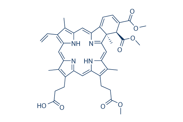
Chemical Structure
Molecular Weight: 718.79
Quality Control
Cell Culture, Treatment & Working Concentration
| Cell Lines | Assay Type | Concentration | Incubation Time | Formulation | Activity Description | PMID |
|---|---|---|---|---|---|---|
| HL-60 | Function assay | ~100 ng/mL | DMSO | increases DNA fragmentation levels | 10607710 | |
| HL-60 | cytotoxicity assay | ~100 ng/mL | DMSO | inhibits cell viability | 10607710 | |
| Jurkat | Apoptosis assay | ~280 nM | DMSO | induces a Bcl-2-dependent apoptosis | 11245415 | |
| RIF-1 | Function assay | 1 μg/ml | DMSO | decreases oxygen consumption | 12615718 | |
| RIF-1 | cytotoxicity assay | 1 μg/ml | DMSO | decrease to 20 ± 5% cell survival | 12615718 | |
| SVEC4-10 | Function assay | 200 ng/ml | DMSO | induces microtubule depolymerization | 16467106 | |
| SVEC4-10 | Function assay | 200 ng/ml | DMSO | induces stress actin fiber formation | 16467106 | |
| ARPE-19 | cytotoxicity assay | ~0.1 μg/ml | DMSO | shows a dose-dependent toxicity | 16987905 | |
| ARPE-19 | Function assay | 0.01 μg/ml | DMSO | increases VEGF and reduces PEDF expression | 16987905 | |
| Y-79 | Growth inhibitory assay | ~1 μg/ml | DMSO | decreases retinoblastoma cell proliferation | 18579764 | |
| WERI-Rb1 | Growth inhibitory assay | ~1 μg/ml | DMSO | decreases retinoblastoma cell proliferation | 18579764 | |
| RB247C3 | Growth inhibitory assay | ~1 μg/ml | DMSO | decreases retinoblastoma cell proliferation | 18579764 | |
| RB355 | Growth inhibitory assay | ~1 μg/ml | DMSO | decreases retinoblastoma cell proliferation | 18579764 | |
| RB383 | Growth inhibitory assay | ~1 μg/ml | DMSO | decreases retinoblastoma cell proliferation | 18579764 | |
| hFibro | cytotoxicity assay | 0.5 µg/ml | DMSO | decreases viability by 86,5% | 23441114 | |
| pTMC | cytotoxicity assay | 0.5 µg/ml | DMSO | decreases viability by 92.9% | 23441114 | |
| hTMC | cytotoxicity assay | 0.5 µg/ml | DMSO | decreases viability by 88.9% | 23441114 | |
| ARPE-19 | cytotoxicity assay | 0.5 µg/ml | DMSO | decreases viability by 55.5% | 23441114 | |
| Panc-1 | Growth inhibitory assay | 10 μM | DMSO | inhibits cell proliferation | 24069069 | |
| MIA PaCa-2 | Growth inhibitory assay | 10 μM | DMSO | inhibits cell proliferation | 24069069 | |
| BxPC-3 | Growth inhibitory assay | 10 μM | DMSO | inhibits cell proliferation completely | 24069069 | |
| SU86.86 | Growth inhibitory assay | 10 μM | DMSO | inhibits cell proliferation completely | 24069069 | |
| MCF-7 | Autophagy assay | 10 μM | DMSO | inhibits gemcitabine-induced autophagy | 24069069 | |
| WERI | Growth inhibitory assay | ~10 μg/ml | DMSO | inhibits growth of retinoblastoma cells | 24837142 | |
| WERI | Function assay | ~10 μg/ml | DMSO | blocks cell cycle progression | 24837142 | |
| Y-79 | Function assay | ~10 μg/ml | DMSO | blocks cell cycle progression | 24837142 | |
| Y-79 | Function assay | ~10 μg/ml | DMSO | affects YAP-TEAD proto-oncogene pathway | 24837142 | |
| Y-79 | Function assay | ~10 μg/ml | DMSO | down-regulates pluripotency marker OCT-4 | 24837142 | |
| Phototoxicity assay | B16F10 | 24 hrs | IC50 = 1.07 μM | 27136389 | ||
| Phototoxicity assay | B16F10 | 24 hrs | IC50 = 1.2 μM | 27136389 | ||
| Phototoxicity assay | A375 | 24 hrs | IC50 = 2.06 μM | 27136389 | ||
| Dark toxicity assay | B16F10 | 48 hrs | IC50 = 24.92 μM | 27136389 | ||
| Dark toxicity assay | B16F10 | 48 hrs | IC50 = 25.03 μM | 27136389 | ||
| Dark toxicity assay | A375 | 48 hrs | IC50 = 36.33 μM | 27136389 | ||
| qHTS assay | TC32 | qHTS of pediatric cancer cell lines to identify multiple opportunities for drug repurposing: Primary screen for TC32 cells | 29435139 | |||
| qHTS assay | U-2 OS | qHTS of pediatric cancer cell lines to identify multiple opportunities for drug repurposing: Primary screen for U-2 OS cells | 29435139 | |||
| qHTS assay | A673 | qHTS of pediatric cancer cell lines to identify multiple opportunities for drug repurposing: Primary screen for A673 cells | 29435139 | |||
| qHTS assay | DAOY | qHTS of pediatric cancer cell lines to identify multiple opportunities for drug repurposing: Primary screen for DAOY cells | 29435139 | |||
| qHTS assay | Saos-2 | qHTS of pediatric cancer cell lines to identify multiple opportunities for drug repurposing: Primary screen for Saos-2 cells | 29435139 | |||
| qHTS assay | BT-37 | qHTS of pediatric cancer cell lines to identify multiple opportunities for drug repurposing: Primary screen for BT-37 cells | 29435139 | |||
| qHTS assay | RD | qHTS of pediatric cancer cell lines to identify multiple opportunities for drug repurposing: Primary screen for RD cells | 29435139 | |||
| qHTS assay | SK-N-SH | qHTS of pediatric cancer cell lines to identify multiple opportunities for drug repurposing: Primary screen for SK-N-SH cells | 29435139 | |||
| qHTS assay | BT-12 | qHTS of pediatric cancer cell lines to identify multiple opportunities for drug repurposing: Primary screen for BT-12 cells | 29435139 | |||
| qHTS assay | MG 63 (6-TG R) | qHTS of pediatric cancer cell lines to identify multiple opportunities for drug repurposing: Primary screen for MG 63 (6-TG R) cells | 29435139 | |||
| qHTS assay | OHS-50 | qHTS of pediatric cancer cell lines to identify multiple opportunities for drug repurposing: Primary screen for OHS-50 cells | 29435139 | |||
| qHTS assay | Rh41 | qHTS of pediatric cancer cell lines to identify multiple opportunities for drug repurposing: Primary screen for Rh41 cells | 29435139 | |||
| qHTS assay | SJ-GBM2 | qHTS of pediatric cancer cell lines to identify multiple opportunities for drug repurposing: Primary screen for SJ-GBM2 cells | 29435139 | |||
| qHTS assay | SK-N-MC | qHTS of pediatric cancer cell lines to identify multiple opportunities for drug repurposing: Primary screen for SK-N-MC cells | 29435139 | |||
| qHTS assay | LAN-5 | qHTS of pediatric cancer cell lines to identify multiple opportunities for drug repurposing: Primary screen for LAN-5 cells | 29435139 | |||
| Antitumor assay | B16F10 | 2 mg/kg | 2 hrs | Antitumor activity against B16F10 cells implanted in C57BL/6 mouse assessed as tumor growth inhibition at 2 mg/kg, iv administered for 2 hrs followed by irradiation with laser at 150 J/cm'2 for 10 mins | 27136389 | |
| Click to View More Cell Line Experimental Data | ||||||
Solubility
|
In vitro |
DMSO
: 100 mg/mL
(139.12 mM)
Water : Insoluble Ethanol : Insoluble |
Molarity Calculator
|
In vivo |
|||||
In vivo Formulation Calculator (Clear solution)
Step 1: Enter information below (Recommended: An additional animal making an allowance for loss during the experiment)
Step 2: Enter the in vivo formulation (This is only the calculator, not formulation. Please contact us first if there is no in vivo formulation at the solubility Section.)
Calculation results:
Working concentration: mg/ml;
Method for preparing DMSO master liquid: mg drug pre-dissolved in μL DMSO ( Master liquid concentration mg/mL, Please contact us first if the concentration exceeds the DMSO solubility of the batch of drug. )
Method for preparing in vivo formulation: Take μL DMSO master liquid, next addμL PEG300, mix and clarify, next addμL Tween 80, mix and clarify, next add μL ddH2O, mix and clarify.
Method for preparing in vivo formulation: Take μL DMSO master liquid, next add μL Corn oil, mix and clarify.
Note: 1. Please make sure the liquid is clear before adding the next solvent.
2. Be sure to add the solvent(s) in order. You must ensure that the solution obtained, in the previous addition, is a clear solution before proceeding to add the next solvent. Physical methods such
as vortex, ultrasound or hot water bath can be used to aid dissolving.
Chemical Information, Storage & Stability
| Molecular Weight | 718.79 | Formula | C41H42N4O8 |
Storage (From the date of receipt) | 3 years-20°C (in the dark)powder |
|---|---|---|---|---|---|
| CAS No. | 129497-78-5 | Download SDF | Storage of Stock Solutions |
|
|
| Synonyms | CL 318952 | Smiles | COC(=O)CCC1=C(C)C2=CC3=NC(=CC4=C(C)C(=C([NH]4)C=C5N=C(C=C1[NH]2)C(=C5C)CCC(O)=O)C=C)C6=CC=C(C(C(=O)OC)C36C)C(=O)OC | ||
Mechanism of Action
| Targets/IC50/Ki |
VDA
(Endothelial cells) YAP/TEAD interaction
|
|---|---|
| In vitro |
Verteporfin is about four times more efficient in absorbing light at wavelengths that penetrate tissues best (i.e., around 700 nm) and thus provides a much higher cytotoxic effect than hematoporphyrin (10 times more in human adherent cell lines). This compound is lipophilic and is more readily taken up by malignant or activated cells, compared with normal or resting cells. It binds with LDL to form a complex, which is then taken up into proliferating cells (e.g., neovascular endothelial cells) probably via LDL receptors and endocytosis. This therapy achieves complete angiographic occlusion of the neovascular compartment by thrombosis of vascular channels, following selective endothelial damage. It selectively induces reproducible and isolated choriocapillary occlusion without alteration of overlying photoreceptors or ganglion cells, as shown by light and electron microscopy. This chemical conbined with light rapidly exhibits apoptotic changes reflected by caspase-3 and caspase-9 activation and PARP cleavage in HL-60 cells, changes that are blocked by the general caspase inhibitor ZVAD.fmk. |
| In vivo |
Verteporfin can be used for angiographic visualization of choroidal vessels and CNV, which demonstrates that the photosensitizer accumulates rapidly in experimental CNV in monkeys. This compound accumulates rapidly in the established vasculature of the choroid, RPE, and photoreceptors of rabbit eyes. It reaches maximal tissue levels within 3 hours of intravenous injection, followed by a rapid decline within 24 hours in mice. This chemical is metabolized to a less active form in vivo and is cleared very rapidly, predominantly in the feces and a very small proportion excreted in urine. The therapy effectively and selectively prevents fluorescein dye leakage from experimentally induced CNV in monkeys. |
References |
|
Applications
| Methods | Biomarkers | Images | PMID |
|---|---|---|---|
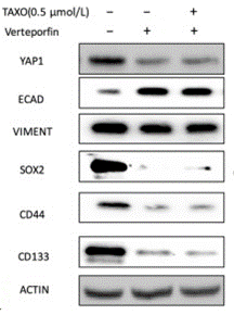
| Western blot | ECAD / Vimentin / Sox2 / CD44 / CD133 c-Myc / Bcl-2 p-S6(S240/244) / p-4EBP1(S65) beta-catenin |
|
30467925 |
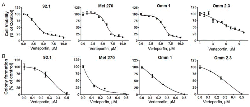
| Growth inhibition assay | Cell viability |
|
28042502 |
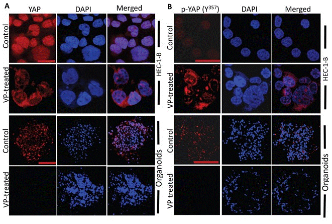
| Immunofluorescence | p-YAP(Y357) Calreticulin YAP1 |
|
28404908 |
Clinical Trial Information
(data from https://clinicaltrials.gov, updated on 2024-05-22)
| NCT Number | Recruitment | Conditions | Sponsor/Collaborators | Start Date | Phases |
|---|---|---|---|---|---|
| NCT04590664 | Recruiting | Glioblastoma|Recurrent Glioblastoma |
Emory University|National Cancer Institute (NCI) |
January 15 2021 | Phase 1|Phase 2 |
| NCT03797547 | Unknown status | Myopic Choroidal Neovascularisation |
Poitiers University Hospital |
June 22 2018 | -- |
| NCT01846273 | Completed | Age-related Macular Degeneration|Polypoidal Choroidal Vasculopathy |
Novartis Pharmaceuticals|Novartis |
August 7 2013 | Phase 4 |
| NCT00423189 | Terminated | Age-Related Macular Degeneration |
David M. Brown M.D.|Novartis Pharmaceuticals|Greater Houston Retina Research |
January 2007 | Phase 4 |
| NCT00403442 | Terminated | Macular Degeneration |
Vitreous -Retina- Macula Consultants of New York|QLT Inc. |
September 2006 | Phase 1 |
Tech Support
Tel: +1-832-582-8158 Ext:3
If you have any other enquiries, please leave a message.
Signaling Pathway Map
Products are for research use only. Not for human use. We do not sell to patients.
©Copyright 2013 Selleck Chemicals. All Rights Reserved.