-
 Australia
-
 Austria
-
 Belgium
-
 Brazil
-
 Canada
-
 China
China
-
 Czech Republic
-
 Denmark
-
 Finland
-
 France
-
 Germany
-
 Greece
-
 Hong Kong
-
 Hungary
-
 Iceland
-
 India
India
-
 Ireland
-
 Israel
-
 Italy
Italy
-
 Japan
Japan
-
 Korea
Korea
-
 Luxembourg
-
 Malaysia
-
 Netherlands
-
 New Zealand
-
 Norway
-
 Poland
-
 Qatar
Qatar
-
 Romania
-
 Saudi Arabia
-
 Singapore
-
 Spain
Spain
-
 Sweden
-
 Switzerland
-
 Taiwan
-
 Turkey
-
 United Kingdom
-
 United States
research use only
Zoledronic acid (Zoledronate) Bone Resorption Inhibitor
Cat.No.S1314
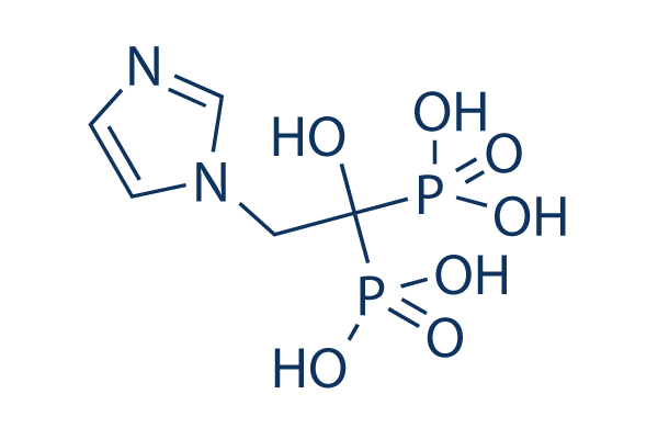
Chemical Structure
Molecular Weight: 272.09
Quality Control
| Related Targets | Bcl-2 Caspase PD-1/PD-L1 Ferroptosis p53 Synthetic Lethality STAT TNF-alpha Ras KRas |
|---|---|
| Other Apoptosis related Inhibitors | Importazole Pitstop 2 Plumbagin Genipin 6-Gingerol Colcemid Sodium propionate Crocin CWI1-2 hydrochloride Necrostatin 2 |
Cell Culture, Treatment & Working Concentration
| Cell Lines | Assay Type | Concentration | Incubation Time | Formulation | Activity Description | PMID |
|---|---|---|---|---|---|---|
| human NCI-H460 cell | Proliferation assay | Antiproliferative activity against human NCI-H460 cell line by MTT assay, IC50=11.7 μM | 16970405 | |||
| human SF-268 cell | Proliferation assay | Antiproliferative activity against human SF-268 cell line by MTT assay, IC50=14.3 μM | 16970405 | |||
| Gamma delta T cells | Function assay | Effective concentration against human Gamma delta T cells, EC50=5.4μM | 15828834 | |||
| HFF cells | Function assay | In vitro inhibitory concentration against the growth of Toxoplasma gondii in human foreskin fibroblast monolayer cells (HFF cells), IC50=0.79μM | 15857119 | |||
| HFF cells | Function assay | Inhibitory concentration against the growth of Toxoplasma gondii overexpressing FPPS enzyme in human foreskin fibroblast monolayer cells; (control = 0.60 uM, experiment 1), IC50=7.8μM | 15857119 | |||
| HFF cells | Function assay | Inhibitory concentration against the growth of Toxoplasma gondii overexpressing FPPS enzyme in human foreskin fibroblast monolayer cells; (control = 0.79 uM, experiment 3), IC50=7.8μM | 15857119 | |||
| HFF cells | Function assay | Inhibitory concentration against the growth of Toxoplasma gondii overexpressing FPPS enzyme in human foreskin fibroblast monolayer cells; (control = 1.1 uM, experiment 2), IC50=8.3μM | 15857119 | |||
| BT-549 | Antitumor assay | Antitumor activity against human BT-549 cells xenografted SCID mouse co-transfected with human gamma delta T lymphocytes assessed as survival time prolongation at 2 ug, ip coadministered with human recombinant IL2 | 18937434 | |||
| BT-549 | Antitumor assay | Antitumor activity against human BT-549 cells xenografted SCID mouse co-transfected with human gamma delta T lymphocytes assessed as survival time prolongation at 5 ug, ip coadministered with human recombinant IL2 | 18937434 | |||
| BL21(DE3) | Function assay | 30 mins | Inhibition of His6-tagged human truncated FPPS (6-353) expressed in Escherichia coli BL21(DE3) cells using geranyl diphosphate and isopentenyl diphosphate as substrate preincubated with enzyme for 30 mins by spectrophotometric analysis, IC50=0.1μM | 23610597 | ||
| BL2-codon plus (DE3) RIL | Function assay | 30 mins | Inhibition of N-terminal His6-tagged Plasmodium vivax GGPPS expressed in Escherichia coli BL2-codon plus (DE3) RIL cells using geranyl diphosphate and isopentenyl diphosphate as substrate preincubated with enzyme for 30 mins by spectrophotometric analysis, IC50=0.13μM | 23610597 | ||
| RPMI8226 | Cytotoxicity assay | 72 hrs | Cytotoxicity against human RPMI8226 cells after 72 hrs by MTT assay, EC50=11μM | 23998921 | ||
| J774 | Cytotoxicity assay | Cytotoxicity against mouse J774 cells assessed as reduction in cell viability, IC50=7.8μM | 24813742 | |||
| MCF7 | Antiproliferative assay | 72 hrs | Antiproliferative activity against human MCF7 cells after 72 hrs by MTT assay, IC50=23μM | 24928399 | ||
| Vgamma9/Vdelta2 T-cells | Function assay | 18 hrs | Binding affinity to butyrophilin 3A1 in human Vgamma9/Vdelta2 T-cells assessed as activation of Vgamma9/Vdelta2 T-cells by upregulation of CD69 and CD25 after 18 hrs, EC50=0.4866μM | 29457898 | ||
| RPMI8226 | Antiproliferative assay | 72 hrs | Antiproliferative activity against human RPMI8226 cells after 72 hrs by MTT assay, EC50=11μM | 30016091 | ||
| RPMI8226 | Function assay | 0.5 uM | Inhibition of GGPPS in human RPMI8226 cells assessed as reduction in Rap1A prenylation at 0.5 uM by Western blot analysis | 30016091 | ||
| J774A.1 | Antiproliferative assay | 100 uM | 72 hrs | Antiproliferative activity against mouse J774A.1 cells assessed as reduction in cell viability at 100 uM after 72 hrs by WST8 assay | 30216851 | |
| RAW264.7 | Antiproliferative assay | 100 uM | 72 hrs | Antiproliferative activity against mouse RAW264.7 cells assessed as reduction in cell viability at 100 uM after 72 hrs by WST8 assay | 30216851 | |
| MG63 | Antiproliferative assay | 100 uM | 72 hrs | Antiproliferative activity against human MG63 cells assessed as reduction in cell viability at 100 uM after 72 hrs by WST8 assay | 30216851 | |
| PC3 | Antiproliferative assay | 100 uM | 72 hrs | Antiproliferative activity against human PC3 cells assessed as reduction in cell viability at 100 uM after 72 hrs by WST8 assay | 30216851 | |
| RAW264.7 | Antiproliferative assay | 100 uM | 72 hrs | Antiproliferative activity against RANKL-differentiated mouse RAW264.7 cells assessed as reduction in cell viability at 100 uM after 72 hrs by CCK8 assay | 30216851 | |
| RAW264.7 | Function assay | 72 hrs | Inhibition of RANKL-induced osteoclastogenesis in mouse RAW264.7 cells after 72 hrs by TRAP staining based microscopic analysis | 30216851 | ||
| MC3T3-E1 | Function assay | 30 to 50 nM | 10 to 15 days | Induction of mineralization in mouse MC3T3-E1 cells at 30 to 50 nM after 10 to 15 days by alizarin red dye based assay | 30216851 | |
| C57BL mouse bone marrow cells | Function assay | 50 nM | 10 to 15 days | Induction of mineralization in C57BL mouse bone marrow cells at 50 nM supplemented with fresh medium containing compound every 3 days for 10 to 15 days by alizarin red dye based assay | 30216851 | |
| C57BL mouse bone marrow cells/human PC3 cells | Function assay | 50 to 100 nM | 10 to 15 days | Induction of mineralization in C57BL mouse bone marrow cells co-cultured with human PC3 cells at 50 to 100 nM supplemented with fresh medium containing compound every 3 days for 10 to 15 days by alizarin red dye based assay | 30216851 | |
| K562 | Function assay | 240 mins | Activation of butyrophilin 3A1 in human K562 cells assessed as interferon-gamma production pretreated for 240 mins followed by HMBPP-treated Vgamma9Vdelta2 T cells addition and measured after 20 hrs by ELISA, EC50=23μM | 31531198 | ||
| MIAPaCa2 | Cytotoxicity assay | 72 hrs | Cytotoxicity against human MIAPaCa2 cells assessed as decrease in cell growth measured after 72 hrs by MTT assay, EC50=13.4μM | 31725297 | ||
| PANC1 | Cytotoxicity assay | 72 hrs | Cytotoxicity against human PANC1 cells assessed as decrease in cell growth measured after 72 hrs by MTT assay, EC50=16.1μM | 31725297 | ||
| Click to View More Cell Line Experimental Data | ||||||
Solubility
|
In vitro |
0.1M NAOH : 25 mg/mL (Ultrasonic and heating for 5 minutes.) Water : 0.5 mg/mL (Warmed with 50℃ water bath; Ultrasonicated)
DMSO
: Insoluble
|
Molarity Calculator
|
In vivo |
|||||
In vivo Formulation Calculator (Clear solution)
Step 1: Enter information below (Recommended: An additional animal making an allowance for loss during the experiment)
Step 2: Enter the in vivo formulation (This is only the calculator, not formulation. Please contact us first if there is no in vivo formulation at the solubility Section.)
Calculation results:
Working concentration: mg/ml;
Method for preparing DMSO master liquid: mg drug pre-dissolved in μL DMSO ( Master liquid concentration mg/mL, Please contact us first if the concentration exceeds the DMSO solubility of the batch of drug. )
Method for preparing in vivo formulation: Take μL DMSO master liquid, next addμL PEG300, mix and clarify, next addμL Tween 80, mix and clarify, next add μL ddH2O, mix and clarify.
Method for preparing in vivo formulation: Take μL DMSO master liquid, next add μL Corn oil, mix and clarify.
Note: 1. Please make sure the liquid is clear before adding the next solvent.
2. Be sure to add the solvent(s) in order. You must ensure that the solution obtained, in the previous addition, is a clear solution before proceeding to add the next solvent. Physical methods such
as vortex, ultrasound or hot water bath can be used to aid dissolving.
Chemical Information, Storage & Stability
| Molecular Weight | 272.09 | Formula | C5H10N2O7P2 |
Storage (From the date of receipt) | |
|---|---|---|---|---|---|
| CAS No. | 118072-93-8 | Download SDF | Storage of Stock Solutions |
|
|
| Synonyms | ZA, CGP-4244, GP42446A, ZOL 446 | Smiles | C1=CN(C=N1)CC(O)(P(=O)(O)O)P(=O)(O)O | ||
Mechanism of Action
| Targets/IC50/Ki |
Rho
(Cell-free assay) Ras
|
|---|---|
| In vitro |
Zoledronic acid (Zoledronate) (10 µM and 100 µM) causes a significant reduction in the proportions of MCF-7 cells (49.54% and 23.55% of control, respectively) (P < 0.05). It has little effect on MDA-MB-231 cells at concentrations of 0.1–10 µM, whereas the 100 µM concentration results in a significant reduction in cell number. This compound (100 µM) results in a 63.5% reduction in MCF-7 cell number at 72 hours and an 87.1% reduction at 96 hours. It (10 µM) results in a greater than 4-fold increase in MCF-7 cell apoptosis whereas the 100 µM concentration induces a 6-fold increase in the proportion of apoptotic cells. When combined with paclitaxel (2 µM), it (10 µM) results in a 5-fold increase in apoptosis (774.8% of control) compared to zoledronic acid alone (155.71%) and a 4-fold increase compared to paclitaxel alone (189.68%). Its induced apoptosis of MCF-7 breast cancer cells can be inhibited by addition of intermediates of the mevalonate pathway, which is consistent with observations in osteoclasts, macrophages and myeloma cells. It enhances OPG gene expression and protein secretion by human osteoblasts (hOB) in a dose-dependent fashion with a maximum effect at 10 nM after 72 hours, consistent with the higher biological potency of Zoledronic acid. This compound prevents the inhibitory effects of the glucocorticoid dexamethasone on OPG mRNA and protein production in human osteoblasts. It induces type I collagen secretion and alkaline phosphatase activity by 2- and 4-fold, respectively, in human osteoblasts. |
| In vivo |
Zoledronic acid (Zoledronate) (120 ug/kg, s.c.) prevents the formation of lesions, prevents cancellous bone loss and loss of bone mineral density, and reduces osteoclast perimeter in 5T2MM-bearing mice. It (120 mg/kg, s.c.) also decreases paraprotein concentration, decreases tumor burden, and reduces angiogenesis in 5T2MM-bearing mice. |
References |
|
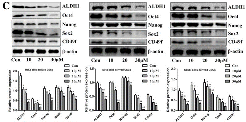
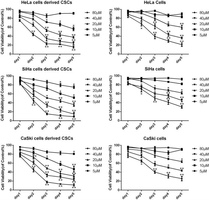
Applications
| Methods | Biomarkers | Images | PMID |
|---|---|---|---|
| Western blot | ALDH1 / Oct4 / Nanog / Sox2 / CD49f N-cadherin / E-cadherin / Vimentin |
|
30791957 |
| Growth inhibition assay | Cell viability |
|
30791957 |
Clinical Trial Information
(data from https://clinicaltrials.gov, updated on 2024-05-22)
| NCT Number | Recruitment | Conditions | Sponsor/Collaborators | Start Date | Phases |
|---|---|---|---|---|---|
| NCT06217718 | Not yet recruiting | Copd|Empowerment|Empowerment Patient|Self Efficacy |
Zahide Aksoy|The Scientific and Technological Research Council of Turkey|Marmara University |
February 15 2024 | Not Applicable |
| NCT05743179 | Recruiting | Hip Fractures|Pneumonia |
The University of Hong Kong|Queen Mary Hospital Hong Kong|Caritas Medical Centre Hong Kong|Prince of Wales Hospital Shatin Hong Kong|United Christian Hospital |
December 5 2022 | Phase 4 |
| NCT02864784 | Withdrawn | Castrate Resistant Prostate Cancer With Bone Metastasis |
Amorphical Ltd. |
June 2022 | Phase 1 |
| NCT04957641 | Completed | Hereditary Angioedema |
Takeda |
April 21 2022 | -- |
| NCT05213286 | Unknown status | Autism Spectrum Disorder|Schizotypal Disorder |
Glostrup University Hospital Copenhagen |
February 1 2022 | Not Applicable |
Tech Support
Tel: +1-832-582-8158 Ext:3
If you have any other enquiries, please leave a message.
Frequently Asked Questions
Question 1:
How can I reconstitute it for in vivo studies?
Answer:
Please dissolve it directly to 30% PEG400+0.5% Tween80+5% Propylene glycol.
Products are for research use only. Not for human use. We do not sell to patients.
©Copyright 2013 Selleck Chemicals. All Rights Reserved.