-
 Australia
-
 Austria
-
 Belgium
-
 Brazil
-
 Canada
-
 China
China
-
 Czech Republic
-
 Denmark
-
 Finland
-
 France
-
 Germany
-
 Greece
-
 Hong Kong
-
 Hungary
-
 Iceland
-
 India
India
-
 Ireland
-
 Israel
-
 Italy
Italy
-
 Japan
Japan
-
 Korea
Korea
-
 Luxembourg
-
 Malaysia
-
 Netherlands
-
 New Zealand
-
 Norway
-
 Poland
-
 Qatar
Qatar
-
 Romania
-
 Saudi Arabia
-
 Singapore
-
 Spain
Spain
-
 Sweden
-
 Switzerland
-
 Taiwan
-
 Turkey
-
 United Kingdom
-
 United States
research use only
Necrostatin-1 (Nec-1) RIPK1 Inhibitor
Cat.No.S8037
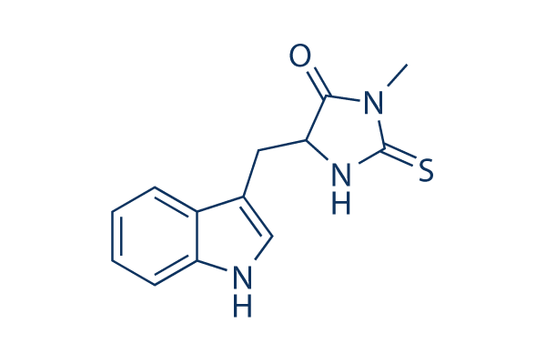
Chemical Structure
Molecular Weight: 259.33
Quality Control
| Related Targets | Bcl-2 Caspase PD-1/PD-L1 Ferroptosis p53 Apoptosis related Synthetic Lethality STAT TNF-alpha Ras |
|---|---|
| Other RIP kinase Inhibitors | Necrostatin 2 racemate (Nec-1s) GSK872 Mito-TEMPO GSK'963 RIPA-56 GSK2982772 GSK583 Resibufogenin ICCB-19 hydrochloride HS-1371 |
Cell Culture, Treatment & Working Concentration
| Cell Lines | Assay Type | Concentration | Incubation Time | Formulation | Activity Description | PMID |
|---|---|---|---|---|---|---|
| HT-22 | Cytotoxicity Assay | 10 μM | 12 h | protects against cell death induced by 5 mmol/L glutamate | 17760869 | |
| Jurkat | Function Assay | 200 μm | 30 min | reduces Naegleria fowleri-induced reactive oxygen species (ROS) generation | 21535020 | |
| Jurkat | Cytotoxicity Assay | 50/ 100/200 μm | 1/3 h | reduces Naegleria fowleri-induced cytotoxicity | 21535020 | |
| SW13 | Cell Viability Assay | 100 μM | 24 h | DMSO | increases cellular survival | 22136818 |
| 8505c | Cell Viability Assay | 100 μM | 24 h | DMSO | increases cellular survival | 22136818 |
| TPC-1 | Cell Viability Assay | 100 μM | 24 h | DMSO | increases cellular survival | 22136818 |
| L929sA | Apoptosis Assay | 10 μM | 1 h | abrogates the interaction of caspase-8 with FADD | 22362767 | |
| L929sA | Apoptosis Assay | 10 μM | 1 h | rescues cells expressing RIPK1ΔID from TNF-induced apoptosis | 22362767 | |
| L929sA | Apoptosis Assay | 10 μM | 1 h | inhibits the apoptotic response to TNF | 22362767 | |
| K562/Adr | Function Assay | 60 μM | 12 h | increases the activity of caspases, caspase 8 and 9 | 22837689 | |
| K562 | Function Assay | 60 μM | 12 h | increases the activity of caspases, caspase 8 and 9 | 22837689 | |
| HL60/Adr | Function Assay | 60 μM | 12 h | increases the activity of caspases, caspase 8 and 9 | 22837689 | |
| HL60 | Function Assay | 60 μM | 12 h | increases the activity of caspases, caspase 8 and 9 | 22837689 | |
| K562/Adr | Function Assay | 60 μM | 12 h | augments the caspase-3 activity | 22837689 | |
| K562 | Function Assay | 60 μM | 12 h | augments the caspase-3 activity | 22837689 | |
| HL60/Adr | Function Assay | 60 μM | 12 h | augments the caspase-3 activity | 22837689 | |
| HL60 | Function Assay | 60 μM | 12 h | augments the caspase-3 activity | 22837689 | |
| K562/Adr | Apoptosis Assay | 60 μM | 12 h | enhances shikonin-induced apoptosis | 22837689 | |
| K562 | Apoptosis Assay | 60 μM | 12 h | enhances shikonin-induced apoptosis | 22837689 | |
| HL60/Adr | Apoptosis Assay | 60 μM | 12 h | enhances shikonin-induced apoptosis | 22837689 | |
| HL60 | Apoptosis Assay | 60 μM | 12 h | enhances shikonin-induced apoptosis | 22837689 | |
| SH-EP | Apoptosis Assay | 10 μM | 72 h | inhibits IAP inhibitor- and Lexatumumab-induced apoptosis | 22890322 | |
| NIH3T3 | Function Assay | 10/50 μM | 1/3 h | ameliorates TNFα-driven complex formation | 23261677 | |
| HT-22 | Function Assay | 25 μM | 0–30 min | DMSO | inhibits ERK Activation induced by glutamate | 23307752 |
| HT-22 | Cell Viability Assay | 10 μM | 12 h | DMSO | protects against glutamate-induced cell death | 23307752 |
| KMS-12-BM | Cytotoxicity Assay | 90 µM | 1 h | blocks BAY 11-7082 induced rapid cell swelling | 23527154 | |
| MM.1S | Cytotoxicity Assay | 90 µM | 1 h | blocks BAY 11-7082 induced rapid cell swelling | 23527154 | |
| ΔN-Karpas 299 | Cytotoxicity Assay | 20 μM | 16 h | inhibits CD30-induced cell death | 23545938 | |
| MEFs | Function Assay | 20 μM | 1/2/4 h | suppresses TNFα-induced RIPK1 phosphorylation | 23727581 | |
| MEFs | Cytotoxicity Assay | 2/6/20 μM | 18 h | inhibits TNFα-induced cell death in RelA KO MEFs | 23727581 | |
| RMS13 | Cell Viability Assay | 40 μg/ml | 24 h | rescues GX15-070-induced loss of cell viability | 23744296 | |
| TE671 | Cell Viability Assay | 40 μg/ml | 24 h | rescues GX15-070-induced loss of cell viability | 23744296 | |
| U87 | Function Assay | 1 mmol/L | 1.5-3 h | suppresses the expression of RIP-1 caused by shikonin | 23840441 | |
| C6 | Function Assay | 1 mmol/L | 1.5-3 h | suppresses the expression of RIP-1 caused by shikonin | 23840441 | |
| U87 | Cytotoxicity Assay | 1 mmol/L | 3 h | blocks shikonin induced necrosis | 23840441 | |
| C6 | Cytotoxicity Assay | 1 mmol/L | 3 h | blocks shikonin induced necrosis | 23840441 | |
| U87 | Cell Viability Assay | 1 mmol/L | 3 h | attenuates Shikonin induced glioma cell death | 23840441 | |
| C6 | Cell Viability Assay | 1 mmol/L | 3 h | attenuates Shikonin induced glioma cell death | 23840441 | |
| L929 | Function Assay | 5 μg/ml | 24 h | blocks zVAD induced necroptosis and autophagy | 23941769 | |
| L929 | Function Assay | 2 μg/ml | 24 h | promots caspase-6 (p20) activity and procaspase-6 cleavage | 23941769 | |
| L929 | Growth Inhibition Assay | 2/5 μg/ml | 24 h | reverses the cell growth inhibition and cell death induced by TNFα alone as well as TNFα + zVAD | 23941769 | |
| L929 | Function Assay | 2/5 μg/ml | 24 h | reversed the autophagy induced by TNFα alone as well as TNFα + zVAD | 23941769 | |
| NRK-52E | Cell Viability Assay | 20 μM | 24 h | inhibits increased Drp1 protein expression after TNF-α Stimulation and ATP Depletion | 24351845 | |
| NRK-52E | Cell Viability Assay | 20 μM | 24 h | increases cell viability after TNF-α Stimulation and ATP Depletion | 24351845 | |
| NRK-52E | Cell Viability Assay | 20 μM | 24 h | protects cells from cell death caused by ischemia injury | 24351845 | |
| AGS | Cell Viability Assay | 60 μm | 1 h | prevents shikonin-induced cell death | 24463199 | |
| L-540 | Cell Viability Assay | 60 μm | 1 h | reduces the Givinostat/Sorafenib-induced cell death | 24561519 | |
| L-540 | Function Assay | 60 μm | 1 h | prevents the mitochondrial membrane depolarization | 24561519 | |
| L-540 | Function Assay | 60 μm | 1 h | prevents the generation of ROS | 24561519 | |
| SK-Hep1 | Function Assay | 60 μM | 18 h | blocks β-lapachone-mediated PAR accumulation and AIF translocation to the cytosol | 24832602 | |
| SK-Hep1 | Function Assay | 60 μM | 18 h | inhibits β-Lapachone-induced leakage of HMGB-1 | 24832602 | |
| SK-Hep1 | Function Assay | 60 μM | 18 h | blocks β-lapachone-induced morphological change, cell death and PI uptake | 24832602 | |
| OHC | Function Assay | 300 μM | DMSO | increases the number of apoptotic OHCs without altering the levels of CC8 after noise exposure | 24874734 | |
| OHC | Function Assay | 300 μM | DMSO | diminishes noise-induced AMPK activation | 24874734 | |
| OHC | Function Assay | 300 μM | DMSO | results in a reduction of noise-induced RIP1 and RIP3 immunofluorescence | 24874734 | |
| OHC | Function Assay | 300 μM | DMSO | decreases noise-induced swollen nuclei | 24874734 | |
| OHC | Function Assay | 300 μM | DMSO | increases noise-induced condensed nuclei | 24874734 | |
| Huh7 | Cell Viability Assay | 50 µM | 24/48 h | DMSO | prevents cell death of rAdHCV co-infected Huh7 cells | 24973240 |
| L929 | Cell Viability Assay | 30 μM | 1 h | inhibits TNF-α-induced cleavage of Topo I | 25095742 | |
| L929 | Cell Viability Assay | 30 μM | 1 h | inhibits TNF-α-induced loss of cell viability | 25095742 | |
| L929-A | Function Assay | 50 μM | 12 h | inhibits the TNFα-induced loss of mitochondrial membrane permeability | 25398540 | |
| L929 | Function Assay | 50 μM | 12 h | inhibits TNFα-induced Bid cleavage | 25398540 | |
| L929-N | Function Assay | 50 μM | 12 h | blocks the cleavage of Caspase-3 and PARP | 25398540 | |
| L929-A | Function Assay | 50 μM | 12 h | blocks the cleavage of Caspase-3 and PARP | 25398540 | |
| L929-N | Cell Viability Assay | 50 μM | 24 h | blocks TNFα-induced cell death | 25398540 | |
| L929-A | Cell Viability Assay | 50 μM | 24 h | blocks TNFα-induced cell death | 25398540 | |
| KMS-12-PE | Cell Viability Assay | 60 μM | 5 h | inhibits SHK-induced cell death | 25530098 | |
| SGC-7901 | Cell Viability Assay | 30 μM | 1 h | suppresses oxaliplatin-mediated cell death | 25767076 | |
| BxPC-3 | Function Assay | 20 μM | 24 h | decreases the early necrotic cells | 26000607 | |
| MiaPaCa-2 | Function Assay | 20 μM | 24 h | decreases the early necrotic cells | 26000607 | |
| NCI-H28 | Cell Viability Assay | 10 μM | 24 h | prevents DAPE-induced reduction of NCI-H28 cell viability | 26004138 | |
| BMDM | Function Assay | 10 μM | 30 min | protects cells from TAKI-induced LDH release | 26381601 | |
| MEFs | Cell Viability Assay | 10 μM | 48 h | DMSO | inhibits zVAD-promoted death of CNOT3-depleted MEFs | 26437789 |
| A549 | Cell Viability Assay | 50 μM | 24 h | inhibits MMS-induced cell death | 26472723 | |
| Jurkat T | Necroptosis assay | Inhibition of TNF-alpha-induced necroptosis in FADD-deficient human Jurkat T cells, EC50 = 0.05 μM. | 18467094 | |||
| Jurkat | Function assay | Inhibition of endogenous RIP1 autophosphorylation in human Jurkat cells, EC50 = 0.182 μM. | 18408713 | |||
| Sf9 | Function assay | 30 mins | Inhibition of recombinant human GST-fused RIPK1 (1 to 497 residues) expressed in baculovirus infected insect Sf9 cells in presence of 32P-gamma-ATP after 30 mins by autoradiogram-based Western blot method, IC50 = 0.182 μM. | 28280261 | ||
| Jurkat T | Necroptosis assay | Effective concentration required for inhibition of necroptosis in FADD deficient Jurkat T cells treated with TNF-alpha, EC50 = 0.49 μM. | 16153840 | |||
| Jurkat | Necroptosis assay | Inhibition of cellular necroptosis in TNFalpha treated FADD deficient human Jurkat cells, EC50 = 0.49 μM. | 18408713 | |||
| Jurkat T | Necroptosis assay | 30 uM | 24 hrs | Inhibition of necroptosis in TNF-alpha-induced human Jurkat T cells assessed as cell viability at 30 uM after 24 hrs | 18467094 | |
| L929 | Necroptosis assay | 30 uM | 24 hrs | Inhibition of necroptosis in zVAD-induced mouse L929 cells assessed as cell viability at 30 uM after 24 hrs | 18467094 | |
| L929 | Necroptosis assay | 30 uM | 24 hrs | Inhibition of necroptosis in TNF-alpha-induced mouse L929 cells assessed as cell viability at 30 uM after 24 hrs | 18467094 | |
| 3T3 | Cell death assay | 24 hrs | Inhibition of death receptor signaling mediated necrotic cell death in mouse 3T3 cells assessed as cell viability after 24 hrs by ATP based viability assay in presence of TNFalpha and zVAD.fmk | 16408008 | ||
| 3T3 | Cell death assay | 24 hrs | Inhibition of death receptor signaling mediated necrotic cell death in mouse 3T3 cells assessed as cell viability after 24 hrs by ATP based viability assay in presence of FasL and zVAD.fmk | 16408008 | ||
| MEF | Cell death assay | 16 hrs | Inhibition of death receptor signaling mediated necrotic cell death in SV40 transformed mouse MEF cells assessed as cell viability after 16 hrs by ATP based viability assay in presence of TNFalpha, CHX and zVAD.fmk | 16408008 | ||
| IEC18 | Cell death assay | Inhibition of death receptor signaling mediated necrotic cell death in rat IEC18 cells assessed as cell viability in presence of TNFalpha and zVAD.fmk | 16408008 | |||
| HL60 | Cell death assay | Inhibition of death receptor signaling mediated necrotic cell death in human HL60 cells assessed as cell viability in presence of TNFalpha and zVAD.fmk | 16408008 | |||
| L929 | Cell death assay | 24 hrs | Inhibition of death receptor signaling mediated necrotic cell death in mouse L929 cells assessed as cell viability after 24 hrs by ATP based viability assay in presence of TNFalpha | 16408008 | ||
| Jurkat | Necrosis assay | Inhibition of necrosis in human Jurkat cells assessed as nuclear condensation by bright field microscopy in presence of FasL, CHX and zVAD-fmk | 16408008 | |||
| Jurkat | Necrosis assay | Inhibition of necrosis in human Jurkat cells assessed as organelle swelling by bright field microscopy in presence of FasL, CHX and zVAD-fmk | 16408008 | |||
| Jurkat | Necrosis assay | Inhibition of necrosis in human Jurkat cells assessed as early loss of plasma membrane integrity by bright field microscopy in presence of FasL, CHX and zVAD-fmk | 16408008 | |||
| Jurkat | Necrosis assay | Inhibition of necrosis in human Jurkat cells assessed as appearance of translucent cytosol in presence of FasL, CHX and zVAD-fmk | 16408008 | |||
| Jurkat | Necrosis assay | Inhibition of necrosis in human Jurkat cells deficient in FADD assessed as inhibition of nuclear condensation by bright field microscopy in presence of TNFalpha | 16408008 | |||
| Jurkat | Necrosis assay | Inhibition of necrosis in human Jurkat cells deficient in FADD assessed as inhibition of organelle swelling by bright field microscopy in presence of TNFalpha | 16408008 | |||
| Jurkat | Necrosis assay | Inhibition of necrosis in human Jurkat cells deficient in FADD assessed as inhibition of early loss of plasma membrane integrity by bright field microscopy in presence of TNFalpha | 16408008 | |||
| Jurkat | Necrosis assay | Inhibition of necrosis in human Jurkat cells deficient in FADD assessed as inhibition of appearance of translucent cytosol in presence of TNFalpha | 16408008 | |||
| U937 | Cell death assay | 48 hrs | Inhibition of death receptor signaling mediated necroptotic cell death in human U937 cells assessed as cell viability after 48 hrs by ATP based viability assay in presence of TNFalpha and zVAD-fmk | 16408008 | ||
| 3T3 | Cell death assay | 24 hrs | Inhibition of death receptor signaling mediated necroptotic cell death in mouse 3T3 cells assessed as cell viability after 24 hrs by ATP based viability assay in presence of TNFalpha and zVAD-fmk | 16408008 | ||
| Jurkat | Cell death assay | 24 hrs | Inhibition of death receptor signaling mediated necroptotic cell death in human Jurkat cells deficient in FADD assessed as decreased levels of PE-conjugated LC3-II (autophagy marker) after 24 hrs by Western blot method in presence of TNFalpha | 16408008 | ||
| L929 | Cell death assay | 24 hrs | Inhibition of death receptor signaling mediated necroptotic cell death in mouse L929 cells assessed as decreased levels of PE-conjugated autophagy marker LC3-II after 24 hrs by Western blot method in presence of TNFalpha | 16408008 | ||
| 3T3 | Cell death assay | 24 hrs | Inhibition of death receptor signaling mediated necroptotic cell death in mouse 3T3 cells assessed as decreased levels of PE-conjugated autophagy marker LC3-II after 24 hrs by Western blot method in presence of TNFalpha and zVAD-fmk | 16408008 | ||
| 3T3 | Cell death assay | 24 hrs | Inhibition of death receptor signaling mediated necroptotic cell death in mouse 3T3 cells assessed as decreased levels of PE-conjugated autophagy marker LC3-II after 24 hrs by Western blot method in presence of FasL and zVAD-fmk | 16408008 | ||
| 3T3 | Cell death assay | 24 hrs | Inhibition of death receptor signaling mediated necroptotic cell death in mouse 3T3 cells assessed as decreased levels of PE-conjugated autophagy marker LC3-II after 24 hrs by Western blot method in presence of rapamycin | 16408008 | ||
| Jurkat | Cell death assay | 48 hrs | Inhibition of death receptor signaling mediated necroptotic cell death in human Jurkat cells deficient in FADD and expressing FKBP12-based dimerization domain assessed as cell viability after 48 hrs by FACS in presence of AP1510, zVAD-fmk | 16408008 | ||
| Jurkat | Cell death assay | 48 hrs | Inhibition of death receptor signaling mediated necroptotic cell death in human Jurkat cells deficient in FADD and expressing RIP kinase assessed as cell viability after 48 hrs by FACS in presence of AP1510, zVAD-fmk | 16408008 | ||
| Jurkat | Cell death assay | 48 hrs | Inhibition of death receptor signaling mediated necroptotic cell death in human Jurkat cells deficient in FADD and expressing RIP K45M mutant assessed as cell viability after 48 hrs by FACS in presence of AP1510, zVAD-fmk | 16408008 | ||
| Jurkat | Cell death assay | 48 hrs | Inhibition of death receptor signaling mediated necroptotic cell death in human Jurkat cells deficient in FADD and expressing RIP kinase domain assessed as cell viability after 48 hrs by FACS in presence of AP1510, zVAD-fmk | 16408008 | ||
| Jurkat | Cell death assay | 48 hrs | Inhibition of death receptor signaling mediated necroptotic cell death in human Jurkat cells deficient in FADD and expressing FKBP12-based dimerization domain assessed as cell viability after 48 hrs by FACS in presence of AP1510 | 16408008 | ||
| Jurkat | Cell death assay | 48 hrs | Inhibition of death receptor signaling mediated necroptotic cell death in human Jurkat cells deficient in FADD and expressing RIP kinase assessed as cell viability after 48 hrs by FACS in presence of AP1510 | 16408008 | ||
| Jurkat | Cell death assay | 48 hrs | Inhibition of death receptor signaling mediated necroptotic cell death in human Jurkat cells deficient in FADD and expressing RIP K45M mutant assessed as cell viability after 48 hrs by FACS in presence of AP1510 | 16408008 | ||
| Jurkat | Cell death assay | 48 hrs | Inhibition of death receptor signaling mediated necroptotic cell death in human Jurkat cells deficient in FADD and expressing RIP kinase domain assessed as cell viability after 48 hrs by FACS in presence of AP1510 | 16408008 | ||
| Sf9 | Function assay | Inhibition of human RIP1 K45M mutant autophosphorylation expressed in Sf9 cells | 18408713 | |||
| Jurkat | Cytoprotective assay | 30 uM | 1 hr | Cytoprotective activity against FasL-induced necroptosis in human Jurkat cells assessed as increase in cell viability at 30 uM incubated for 1 hr followed by FasL stimulation measured after 20 hrs by Alamar blue assay | 29541357 | |
| Jurkat | Cytoprotective assay | 30 uM | 1 hr | Cytoprotective activity against CHX-induced necroptosis in human Jurkat cells assessed as increase in cell viability at 30 uM incubated for 1 hr followed by CHX stimulation by Alamar blue assay | 29541357 | |
| Jurkat | Cytoprotective assay | 30 uM | 1 hr | Cytoprotective activity against Z-VAD-induced necroptosis in human Jurkat cells assessed as increase in cell viability at 30 uM incubated for 1 hr followed by Z-VAD stimulation by Alamar blue assay | 29541357 | |
| Jurkat | Cytoprotective assay | 30 uM | 1 hr | Cytoprotective activity against FasL-induced necroptosis in human Jurkat cells assessed as increase in cell viability at 30 uM incubated for 1 hr followed by FasL stimulation measured after 20 hrs by phase contrast microscopy | 29541357 | |
| Jurkat | Cytoprotective assay | 30 uM | 1 hr | Cytoprotective activity against CHX-induced necroptosis in human Jurkat cells assessed as increase in cell viability at 30 uM incubated for 1 hr followed by CHX stimulation by phase contrast microscopy | 29541357 | |
| Jurkat | Cytoprotective assay | 30 uM | 1 hr | Cytoprotective activity against Z-VAD-induced necroptosis in human Jurkat cells assessed as increase in cell viability at 30 uM incubated for 1 hr followed by Z-VAD stimulation by phase contrast microscopy | 29541357 | |
| Click to View More Cell Line Experimental Data | ||||||
Solubility
|
In vitro |
DMSO
: 57 mg/mL
(219.79 mM)
Water : Insoluble Ethanol : Insoluble |
Molarity Calculator
|
In vivo |
|||||
In vivo Formulation Calculator (Clear solution)
Step 1: Enter information below (Recommended: An additional animal making an allowance for loss during the experiment)
Step 2: Enter the in vivo formulation (This is only the calculator, not formulation. Please contact us first if there is no in vivo formulation at the solubility Section.)
Calculation results:
Working concentration: mg/ml;
Method for preparing DMSO master liquid: mg drug pre-dissolved in μL DMSO ( Master liquid concentration mg/mL, Please contact us first if the concentration exceeds the DMSO solubility of the batch of drug. )
Method for preparing in vivo formulation: Take μL DMSO master liquid, next addμL PEG300, mix and clarify, next addμL Tween 80, mix and clarify, next add μL ddH2O, mix and clarify.
Method for preparing in vivo formulation: Take μL DMSO master liquid, next add μL Corn oil, mix and clarify.
Note: 1. Please make sure the liquid is clear before adding the next solvent.
2. Be sure to add the solvent(s) in order. You must ensure that the solution obtained, in the previous addition, is a clear solution before proceeding to add the next solvent. Physical methods such
as vortex, ultrasound or hot water bath can be used to aid dissolving.
Chemical Information, Storage & Stability
| Molecular Weight | 259.33 | Formula | C13H13N3OS |
Storage (From the date of receipt) | |
|---|---|---|---|---|---|
| CAS No. | 4311-88-0 | Download SDF | Storage of Stock Solutions |
|
|
| Synonyms | Nec-1 | Smiles | CN1C(=O)C(NC1=S)CC2=CNC3=CC=CC=C32 | ||
Mechanism of Action
| Features |
A powerful tool for characterizing the role of necroptosis with characterized primary target.
|
|---|---|
| Targets/IC50/Ki |
IDO
RIP1
(293T cells) 490 nM(EC50)
|
| In vitro |
Necrostatin-1 (1-100 μM) inhibits the autophosphorylation of overexpressed and endogenous RIP1.It is found RIP1 is the primary cellular target responsible for the antinecroptosis activity of this compound. This chemical efficiently suppresses necroptotic cell death triggered by an array of stimuli in a variety of cell types. It, previously identified as small-molecule inhibitor of necroptosis, inhibits RIP kinase-induced necroptosis and inhibits TNF-α-induced necroptosis in jurkat cells with EC50 of 490 nM. |
| Kinase Assay |
RIP1 kinase assay
|
|
Phosphorylation of RIP1 requires its kinase activity. Expression constructs of FLAGtagged wild-type (WT) or a kinase-inactive pointmutant of RIP1 (K45M) are are transfected into 293T cells and RIP1 kinase assay is performed as described in the Methods in the presence of [γ-32P]ATP for 30 min at 30℃. Samples are subjected to SDS-PAGE and RIP1 band is visualized by autoradiography. Relative intensities of radioactive bands are quantified and are shown (ratio) in this and all other autoradiographs. In parallel to kinase reactions, a sample of beads is subjected to western blot analysis using anti-RIP1 antibody to ensure equal protein amounts in kinase reactions.
|
|
| In vivo |
Necrostatin-1 (Nec-1) is a specific small molecule inhibitor of receptor-interacting protein kinase 1 (RIPK1) that specifically inhibits phosphorylation of this compound. |
References |
|
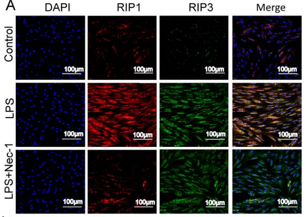
Applications
| Methods | Biomarkers | Images | PMID |
|---|---|---|---|
| Immunofluorescence | RIP1 / RIP3 |
|
30462730 |
Tech Support
Tel: +1-832-582-8158 Ext:3
If you have any other enquiries, please leave a message.
Products are for research use only. Not for human use. We do not sell to patients.
©Copyright 2013 Selleck Chemicals. All Rights Reserved.