-
 Australia
-
 Austria
-
 Belgium
-
 Brazil
-
 Canada
-
 China
China
-
 Czech Republic
-
 Denmark
-
 Finland
-
 France
-
 Germany
-
 Greece
-
 Hong Kong
-
 Hungary
-
 Iceland
-
 India
India
-
 Ireland
-
 Israel
-
 Italy
Italy
-
 Japan
Japan
-
 Korea
Korea
-
 Luxembourg
-
 Malaysia
-
 Netherlands
-
 New Zealand
-
 Norway
-
 Poland
-
 Qatar
Qatar
-
 Romania
-
 Saudi Arabia
-
 Singapore
-
 Spain
Spain
-
 Sweden
-
 Switzerland
-
 Taiwan
-
 Turkey
-
 United Kingdom
-
 United States
research use only
Anisomycin (Flagecidin) Protein Synthesis Inhibitor
Cat.No.S7409
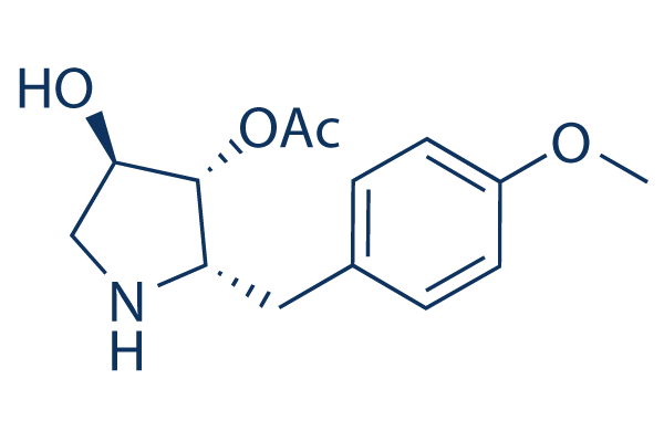
Chemical Structure
Molecular Weight: 265.3
Quality Control
| Related Targets | ERK p38 MAPK Raf MEK Ras KRas S6 Kinase MAP4K TAK1 Mixed Lineage Kinase |
|---|---|
| Other JNK Inhibitors | CC-90001 SP600125 JNK-IN-8 JNK Inhibitor IX Tanzisertib Hydrochloride (CC-930) JNK Inhibitor VIII Bentamapimod (AS602801) Polyphyllin I BI-78D3 DTP3 |
Cell Culture, Treatment & Working Concentration
| Cell Lines | Assay Type | Concentration | Incubation Time | Formulation | Activity Description | PMID |
|---|---|---|---|---|---|---|
| HEK293 | Function assay | Inhibitory concentration required to produce cytotoxicity against HEK293 cells, IC50=0.02μM. | 16005213 | |||
| HeLa | Function assay | 10 uM | Inhibition of translation in human HeLa cells at 10 uM by 35S-methionine metabolic labeling study | 15165136 | ||
| HEK293 | Function assay | 100 uM | 15 mins | Increase of JNK phosphorylation in U50488 treated untransfected HEK293 cells at 100 uM after 15 mins | 17702750 | |
| HEK293 | Function assay | 50 uM | 15 mins | Increase of U50488-induced JNK phosphorylation in untransfected HEK293 cells at 50 uM after 15 mins | 17702750 | |
| RAW264.7 | Function assay | 5 uM | 30 mins | Activation of p38MAPK in mouse RAW264.7 cells assessed as phosphorylation at Thr180/Tyr182 at 5 uM after 30 mins by Western blotting analysis | 23294286 | |
| Sf9 | Function assay | 10 uM | 6 to 24 hr | Increase of ATP level in Spodoptera frugiperda (fall armyworm) Sf9 cells at 10 uM after 6 to 24 hr by luminescent cell viability assay | ChEMBL | |
| Sf9 | Cytotoxicity assay | 10 uM | 72 hr | Cytotoxicity against Spodoptera frugiperda (fall armyworm) Sf9 cells at 10 uM after 72 hr by trypan blue dye exclusion test | ChEMBL | |
| VERO-E6 | Function assay | 48 hrs | Determination of IC50 values for inhibition of SARS-CoV-2 induced cytotoxicity of VERO-E6 cells after 48 hours exposure to 0.01 MOI SARS CoV-2 virus by high content imaging, IC50=0.09μM. | ChEMBL | ||
| VERO-E6 | Function assay | 48 hrs | Toxicity CC50 against VERO-E6 cells determined at 48 hours by high content imaging (same conditions as 2_LEY without exposure to 0.01 MOI SARS CoV-2 virus), CC50=0.1μM. | ChEMBL | ||
| Vero E6 | Function assay | CC50 determination at MOI 0.004 using CellTiter- Glo (CTG) assay, performed 3 days post-infection in Vero E6 cells, CC50<0.39μM. | ChEMBL | |||
| Vero E6 | Function assay | CC50 determination at MOI 0.01 using CellTiter- Glo (CTG) assay, performed 3 days post-infection in Vero E6 cells, CC50<0.39μM. | ChEMBL | |||
| Click to View More Cell Line Experimental Data | ||||||
Solubility
|
In vitro |
DMSO
: 53 mg/mL
(199.77 mM)
Ethanol : 16 mg/mL Water : Insoluble |
Molarity Calculator
|
In vivo |
|||||
In vivo Formulation Calculator (Clear solution)
Step 1: Enter information below (Recommended: An additional animal making an allowance for loss during the experiment)
Step 2: Enter the in vivo formulation (This is only the calculator, not formulation. Please contact us first if there is no in vivo formulation at the solubility Section.)
Calculation results:
Working concentration: mg/ml;
Method for preparing DMSO master liquid: mg drug pre-dissolved in μL DMSO ( Master liquid concentration mg/mL, Please contact us first if the concentration exceeds the DMSO solubility of the batch of drug. )
Method for preparing in vivo formulation: Take μL DMSO master liquid, next addμL PEG300, mix and clarify, next addμL Tween 80, mix and clarify, next add μL ddH2O, mix and clarify.
Method for preparing in vivo formulation: Take μL DMSO master liquid, next add μL Corn oil, mix and clarify.
Note: 1. Please make sure the liquid is clear before adding the next solvent.
2. Be sure to add the solvent(s) in order. You must ensure that the solution obtained, in the previous addition, is a clear solution before proceeding to add the next solvent. Physical methods such
as vortex, ultrasound or hot water bath can be used to aid dissolving.
Chemical Information, Storage & Stability
| Molecular Weight | 265.3 | Formula | C14H19NO4 |
Storage (From the date of receipt) | |
|---|---|---|---|---|---|
| CAS No. | 22862-76-6 | Download SDF | Storage of Stock Solutions |
|
|
Mechanism of Action
| Targets/IC50/Ki |
JNK
|
|---|---|
| In vitro |
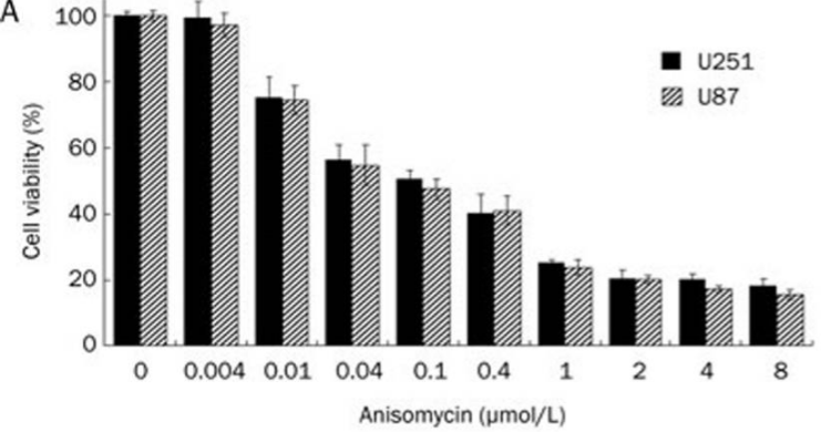
Anisomycin (3 μM) decreases protein synthesis in MDA16 and MDA-MB-468 cells, and reduces colony formation by MDA-MB-468 cells. This compound causes an increase in the number of apoptotic cells in MDA-MB-468 cultures, but not in MDA16 cultures. It actives JNK phosphorylation in MDA-MB-468 cells. In U251 and U87 cells, this chemical (0.01-8 μM) inhibits the cell growth in time- and concentration-dependent manners with the IC50 (48 h) values of 0.233 and 0.192 μmol/L, respectively. It (4 μM) causes 21.5% and 25.3% of apoptosis proportion in U251 and U87 cells, respectively, and activates p38 MAPK and JNK, while inactivated ERK1/2. This compound (4 μM) reduces the level of PP2A/C subunit in a time-dependent manner in U251 and U87 cells. It inhibits EAC cell proliferation in concentration-dependent manner.
|
| Kinase Assay |
JNK phosphorylation
|
|
500,000 cells/well are seeded in 6-well plates and incubated overnight. Cells are then incubated for 1 h with test compounds or DMSO as vehicle control (final concentration 1% v/v). Puromycin is added (final concentration of 18 μM) and cells incubated for a further 10 min to label nascent polypeptide chains. Background labelling is determined by incubating cells without puromycin. Cells are then washed in HBSS, harvested by scraping and centrifuged (300 g, 5 min). Cells are resuspended in 0.5 mL 50 mM DTT containing phosphatase inhibitors and incubated at 95℃ for 10 min. Samples are then snap frozen in liquid nitrogen and stored at -20℃ until blotted. Samples (20–30 μg protein/sample) are blotted onto a PVDF membrane. The membrane is blocked and incubated with anti-phospho-Thr183/Tyr185-JNK antibody overnight at 4℃. Secondary antibodies are used to label the primary antibody and detected using an infrared scanner. The intensity of the fluorescence signal for anti-phospho-JNK antibody is background corrected and normalized for loading.
|
|
| In vivo |
Peritumoral administration of anisomycin (5 mg/kg) significantly suppresses Ehrlich ascites carcinoma (EAC) growth resulting in the survival of approximately 60% of the mice 90 days after EAC inoculation.
|
References |
|
Applications
| Methods | Biomarkers | Images | PMID |
|---|---|---|---|
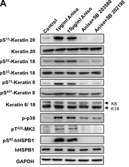
| Western blot | p-Keratin 20 / Keratin 20 / p-Keratin 18 / Keratin 18 / p-Keratin 8 / Keratin 8 / p-MK2 / p-hHSPB1 / hHSPB1 PP2A / PP2C p-ERK / ERK / p-JNK / JNK / p-p38 / ATF3 |
|
20724476 |
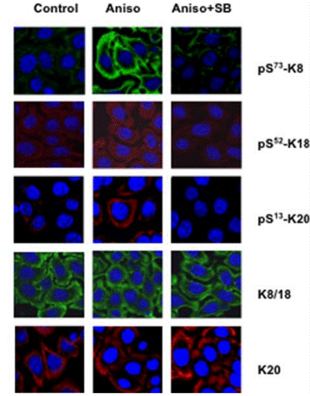
| Immunofluorescence | p-Keratin 8 / p-Keratin 18 / p-Keratin 20 |
|
20724476 |
| Growth inhibition assay | Cell viability |
|
22684030 |
Tech Support
Tel: +1-832-582-8158 Ext:3
If you have any other enquiries, please leave a message.
Signaling Pathway Map
Products are for research use only. Not for human use. We do not sell to patients.
©Copyright 2013 Selleck Chemicals. All Rights Reserved.