- Inhibitors
- By product type
- Natural Products
- Inducing Agents
- Peptides
- Antibiotics
- Antibody-drug Conjugates(ADC)
- PROTAC
- Hydrotropic Agents
- Dyes
- By Signaling Pathways
- PI3K/Akt/mTOR
- Epigenetics
- Methylation
- Immunology & Inflammation
- Protein Tyrosine Kinase
- Angiogenesis
- Apoptosis
- Autophagy
By research - Antibodies
- Compound Libraries
- Popular Compound Libraries
- Customize Library
- Clinical and FDA-approved Related
- Bioactive Compound Libraries
- Inhibitor Related
- Natural Product Related
- Metabolism Related
- Cell Death Related
- By Signaling Pathway
- By Disease
- Anti-infection and Antiviral Related
- Neuronal and Immunology Related
- Fragment and Covalent Related
- FDA-approved Drug Library
- FDA-approved & Passed Phase I Drug Library
- Preclinical/Clinical Compound Library
- Bioactive Compound Library-I
- Bioactive Compound Library-Ⅱ
- Kinase Inhibitor Library
- Express-Pick Library
- Natural Product Library
- Human Endogenous Metabolite Compound Library
- Alkaloid Compound LibraryNew
- Angiogenesis Related compound Library
- Anti-Aging Compound Library
- Anti-alzheimer Disease Compound Library
- Antibiotics compound Library
- Anti-cancer Compound Library
- Anti-cancer Compound Library-Ⅱ
- Anti-cancer Metabolism Compound Library
- Anti-Cardiovascular Disease Compound Library
- Anti-diabetic Compound Library
- Anti-infection Compound Library
- Antioxidant Compound Library
- Anti-parasitic Compound Library
- Antiviral Compound Library
- Apoptosis Compound Library
- Autophagy Compound Library
- Calcium Channel Blocker LibraryNew
- Cambridge Cancer Compound Library
- Carbohydrate Metabolism Compound LibraryNew
- Cell Cycle compound library
- CNS-Penetrant Compound Library
- Covalent Inhibitor Library
- Cytokine Inhibitor LibraryNew
- Cytoskeletal Signaling Pathway Compound Library
- DNA Damage/DNA Repair compound Library
- Drug-like Compound Library
- Endoplasmic Reticulum Stress Compound Library
- Epigenetics Compound Library
- Exosome Secretion Related Compound LibraryNew
- FDA-approved Anticancer Drug LibraryNew
- Ferroptosis Compound Library
- Flavonoid Compound Library
- Fragment Library
- Glutamine Metabolism Compound Library
- Glycolysis Compound Library
- GPCR Compound Library
- Gut Microbial Metabolite Library
- HIF-1 Signaling Pathway Compound Library
- Highly Selective Inhibitor Library
- Histone modification compound library
- HTS Library for Drug Discovery
- Human Hormone Related Compound LibraryNew
- Human Transcription Factor Compound LibraryNew
- Immunology/Inflammation Compound Library
- Inhibitor Library
- Ion Channel Ligand Library
- JAK/STAT compound library
- Lipid Metabolism Compound LibraryNew
- Macrocyclic Compound Library
- MAPK Inhibitor Library
- Medicine Food Homology Compound Library
- Metabolism Compound Library
- Methylation Compound Library
- Mouse Metabolite Compound LibraryNew
- Natural Organic Compound Library
- Neuronal Signaling Compound Library
- NF-κB Signaling Compound Library
- Nucleoside Analogue Library
- Obesity Compound Library
- Oxidative Stress Compound LibraryNew
- Plant Extract Library
- Phenotypic Screening Library
- PI3K/Akt Inhibitor Library
- Protease Inhibitor Library
- Protein-protein Interaction Inhibitor Library
- Pyroptosis Compound Library
- Small Molecule Immuno-Oncology Compound Library
- Mitochondria-Targeted Compound LibraryNew
- Stem Cell Differentiation Compound LibraryNew
- Stem Cell Signaling Compound Library
- Natural Phenol Compound LibraryNew
- Natural Terpenoid Compound LibraryNew
- TGF-beta/Smad compound library
- Traditional Chinese Medicine Library
- Tyrosine Kinase Inhibitor Library
- Ubiquitination Compound Library
-
Cherry Picking
You can personalize your library with chemicals from within Selleck's inventory. Build the right library for your research endeavors by choosing from compounds in all of our available libraries.
Please contact us at info@selleckchem.com to customize your library.
You could select:
- Bioreagents
- qPCR
- 2x SYBR Green qPCR Master Mix
- 2x SYBR Green qPCR Master Mix(Low ROX)
- 2x SYBR Green qPCR Master Mix(High ROX)
- Protein Assay
- Protein A/G Magnetic Beads for IP
- Anti-Flag magnetic beads
- Anti-Flag Affinity Gel
- Anti-Myc magnetic beads
- Anti-HA magnetic beads
- Poly DYKDDDDK Tag Peptide lyophilized powder
- Protease Inhibitor Cocktail
- Protease Inhibitor Cocktail (EDTA-Free, 100X in DMSO)
- Phosphatase Inhibitor Cocktail (2 Tubes, 100X)
- Cell Biology
- Cell Counting Kit-8 (CCK-8)
- Animal Experiment
- Mouse Direct PCR Kit (For Genotyping)
- Featured Products
- MRTX1133
- Nab-Paclitaxel
- KP-457
- IAG933
- RMC-6236 (Daraxonrasib)
- RMC-7977
- Zoldonrasib (RMC-9805)
- GsMTx4
- Navitoclax (ABT-263)
- TSA (Trichostatin A)
- Y-27632 Dihydrochloride
- SB431542
- SB202190
- MK-2206 Dihydrochloride
- LY294002
- Alisertib (MLN8237)
- XAV-939
- CHIR-99021 (Laduviglusib)
- Bafilomycin A1 (Baf-A1)
- Thiazovivin (TZV)
- CP-673451
- Verteporfin
- DAPT
- Galunisertib (LY2157299)
- MG132
- SBE-β-CD
- Tween 80
- Bavdegalutamide (ARV-110)
- Z-VAD-FMK
- Wnt-C59 (C59)
- IWR-1-endo
- (+)-JQ1
- 3-Deazaneplanocin A (DZNep) Hydrochloride
- RepSox (E-616452)
- Erastin
- Q-VD-Oph
- Puromycin Dihydrochloride
- Cycloheximide
- Telaglenastat (CB-839)
- A-83-01
- Ceralasertib (AZD6738)
- Liproxstatin-1
- Emricasan (IDN-6556)
- PMA (Phorbol 12-myristate 13-acetate)
- Dibutyryl cAMP (Bucladesine) sodium
- Nedisertib (M3814)
- PLX5622
- IKE (Imidazole Ketone Erastin)
- STM2457
- Saruparib (AZD5305)
- New Products
- Contact Us
research use only
(+)-JQ1 BET inhibitor
Cat.No.S7110

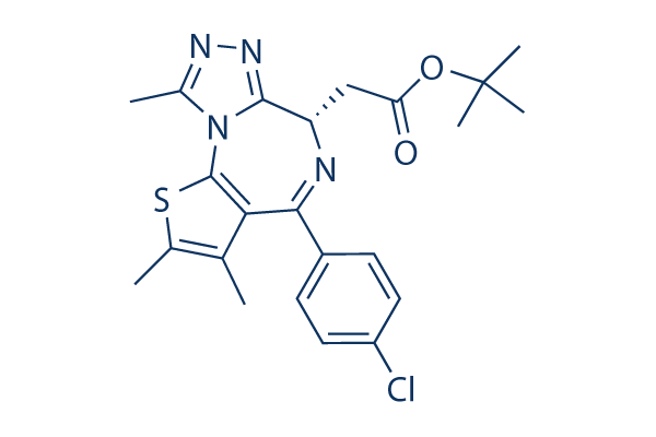
Chemical Structure
Molecular Weight: 456.99
Quality Control
Batch:
Purity:
99.99%
99.99
| Related Targets | Epigenetic Reader Domain HDAC JAK Histone Methyltransferase AMPK PKC PARP HIF Histone Acetyltransferase Aurora Kinase Sirtuin PRMT Histone Demethylase EZH1/2 DNA Methyltransferase Pim LSD1 MLL JMJD G9a/GLP NSD FTO MicroRNA PAD SETD SMYD DOT1 COMT WDR5 HNMT | |
|---|---|---|
| Related Products | Quizartinib (AC220) BI-4464 |
Cell Culture, Treatment & Working Concentration
| Cell Lines | Assay Type | Concentration | Incubation Time | Formulation | Activity Description | PMID |
|---|---|---|---|---|---|---|
| K1 | Cell Viability Assay | 250/500/1000 nM | 24/48/72 h | DMSO | inhibits cell viability in both dose- and time- dependent manner | 26707881 |
| BCPAP | Cell Viability Assay | 250/500/1000 nM | 24/48/72 h | DMSO | inhibits cell viability in both dose- and time- dependent manner | 26707881 |
| K1 | Cell Cycle Assay | 250/500/1000 nM | 72 h | DMSO | arrests cell cycle at G0/G1 phase | 26707881 |
| BCPAP | Cell Cycle Assay | 250/500/1000 nM | 72 h | DMSO | arrests cell cycle at G0/G1 phase | 26707881 |
| Hep3B | Growth Inhibition Assay | 0-10 μM | 5 d | DMSO | IC50=0.08 μM | 26575167 |
| HCCLM3 | Growth Inhibition Assay | 0-10 μM | 5 d | DMSO | IC50=0.14 μM | 26575167 |
| HuH7 | Growth Inhibition Assay | 0-10 μM | 5 d | DMSO | IC50=0.21 μM | 26575167 |
| HepG2 | Growth Inhibition Assay | 0-10 μM | 5 d | DMSO | IC50=0.34 μM | 26575167 |
| SMMC7721 | Growth Inhibition Assay | 0-10 μM | 5 d | DMSO | IC50=0.41 μM | 26575167 |
| BEL7402 | Growth Inhibition Assay | 0-10 μM | 5 d | DMSO | IC50=0.47 μM | 26575167 |
| MHCC97H | Growth Inhibition Assay | 0-10 μM | 5 d | DMSO | IC50=0.41 μM | 26575167 |
| Hep3B | Cell Cycle Assay | 0.1/0.5/2.5 μM | 48 h | DMSO | leads to a substantial accumulation of HCC cells in sub-G1 phase | 26575167 |
| HCCLM3 | Cell Cycle Assay | 0.1/0.5/2.5 μM | 48 h | DMSO | leads to a substantial accumulation of HCC cells in sub-G1 phase | 26575167 |
| Hep3B | Apoptosis Assay | 0.1/0.5/2.5 μM | 48 h | DMSO | activates caspase-3 and caspase-9 expression and induced PARP cleavage as well as cytochrome c release into the cytoplasm from mitochondria | 26575167 |
| HCCLM3 | Apoptosis Assay | 0.1/0.5/2.5 μM | 48 h | DMSO | activates caspase-3 and caspase-9 expression and induced PARP cleavage as well as cytochrome c release into the cytoplasm from mitochondria | 26575167 |
| A549 | Growth Inhibition Assay | 0.1-10 μM | 72 h | inhibits cell growth in a dose-dependent manner | 26415225 | |
| H157 | Growth Inhibition Assay | 0.1-10 μM | 72 h | inhibits cell growth in a dose-dependent manner | 26415225 | |
| H1299 | Growth Inhibition Assay | 0.1-10 μM | 72 h | inhibits cell growth in a dose-dependent manner | 26415225 | |
| A549 | Function Assay | 1/2.5/5 μM | 12 h | weakly decreased Bcl-2 levels | 26415225 | |
| H1299 | Function Assay | 1/2.5/5 μM | 12 h | weakly decreased Bcl-2 levels | 26415225 | |
| H157 | Function Assay | 1/2.5/5 μM | 12 h | decreased DR4 expression | 26415225 | |
| H1299 | Function Assay | 1/2.5/5 μM | 12 h | decreased DR4 expression | 26415225 | |
| C8161 | Cell Viability Assay | 0-2 μM | 4 d | DMSO | decreases cell viability in a dose-dependent manner | 26397223 |
| Mel285 | Cell Viability Assay | 0-2 μM | 4 d | DMSO | decreases cell viability in a dose-dependent manner | 26397223 |
| Mel290 | Cell Viability Assay | 0-2 μM | 4 d | DMSO | decreases cell viability in a dose-dependent manner | 26397223 |
| 92.1 | Cell Viability Assay | 0-2 μM | 4 d | DMSO | decreases cell viability in a dose-dependent manner | 26397223 |
| Omm1.3 | Cell Viability Assay | 0-2 μM | 4 d | DMSO | decreases cell viability in a dose-dependent manner | 26397223 |
| Mel202 | Cell Viability Assay | 0-2 μM | 4 d | DMSO | decreases cell viability in a dose-dependent manner | 26397223 |
| Mel270 | Cell Viability Assay | 0-2 μM | 4 d | DMSO | decreases cell viability in a dose-dependent manner | 26397223 |
| Omm1 | Cell Viability Assay | 0-2 μM | 4 d | DMSO | decreases cell viability in a dose-dependent manner | 26397223 |
| 92.1 | Apoptosis Assay | 500 nM | 48 h | DMSO | induces apoptosis | 26397223 |
| Omm1.3 | Apoptosis Assay | 500 nM | 48 h | DMSO | induces apoptosis | 26397223 |
| 92.1 | Cell Cycle Assay | 500 nM | 24/48/72 h | DMSO | induces the cell accumulation at sub-G1 | 26397223 |
| Omm1.3 | Cell Cycle Assay | 500 nM | 24/48/72 h | DMSO | induces the cell accumulation at sub-G1 | 26397223 |
| A549 | Function Assay | 100/400/1000 nM | 24 h | upregulates and activates SIRT1 | 26212199 | |
| MCF-7 | Function Assay | 100/400/1000 nM | 24 h | upregulates and activates SIRT1 | 26212199 | |
| HEK293 | Function Assay | 100/400/1000 nM | 24 h | upregulates and activates SIRT1 | 26212199 | |
| 858 | Cell Viability Assay | 0-1 μM | 5 d | DMSO | decreases cell viability in a dose-dependent manner | 26206333 |
| DDR2L63V | Cell Viability Assay | 0-1 μM | 5 d | DMSO | decreases cell viability in a dose-dependent manner | 26206333 |
| BE(2)-C | Cell Viability Assay | 1 μM | 1-4 d | decreases cell viability significantly | 26067464 | |
| IMR-32 | Cell Viability Assay | 1 μM | 1-4 d | decreases cell viability significantly | 26067464 | |
| JF | Cell Viability Assay | 1 μM | 1-4 d | decreases cell viability significantly | 26067464 | |
| BE(2)-M17 | Cell Viability Assay | 1 μM | 1-4 d | decreases cell viability significantly | 26067464 | |
| SK-N-SH | Cell Viability Assay | 1 μM | 1-4 d | decreases cell viability significantly | 26067464 | |
| SK-N-DZ | Cell Viability Assay | 1 μM | 1-4 d | decreases cell viability significantly | 26067464 | |
| HMC-1.1 | Growth Inhibition Assay | 5-5000 nM | 48 h | DMSO | inhibits cell growth in a dose-dependent manner | 26055303 |
| HMC-1.2 | Growth Inhibition Assay | 5-5000 nM | 48 h | DMSO | inhibits cell growth in a dose-dependent manner | 26055303 |
| ROSA KIT WT | Growth Inhibition Assay | 5-5000 nM | 48 h | DMSO | inhibits cell growth in a dose-dependent manner | 26055303 |
| ROSA KIT D816V | Growth Inhibition Assay | 5-5000 nM | 48 h | DMSO | inhibits cell growth in a dose-dependent manner | 26055303 |
| HMC-1.1 | Apoptosis Assay | 200-5000 nM | 48 h | DMSO | induces cell apoptosis in a dose-dependent manner | 26055303 |
| HMC-1.2 | Apoptosis Assay | 200-5000 nM | 48 h | DMSO | induces cell apoptosis in a dose-dependent manner | 26055303 |
| ROSA KIT WT | Apoptosis Assay | 200-5000 nM | 48 h | DMSO | induces cell apoptosis in a dose-dependent manner | 26055303 |
| ROSA KIT D816V | Apoptosis Assay | 200-5000 nM | 48 h | DMSO | induces cell apoptosis in a dose-dependent manner | 26055303 |
| 494H | Growth Inhibition Assay | 72 h | DMSO | IC50=0.122±0.004 μM | 25944566 | |
| 493H | Growth Inhibition Assay | 72 h | DMSO | IC50=0.047±0.009 μM | 25944566 | |
| 716H | Growth Inhibition Assay | 72 h | DMSO | IC50=0.212±0.034 μM | 25944566 | |
| 148I | Growth Inhibition Assay | 72 h | DMSO | IC50=0.284±0.035 μM | 25944566 | |
| 98Sc | Growth Inhibition Assay | 72 h | DMSO | IC50=0.115±0.004 μM | 25944566 | |
| 89R | Growth Inhibition Assay | 72 h | DMSO | IC50=0.126±0.003 μM | 25944566 | |
| 494L | Growth Inhibition Assay | 72 h | DMSO | IC50=0.317±0.012 μM | 25944566 | |
| 493L | Growth Inhibition Assay | 72 h | DMSO | IC50=0.050±0.011 μM | 25944566 | |
| 148L | Growth Inhibition Assay | 72 h | DMSO | IC50=0.146±0.017 μM | 25944566 | |
| 98L | Growth Inhibition Assay | 72 h | DMSO | IC50=0.309±0.029 μM | 25944566 | |
| OS17 | Growth Inhibition Assay | 72 h | DMSO | IC50=0.079±0.003 μM | 25944566 | |
| OS9 | Growth Inhibition Assay | 72 h | DMSO | IC50=0.406±0.028 μM | 25944566 | |
| MG63 | Growth Inhibition Assay | 72 h | DMSO | IC50=0.114±0.025 μM | 25944566 | |
| SAOS2 | Growth Inhibition Assay | 72 h | DMSO | IC50=0.217±0.003 μM | 25944566 | |
| U2OS | Growth Inhibition Assay | 72 h | DMSO | IC50=0.198±0.008 μM | 25944566 | |
| SJSA-1 | Growth Inhibition Assay | 72 h | DMSO | IC50=0.100±0.010 μM | 25944566 | |
| 494H | Apoptosis Assay | 0.25/0.5/1.0 μM | 24 h | DMSO | increases levels of cleaved caspase-3 | 25944566 |
| 148I | Apoptosis Assay | 0.25/0.5/1.0 μM | 24 h | DMSO | increases levels of cleaved caspase-3 | 25944566 |
| OS17 | Apoptosis Assay | 0.25/0.5/1.0 μM | 24 h | DMSO | increases levels of cleaved caspase-3 | 25944566 |
| 494H | Apoptosis Assay | 1 μM | 48 h | DMSO | induces cell apoptosis significantly | 25944566 |
| 148I | Apoptosis Assay | 1 μM | 48 h | DMSO | induces cell apoptosis significantly | 25944566 |
| OS17 | Apoptosis Assay | 1 μM | 48 h | DMSO | induces cell apoptosis significantly | 25944566 |
| MOLM13 | Apoptosis Assay | 250 nM | 48 h | DMSO | induces significantly apoptosis cotreatment with quizartinib | 25053825 |
| MV4-11 | Apoptosis Assay | 250 nM | 48 h | DMSO | induces significantly apoptosis cotreatment with quizartinib | 25053825 |
| MOLM13 | Function Assay | 250 nM | 24 h | DMSO | enhances quizartinib-induced more p21, BIM, and cleaved PARP | 25053825 |
| MV4-11 | Function Assay | 250 nM | 24 h | DMSO | enhances quizartinib-induced more p21, BIM, and cleaved PARP | 25053825 |
| MOLM13 | Apoptosis Assay | 250 nM | 48 h | DMSO | induces significantly apoptosis cotreatment with ponatinib | 25053825 |
| MV4-11 | Apoptosis Assay | 250 nM | 48 h | DMSO | induces significantly apoptosis cotreatment with ponatinib | 25053825 |
| Hela | Cell Viability Assay | 0-500 nM | 72 h | DMSO | decreases cell viability in a dose-dependent manner | 25009295 |
| HBL-1 | Cell Viability Assay | 0-500 nM | 72 h | DMSO | decreases cell viability in a dose-dependent manner | 25009295 |
| HLY-1 | Cell Viability Assay | 0-500 nM | 72 h | DMSO | decreases cell viability in a dose-dependent manner | 25009295 |
| OCI-Ly3 | Cell Viability Assay | 0-500 nM | 72 h | DMSO | decreases cell viability in a dose-dependent manner | 25009295 |
| OCI-Ly10 | Cell Viability Assay | 0-500 nM | 72 h | DMSO | decreases cell viability in a dose-dependent manner | 25009295 |
| SU-DHL-4 | Cell Viability Assay | 0-500 nM | 72 h | DMSO | decreases cell viability in a dose-dependent manner | 25009295 |
| SU-DHL-5 | Cell Viability Assay | 0-500 nM | 72 h | DMSO | decreases cell viability in a dose-dependent manner | 25009295 |
| SU-DHL-6 | Cell Viability Assay | 0-500 nM | 72 h | DMSO | decreases cell viability in a dose-dependent manner | 25009295 |
| SU-DHL-10 | Cell Viability Assay | 0-500 nM | 72 h | DMSO | decreases cell viability in a dose-dependent manner | 25009295 |
| RC-K8 | Cell Viability Assay | 0-500 nM | 72 h | DMSO | decreases cell viability in a dose-dependent manner | 25009295 |
| OCI-Ly8 | Cell Viability Assay | 0-500 nM | 72 h | DMSO | decreases cell viability in a dose-dependent manner | 25009295 |
| OCL-Ly18 | Cell Viability Assay | 0-500 nM | 72 h | DMSO | decreases cell viability in a dose-dependent manner | 25009295 |
| OCI-Ly3 | Growth Inhibition Assay | 172/250/500 nM | 2/7 d | DMSO | induces cell-cycle arrest at sub-G1 with minimal cell death | 25009295 |
| OCI-Ly8 | Growth Inhibition Assay | 172/250/500 nM | 2/7 d | DMSO | induces cell-cycle arrest at sub-G1 with minimal cell death | 25009295 |
| SU-DHL-4 | Growth Inhibition Assay | 172/250/500 nM | 2/7 d | DMSO | induces cell-cycle arrest at sub-G1 with minimal cell death | 25009295 |
| SU-DHL-10 | Growth Inhibition Assay | 172/250/500 nM | 2/7 d | DMSO | induces cell-cycle arrest at sub-G1 with minimal cell death | 25009295 |
| OCI-Ly3 | Apoptosis Assay | 172/250 nM | 7d | DMSO | increases caspase-3/7 activity significantly | 25009295 |
| OCI-Ly8 | Apoptosis Assay | 172/250 nM | 7d | DMSO | increases caspase-3/7 activity significantly | 25009295 |
| SU-DHL-4 | Apoptosis Assay | 172/250 nM | 7d | DMSO | increases caspase-3/7 activity significantly | 25009295 |
| SU-DHL-10 | Apoptosis Assay | 172/250 nM | 7d | DMSO | increases caspase-3/7 activity significantly | 25009295 |
| Rosetta2 DE3 | Function assay | Kd = 0.0062 μM | 26080064 | |||
| Rosetta2 DE3 | Fluorescence polarization assay | 30 mins | Ki = 0.0066 μM | 26080064 | ||
| Rosetta2 DE3 | Function assay | Kd = 0.0067 μM | 26080064 | |||
| Rosetta2 DE3 | Fluorescence polarization assay | 30 mins | Ki = 0.0076 μM | 26080064 | ||
| Rosetta2 DE3 | Fluorescence polarization assay | 30 mins | Ki = 0.0089 μM | 26080064 | ||
| Rosetta2 DE3 | Fluorescence polarization assay | 30 mins | Ki = 0.0107 μM | 26080064 | ||
| Rosetta2 DE3 | Function assay | Kd = 0.0117 μM | 26080064 | |||
| MV4-11 | Antiproliferative activity assay | 72 h | IC50 = 0.012 μM | 26731490 | ||
| Rosetta2 DE3 | Fluorescence polarization assay | 30 mins | Ki = 0.012 μM | 29758518 | ||
| VCaP | Antiproliferative activity assay | 12 h | IC50 = 0.012 μM | 28463487 | ||
| Rosetta2 DE3 | Fluorescence polarization assay | 30 mins | Ki = 0.0125 μM | 26080064 | ||
| Rosetta2 DE3 | Function assay | Kd = 0.0128 μM | 26080064 | |||
| Rosetta2 DE3 | Fluorescence polarization assay | 30 mins | Ki = 0.0132 μM | 26080064 | ||
| Rosetta2 DE3 | Function assay | Kd = 0.0136 μM | 26080064 | |||
| Rosetta2 DE3 | Function assay | Kd = 0.0147 μM | 26080064 | |||
| Rosetta2 DE3 | Fluorescence polarization assay | 30 mins | Ki = 0.0149 μM | 28463487 | ||
| TY82 | Antiproliferative activity assay | 72 h | IC50 = 0.018 μM | 28586718 | ||
| MM1S | Antiproliferative activity assay | 72 h | IC50 = 0.019 μM | 28586718 | ||
| MM1S | Cytotoxicity assay | 72 h | IC50 = 0.02 μM | 29758518 | ||
| HT-29 | Antiproliferative activity assay | 12 h | IC50 = 0.02 μM | 28535045 | ||
| MV4-11 | Growth inhibition assay | 72 h | IC50 = 0.023 μM | 25559428 | ||
| MV4-11 | Cytotoxicity assay | 4 days | IC50 = 0.024 μM | 28463487 | ||
| MV4-11 | Growth inhibition assay | 4 days | IC50 = 0.024 μM | 26080064 | ||
| Rosetta2 DE3 | Function assay | 30 mins | IC50 = 0.0287 μM | 26080064 | ||
| NALM16 | Cytotoxicity assay | 5 days | EC50 = 0.03 μM | 29170024 | ||
| BL21(DE3)-R3-pRARE2 | Function assay | 30 mins | IC50 = 0.033 μM | 28195723 | ||
| BL21(DE3) | Function assay | Kd = 0.034 μM | 26731490 | |||
| Rosetta2 DE3 | Fluorescence polarization assay | 30 mins | IC50 = 0.0357 μM | 26080064 | ||
| Rosetta2 DE3 | Fluorescence polarization assay | 30 mins | IC50 = 0.0422 μM | 28463487 | ||
| Rosetta2 DE3 | Fluorescence polarization assay | 30 mins | IC50 = 0.0467 μM | 28463487 | ||
| MOLM13 | Cytotoxicity assay | 4 days | IC50 = 0.056 μM | 28463487 | ||
| MOLM13 | Growth inhibition assay | 4 days | IC50 = 0.056 μM | 26080064 | ||
| HL60 | Antiproliferative activity assay | 72 h | IC50 = 0.06 μM | 29170024 | ||
| HL60 | Growth inhibition assay | 3 days | GC50 = 0.06 μM | 29657099 | ||
| NALM6 | Cytotoxicity assay | 5 days | EC50 = 0.06 μM | 28549889 | ||
| Raji | Function assay | IC50 = 0.06 μM | 26731490 | |||
| Raji | Function assay | 4 h | IC50 = 0.069 μM | 24900758 | ||
| MM1S | Antiproliferative activity assay | 72 h | IC50 = 0.0691 μM | 29525435 | ||
| BL21 (DE3)-codon plus-RIL | Fluorescence polarization assay | by fluorescence anisotropy assay | IC50 = 0.07 μM | 28586718 | ||
| 22Rv1 | Antiproliferative activity assay | 96 h | IC50 = 0.071 μM | 29758518 | ||
| 22Rv1 | Antiproliferative activity assay | 12 h | IC50 = 0.071 μM | 29541371 | ||
| MV4-11 | Antiproliferative activity assay | IC50 = 0.072 μM | 28195723 | |||
| BL21(DE3)-R3-pRARE2 | Function assay | 30 mins | IC50 = 0.077 μM | 26731490 | ||
| BL21(DE3)-R3-pRARE2 | Function assay | 30 mins | IC50 = 0.077 μM | 28195723 | ||
| BL21(DE3)-R3-pRARE2 | Function assay | 30 mins | IC50 = 0.077 μM | 29776834 | ||
| MV4-11 | Cytotoxicity assay | 24 h | GI50 = 0.08 μM | 26191363 | ||
| MV411 | Antiproliferative activity assay | 72 h | IC50 = 0.08 μM | 28314513 | ||
| TY82 | Antiproliferative activity assay | 72 h | IC50 = 0.0808 μM | 29525435 | ||
| HL60 | Function assay | 24 h | IC50 = 0.086 μM | 28549889 | ||
| 697 | Cytotoxicity assay | 5 days | EC50 = 0.09 μM | 29170024 | ||
| Loucy | Cytotoxicity assay | 5 days | EC50 = 0.09 μM | 29170024 | ||
| BL21(DE3) | Function assay | Kd = 0.092 μM | 29541371 | |||
| BL21(DE3)-R3-pRARE2 | Function assay | Kd = 0.1 μM | 28595007 | |||
| HT-29 | Growth inhibition assay | 72 h | IC50 = 0.104 μM | 25559428 | ||
| MM1S | Growth inhibition assay | 72 h | IC50 = 0.109 μM | 25559428 | ||
| LNCAP cells | Antiproliferative activity assay | IC50 = 0.1096 μM | 29758518 | |||
| T cells | Function assay | 24 h | IC50 = 0.11 μM | 28314513 | ||
| HL60 | Antiproliferative activity assay | 72 h | IC50 = 0.11 μM | 26869194 | ||
| BL21(DE3) | Function assay | IC50 = 0.12 μM | 26731490 | |||
| BL21(DE3) | Function assay | 2.5 h | IC50 = 0.12 μM | 29541371 | ||
| H1299 | Function assay | 24 h | EC50 = 0.153 μM | 28949521 | ||
| HD-MB03 | Cytotoxicity assay | 5 days | EC50 = 0.16 μM | 29758518 | ||
| LNCAP | Antiproliferative activity assay | 96 h | IC50 = 0.16 μM | 29758518 | ||
| Hs578T | Antiproliferative activity assay | 12 h | IC50 = 0.16 μM | 29758518 | ||
| MV4-11 | Antiproliferative activity assay | 12 h | IC50 = 0.16 μM | 29170024 | ||
| LNCAP | Antiproliferative activity assay | 12 h | IC50 = 0.16 μM | 29541371 | ||
| C4-2B | Antiproliferative activity assay | 96 h | IC50 = 0.19 μM | 29758518 | ||
| C4-2B | Antiproliferative activity assay | 12 h | IC50 = 0.19 μM | 29541371 | ||
| Vero E6 | Antiviral activity assay | 48 h | IC50 = 0.19275 μM | 32353859 | ||
| MCF7 | Antiproliferative activity assay | 12 h | IC50 = 0.2 μM | 29758518 | ||
| MV4-11 | Antiproliferative activity assay | IC50 = 0.24 μM | 27142751 | |||
| MV4-11 | Cytotoxicity assay | 72 h | IC50 = 0.242 μM | 23517011 | ||
| MX1 | Antiproliferative activity assay | 72 h | EC50 = 0.254 μM | 28949521 | ||
| HT-29 | Antiproliferative activity assay | 72 h | IC50 = 0.28 μM | 26731490 | ||
| HFL1 | Antiproliferative activity assay | 12 h | IC50 = 0.29 μM | 29758518 | ||
| MDA-MB-231 | Growth inhibition assay | 3 days | GC50 = 0.3 μM | 28549889 | ||
| MV4-11 | Antiproliferative activity assay | 48 h | EC50 = 0.33113 μM | 28595007 | ||
| K562 | Antiproliferative activity assay | IC50 = 0.64 μM | 27142751 | |||
| HL60 | Antiproliferative activity assay | 48 h | EC50 = 0.74131 μM | 28595007 | ||
| NCI-H1975 | Antiproliferative activity assay | 12 h | IC50 = 1.23 μM | 29758518 | ||
| SAE | Function assay | 4 h | IC50 = 1.38 μM | 29649741 | ||
| SAE | Function assay | 4 h | IC50 = 1.49 μM | 29649741 | ||
| SAE | Function assay | 4 h | IC50 = 1.51 μM | 29649741 | ||
| U2OS | Antiproliferative activity assay | 12 h | IC50 = 1.62 μM | 29758518 | ||
| SAE | Function assay | 4 h | IC50 = 1.63 μM | 29649741 | ||
| A549 | Antiproliferative activity assay | 12 h | IC50 = 1.67 μM | 29758518 | ||
| MCF7 | Growth inhibition assay | 3 days | GC50 = 1.7 μM | 28549889 | ||
| DU145 | Antiproliferative activity assay | 96 h | IC50 = 2.52 μM | 29758518 | ||
| DU145 | Antiproliferative activity assay | 12 h | IC50 = 2.52 μM | 29541371 | ||
| T47D | Growth inhibition assay | 3 days | GC50 = 2.8 μM | 28549889 | ||
| PC3 | Antiproliferative activity assay | 96 h | IC50 = 3.01 μM | 29758518 | ||
| PC3 | Antiproliferative activity assay | 12 h | IC50 = 3.01 μM | 29541371 | ||
| HeLa | Antiproliferative activity assay | 12 h | IC50 = 3.76 μM | 29758518 | ||
| K562 | Growth inhibition assay | 3 days | GC50 = 3.8 μM | 28549889 | ||
| A2780 | Growth inhibition assay | 3 days | GC50 = 4 μM | 28549889 | ||
| HL60 | Cytotoxicity assay | 24 h | IC50 = 5.3 μM | 27266999 | ||
| MV4-11 | Cytotoxicity assay | 24 h | IC50 = 6.4 μM | 27266999 | ||
| K562 | Antiproliferative activity assay | 72 h | IC50 = 9.12 μM | 28314513 | ||
| OVCAR5 | Growth inhibition assay | 3 days | GC50 = 12 μM | 28549889 | ||
| Click to View More Cell Line Experimental Data | ||||||
Chemical Information, Storage & Stability
| Molecular Weight | 456.99 | Formula | C23H25ClN4O2S |
Storage (From the date of receipt) | |
|---|---|---|---|---|---|
| CAS No. | 1268524-70-4 | Download SDF | Storage of Stock Solutions |
|
|
| Synonyms | N/A | Smiles | CC1=C(SC2=C1C(=NC(C3=NN=C(N32)C)CC(=O)OC(C)(C)C)C4=CC=C(C=C4)Cl)C | ||
Solubility
|
In vitro |
DMSO
: 91 mg/mL
(199.12 mM)
Ethanol : 91 mg/mL Water : Insoluble |
Molarity Calculator
|
In vivo |
|||||
In vivo Formulation Calculator (Clear solution)
Step 1: Enter information below (Recommended: An additional animal making an allowance for loss during the experiment)
mg/kg
g
μL
Step 2: Enter the in vivo formulation (This is only the calculator, not formulation. Please contact us first if there is no in vivo formulation at the solubility Section.)
% DMSO
%
% Tween 80
% ddH2O
%DMSO
%
Calculation results:
Working concentration: mg/ml;
Method for preparing DMSO master liquid: mg drug pre-dissolved in μL DMSO ( Master liquid concentration mg/mL, Please contact us first if the concentration exceeds the DMSO solubility of the batch of drug. )
Method for preparing in vivo formulation: Take μL DMSO master liquid, next addμL PEG300, mix and clarify, next addμL Tween 80, mix and clarify, next add μL ddH2O, mix and clarify.
Method for preparing in vivo formulation: Take μL DMSO master liquid, next add μL Corn oil, mix and clarify.
Note: 1. Please make sure the liquid is clear before adding the next solvent.
2. Be sure to add the solvent(s) in order. You must ensure that the solution obtained, in the previous addition, is a clear solution before proceeding to add the next solvent. Physical methods such
as vortex, ultrasound or hot water bath can be used to aid dissolving.
Mechanism of Action
| Features |
(+)-JQ1 is more effective than (-)-JQ1.
|
|---|---|
| Targets/IC50/Ki | |
| In vitro |
(+)-JQ1 enantiomer binds directly into the Kac binding site of BET bromodomains. This compound (500 nM) binds BRD4 competitively with chromatin resulting in differentiation and growth arrest of NMC cells. This chemical (500 nM) attenuates rapid proliferation of NMC 797 and Per403 cell lines as demonstrated by reduced Ki67 staining. This compound (500 nM) potently decreases expression of both BRD4 target genes in NMC 797 cells. It inhibits cellular viability with IC50 of 4 nM in NMC 11060 cells. [1] It results in robust inhibition of MYC expression in MM cell lines. This compound inhibits proliferating of KMS-34 and LR5 with IC50 of 68 nM and 98 nM, respectively. This chemical (500 nM)-treated MM.1S cells results in a pronounced decrease in the proportion of cells in S-phase, with a concomitant increase in cells arrested in G0/G1. It (500 nM) results in pronounced cellular senescence by beta-galactosidase staining. This compound (800 nM) exposure leads to a significant reduction in cell viability among the majority of CD138+ patient-derived MM samples tested. [2] It inhibits growth of LP-1 cells with GI50 of 98 nM. This chemical (625 nM) results in an increase in the percentage of LP-1 cells in G0/G1. It (500 nM) suppresses the expression of MYC, BRD4 and CDK9 in LP-1 cells. [3] This compound (1 μM) activates HIV transcription in latently infected Jurkat T cells. It (50 μM) stimulates predominantly Tat-dependent HIV transcription in both Jurkat and HeLa cells. This chemical (5 μM) induces Brd4 dissociation enables Tat to recruit SEC to HIV promoter and induce Pol II CTD phosphorylation and viral transcription in J-Lat A2 cells. It enables Tat to increase CDK9 T-loop phosphorylation and partially dissociates P-TEFb from 7SK snRNP in Jurkat T cells. [4]
|
| In vivo |
(+)-JQ1 (50 mg/kg) inhibits tumors growth in mice with NMC 797 xenografts. This compound results in effacement of NUT nuclear speckles in mice with NMC 797 xenografts, consistent with competitive binding to nuclear chromatin. It induces strong (grade 31) keratin expression in NMC 797 xenografts. This chemical promotes differentiation, tumor regression and prolonged survival in mice models of NMC xenografts. [1] It results in a significant prolongation in overall survival of SCID-beige mice orthotopically xenografted after intravenous injection with MM.1S-luc+ cells compared to vehicle-treated animals. [2] This compound leads to a highly significant increase in survival of mice bearing Raji xenografts. [3]
|
References |
|
Applications
| Methods | Biomarkers | Images | PMID |
|---|---|---|---|
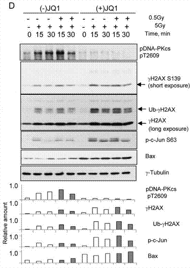
| Western blot | pDNA-PKcs / γH2AX / Ub-γH2AX / p-c-Jun S63 / Bax c-Myc p27 |

|
26119999 |
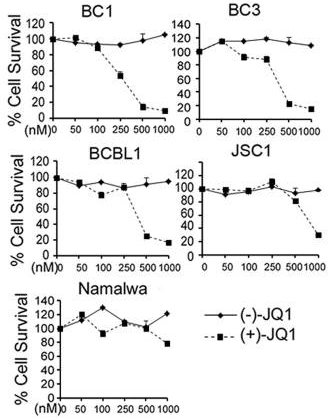
| Growth inhibition assay | Cell viability |

|
23792448 |
| Immunofluorescence | GM130 MHC / EdU |

|
29074567 |
Tech Support
Tel: +1-832-582-8158 Ext:3
If you have any other enquiries, please leave a message.
Frequently Asked Questions
Question 1:
How can I reconstitute it for in vivo injection?
Answer:
It does not dissolve in water/PBS. The vehicle we recommend is 2% DMSO+30% PEG 300+5% Tween 80+ddH2O. This compound can be dissolved in the vehicle at 5mg/ml and you can use it for IV injection.


