- Inhibitors
- By product type
- Natural Products
- Inducing Agents
- Peptides
- Antibiotics
- Antibody-drug Conjugates(ADC)
- PROTAC
- Hydrotropic Agents
- Dyes
- By Signaling Pathways
- PI3K/Akt/mTOR
- Epigenetics
- Methylation
- Immunology & Inflammation
- Protein Tyrosine Kinase
- Angiogenesis
- Apoptosis
- Autophagy
By research - Antibodies
- Compound Libraries
- Popular Compound Libraries
- Customize Library
- Clinical and FDA-approved Related
- Bioactive Compound Libraries
- Inhibitor Related
- Natural Product Related
- Metabolism Related
- Cell Death Related
- By Signaling Pathway
- By Disease
- Anti-infection and Antiviral Related
- Neuronal and Immunology Related
- Fragment and Covalent Related
- FDA-approved Drug Library
- FDA-approved & Passed Phase I Drug Library
- Preclinical/Clinical Compound Library
- Bioactive Compound Library-I
- Bioactive Compound Library-Ⅱ
- Kinase Inhibitor Library
- Express-Pick Library
- Natural Product Library
- Human Endogenous Metabolite Compound Library
- Alkaloid Compound LibraryNew
- Angiogenesis Related compound Library
- Anti-Aging Compound Library
- Anti-alzheimer Disease Compound Library
- Antibiotics compound Library
- Anti-cancer Compound Library
- Anti-cancer Compound Library-Ⅱ
- Anti-cancer Metabolism Compound Library
- Anti-Cardiovascular Disease Compound Library
- Anti-diabetic Compound Library
- Anti-infection Compound Library
- Antioxidant Compound Library
- Anti-parasitic Compound Library
- Antiviral Compound Library
- Apoptosis Compound Library
- Autophagy Compound Library
- Calcium Channel Blocker LibraryNew
- Cambridge Cancer Compound Library
- Carbohydrate Metabolism Compound LibraryNew
- Cell Cycle compound library
- CNS-Penetrant Compound Library
- Covalent Inhibitor Library
- Cytokine Inhibitor LibraryNew
- Cytoskeletal Signaling Pathway Compound Library
- DNA Damage/DNA Repair compound Library
- Drug-like Compound Library
- Endoplasmic Reticulum Stress Compound Library
- Epigenetics Compound Library
- Exosome Secretion Related Compound LibraryNew
- FDA-approved Anticancer Drug LibraryNew
- Ferroptosis Compound Library
- Flavonoid Compound Library
- Fragment Library
- Glutamine Metabolism Compound Library
- Glycolysis Compound Library
- GPCR Compound Library
- Gut Microbial Metabolite Library
- HIF-1 Signaling Pathway Compound Library
- Highly Selective Inhibitor Library
- Histone modification compound library
- HTS Library for Drug Discovery
- Human Hormone Related Compound LibraryNew
- Human Transcription Factor Compound LibraryNew
- Immunology/Inflammation Compound Library
- Inhibitor Library
- Ion Channel Ligand Library
- JAK/STAT compound library
- Lipid Metabolism Compound LibraryNew
- Macrocyclic Compound Library
- MAPK Inhibitor Library
- Medicine Food Homology Compound Library
- Metabolism Compound Library
- Methylation Compound Library
- Mouse Metabolite Compound LibraryNew
- Natural Organic Compound Library
- Neuronal Signaling Compound Library
- NF-κB Signaling Compound Library
- Nucleoside Analogue Library
- Obesity Compound Library
- Oxidative Stress Compound LibraryNew
- Plant Extract Library
- Phenotypic Screening Library
- PI3K/Akt Inhibitor Library
- Protease Inhibitor Library
- Protein-protein Interaction Inhibitor Library
- Pyroptosis Compound Library
- Small Molecule Immuno-Oncology Compound Library
- Mitochondria-Targeted Compound LibraryNew
- Stem Cell Differentiation Compound LibraryNew
- Stem Cell Signaling Compound Library
- Natural Phenol Compound LibraryNew
- Natural Terpenoid Compound LibraryNew
- TGF-beta/Smad compound library
- Traditional Chinese Medicine Library
- Tyrosine Kinase Inhibitor Library
- Ubiquitination Compound Library
-
Cherry Picking
You can personalize your library with chemicals from within Selleck's inventory. Build the right library for your research endeavors by choosing from compounds in all of our available libraries.
Please contact us at info@selleckchem.com to customize your library.
You could select:
- Bioreagents
- qPCR
- 2x SYBR Green qPCR Master Mix
- 2x SYBR Green qPCR Master Mix(Low ROX)
- 2x SYBR Green qPCR Master Mix(High ROX)
- Protein Assay
- Protein A/G Magnetic Beads for IP
- Anti-Flag magnetic beads
- Anti-Flag Affinity Gel
- Anti-Myc magnetic beads
- Anti-HA magnetic beads
- Poly DYKDDDDK Tag Peptide lyophilized powder
- Protease Inhibitor Cocktail
- Protease Inhibitor Cocktail (EDTA-Free, 100X in DMSO)
- Phosphatase Inhibitor Cocktail (2 Tubes, 100X)
- Cell Biology
- Cell Counting Kit-8 (CCK-8)
- Animal Experiment
- Mouse Direct PCR Kit (For Genotyping)
- Featured Products
- MRTX1133
- Nab-Paclitaxel
- KP-457
- IAG933
- RMC-6236 (Daraxonrasib)
- RMC-7977
- Zoldonrasib (RMC-9805)
- GsMTx4
- Navitoclax (ABT-263)
- TSA (Trichostatin A)
- Y-27632 Dihydrochloride
- SB431542
- SB202190
- MK-2206 Dihydrochloride
- LY294002
- Alisertib (MLN8237)
- XAV-939
- CHIR-99021 (Laduviglusib)
- Bafilomycin A1 (Baf-A1)
- Thiazovivin (TZV)
- CP-673451
- Verteporfin
- DAPT
- Galunisertib (LY2157299)
- MG132
- SBE-β-CD
- Tween 80
- Bavdegalutamide (ARV-110)
- Z-VAD-FMK
- Wnt-C59 (C59)
- IWR-1-endo
- (+)-JQ1
- 3-Deazaneplanocin A (DZNep) Hydrochloride
- RepSox (E-616452)
- Erastin
- Q-VD-Oph
- Puromycin Dihydrochloride
- Cycloheximide
- Telaglenastat (CB-839)
- A-83-01
- Ceralasertib (AZD6738)
- Liproxstatin-1
- Emricasan (IDN-6556)
- PMA (Phorbol 12-myristate 13-acetate)
- Dibutyryl cAMP (Bucladesine) sodium
- Nedisertib (M3814)
- PLX5622
- IKE (Imidazole Ketone Erastin)
- STM2457
- Saruparib (AZD5305)
- New Products
- Contact Us
research use only
SU5402 VEGFR inhibitor
Cat.No.S7667

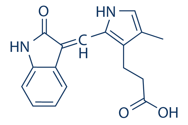
Chemical Structure
Molecular Weight: 296.32
Quality Control
Batch:
Purity:
99.12%
99.12
Cell Culture, Treatment & Working Concentration
| Cell Lines | Assay Type | Concentration | Incubation Time | Formulation | Activity Description | PMID |
|---|---|---|---|---|---|---|
| HUVEC or NIH3T3 | Function assay | Evaluated for the inhibition of cell proliferation induced by VEGF in HUVEC or NIH3T3 cells, IC50=0.05μM | 10893303 | |||
| HUVEC or NIH3T3 | Function assay | Evaluated for the inhibition of cell proliferation induced by FGF in HUVEC or NIH3T3 cells, IC50=2.8μM | 10893303 | |||
| NIH/3T3 | Function assay | Inhibition of acidic FGF-stimulated FGFR1 tyrosine autophosphorylation in mouse NIH/3T3 cells by immunoblotting, IC50=10μM | 9139660 | |||
| NIH 3T3 | Function assay | 5 mins | Inhibition of FGF induced FGFR1 autophosphorylation in mouse NIH 3T3 cells preincubated for 5 mins followed by FGF-stimulation for 5 mins in presence of [gamma-32P]ATP by SDS-PAGE based autoradiography, IC50=10μM | 27914362 | ||
| HUVEC or NIH3T3 | Function assay | Evaluated for the inhibition of cell proliferation induced by PDGF in HUVEC or NIH3T3 cells, IC50=28.4μM | 10893303 | |||
| NIH/3T3 | Growth inhibition assay | Growth inhibition of mouse NIH/3T3 cells assessed as inhibition of acidic FGF-stimulated [3H]thymidine incorporation | 9139660 | |||
| NIH/3T3 | Function assay | Inhibition of FGFR1 in mouse NIH/3T3 cells assessed as inhibition of aFGF-stimulated pp90 phosphoprotein phosphorylation by immunoblotting | 9139660 | |||
| NIH/3T3 | Function assay | Inhibition of FGFR1 in mouse NIH/3T3 cells assessed as inhibition of acidic FGF-stimulated ERK1 phosphorylation by immunoblotting | 9139660 | |||
| NIH/3T3 | Function assay | Inhibition of FGFR1 in mouse NIH/3T3 cells assessed as inhibition of acidic FGF-stimulated ERK2 phosphorylation by immunoblotting | 9139660 | |||
| NIHIR | Function assay | Inhibition of insulin-stimulated insulin receptor beta subunit autophosphorylation in mouse NIHIR cells | 9139660 | |||
| HER14 | Function assay | Inhibition of EGF-stimulated EGFR phosphorylation in mouse HER14 cells by immunoblotting | 9139660 | |||
| HER14 | Function assay | Inhibition of EGFR in mouse HER14 cells assessed as inhibition of EGF-stimulated Shc phosphorylation by immunoblotting | 9139660 | |||
| HER14 | Function assay | Inhibition of EGFR in mouse HER14 cells assessed as inhibition of EGF-stimulated ERK2 activation by immunoblotting | 9139660 | |||
| NIHIR | Function assay | Inhibition of insulin receptor in mouse NIHIR cells assessed as inhibition of insulin-stimulated IRS1 phosphorylation | 9139660 | |||
| NIH/3T3 | Function assay | Inhibition of PDGFR in mouse NIH/3T3 cells assessed as inhibition of PDGF-stimulated tyrosine autophosphorylation by immunoblotting | 9139660 | |||
| NIH/3T3 | Function assay | Inhibition of PDGFR in mouse NIH/3T3 cells assessed as inhibition of PDGF-stimulated phospholipase C gamma phosphorylation by immunoblotting | 9139660 | |||
| NIH/3T3 | Function assay | Inhibition of PDGFR in mouse NIH/3T3 cells assessed as PDGF-stimulated Erk2 activation by immunoblotting | 9139660 | |||
| Click to View More Cell Line Experimental Data | ||||||
Chemical Information, Storage & Stability
| Molecular Weight | 296.32 | Formula | C17H16N2O3 |
Storage (From the date of receipt) | |
|---|---|---|---|---|---|
| CAS No. | 215543-92-3 | Download SDF | Storage of Stock Solutions |
|
|
| Synonyms | N/A | Smiles | CC1=CNC(=C1CCC(=O)O)C=C2C3=CC=CC=C3NC2=O | ||
Solubility
|
In vitro |
DMSO : 59 mg/mL ( (199.1 mM) Moisture-absorbing DMSO reduces solubility. Please use fresh DMSO.) Water : Insoluble Ethanol : Insoluble |
Molarity Calculator
|
In vivo |
|||||
In vivo Formulation Calculator (Clear solution)
Step 1: Enter information below (Recommended: An additional animal making an allowance for loss during the experiment)
mg/kg
g
μL
Step 2: Enter the in vivo formulation (This is only the calculator, not formulation. Please contact us first if there is no in vivo formulation at the solubility Section.)
% DMSO
%
% Tween 80
% ddH2O
%DMSO
%
Calculation results:
Working concentration: mg/ml;
Method for preparing DMSO master liquid: mg drug pre-dissolved in μL DMSO ( Master liquid concentration mg/mL, Please contact us first if the concentration exceeds the DMSO solubility of the batch of drug. )
Method for preparing in vivo formulation: Take μL DMSO master liquid, next addμL PEG300, mix and clarify, next addμL Tween 80, mix and clarify, next add μL ddH2O, mix and clarify.
Method for preparing in vivo formulation: Take μL DMSO master liquid, next add μL Corn oil, mix and clarify.
Note: 1. Please make sure the liquid is clear before adding the next solvent.
2. Be sure to add the solvent(s) in order. You must ensure that the solution obtained, in the previous addition, is a clear solution before proceeding to add the next solvent. Physical methods such
as vortex, ultrasound or hot water bath can be used to aid dissolving.
Mechanism of Action
| Targets/IC50/Ki | |
|---|---|
| In vitro |
SU5402 inhibits VEGF-, FGF-, PDGF- dependent cell proliferation with IC50 of 0.05 μM, 2.80μM, 28.4 μM, respectively. [1] In HUVECs, this compound selectively inhibits VEGF-driven mitogenesis in a dose-dependent manner with IC50 of 0.04 μM. [2] In nasopharyngeal epithelial cells, it attenuates LMP1-mediated aerobic glycolysis, cellular transformation, cell migration, and invasion. [3] In mouse C3H10T1/2 cells, this chemical diminishes the effect of FGF23 on cell differentiation. [4]
|
| Kinase Assay |
FGF-R1 and Flk-1/KDR kinase assays.
|
|
The catalytic portion of FGF-R1 and Flk-1/KDR are expressed as GST fusion proteins following infection of Spodoptera frugiperda (sf9) cells with engineered baculoviruses. GST-FGFR1 and GST-Flk1 are purified to homogeneity from infected sf9 cell lysates by glutathione sepharose chromatography. The assays are performed in 96-well microtiter plates that had been coated overnight with 2.0 μg of a polyGlu-Tyr peptide (4:1) in 0.1 mL of PBS per well. The purified kinases are diluted in kinase assay buffer (100 mM Hepes pH 7.5, 100 mM NaCl, and 0.1 mM sodium orthovanadate) and added to all test wells at 5 ng of GST fusion protein per 0.05 mL volume buffer. Test compounds are diluted in 4% DMSO and added to test wells (0.025 mL/well). The kinase reaction is initiated by the addition of 0.025 mL of 40 μM ATP/40 mM MnCl2, and plates are shaken for 10 min before stopping the reactions with the addition of 0.025 mL of 0.5 M EDTA. The final ATP concentration was 10 μM, which is twice the experimentally determined Km value for ATP. Negative control wells receive MnCl2 alone without ATP. The plates are washed three times with 10 mM Tris pH 7.4, 150 mM NaCl, and 0.05% Tween-20 (TBST). Rabbit polyclonal anti-phosphotyrosine antiserum is added to the wells at a 1:10000 dilution in TBST for 1 h. The plates are then washed three times with TBST. Goat anti-rabbit antiserum conjugated with horseradish peroxidase was then added to all wells for 1 h. The plates are washed three times with TBST, and the peroxidase reaction is detected with the addition of 2,2‘-azinobis(3-ethylbenzthiazoline-6-sulfonic acid) (ABTS). The color readout of the assay is allowed to develop for 20−30 min and read on a Dynatech MR5000 ELISA plate reader using a 410 nM test filter.
|
|
| In vivo |
In mice, SU5416 (25 mg/kg, i.p.) inhibits subcutaneous growth of a panel of tumor cell lines by inhibiting the angiogenic process associated with tumor growth. [2]
|
References |
|
Tech Support
Tel: +1-832-582-8158 Ext:3
If you have any other enquiries, please leave a message.


