-
 Australia
-
 Austria
-
 Belgium
-
 Brazil
-
 Canada
-
 China
China
-
 Czech Republic
-
 Denmark
-
 Finland
-
 France
-
 Germany
-
 Greece
-
 Hong Kong
-
 Hungary
-
 Iceland
-
 India
India
-
 Ireland
-
 Israel
-
 Italy
Italy
-
 Japan
Japan
-
 Korea
Korea
-
 Luxembourg
-
 Malaysia
-
 Netherlands
-
 New Zealand
-
 Norway
-
 Poland
-
 Qatar
Qatar
-
 Romania
-
 Saudi Arabia
-
 Singapore
-
 Spain
Spain
-
 Sweden
-
 Switzerland
-
 Taiwan
-
 Turkey
-
 United Kingdom
-
 United States
research use only
PF-04691502 PI3K inhibitor
Cat.No.S2743
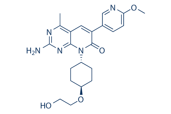
Chemical Structure
Molecular Weight: 425.48
Quality Control
| Related Targets | Akt mTOR GSK-3 ATM/ATR DNA-PK AMPK PDPK1 PTEN PP2A PDK |
|---|---|
| Other PI3K Inhibitors | GDC-0077 (Inavolisib) SAR405 Quercetin (Sophoretin) LY294002 XL147 analogue Tersolisib (STX-478) Buparlisib (BKM120) 740 Y-P (PDGFR 740Y-P) GO-203 TFA Eganelisib (IPI-549) |
Cell Culture, Treatment & Working Concentration
| Cell Lines | Assay Type | Concentration | Incubation Time | Formulation | Activity Description | PMID |
|---|---|---|---|---|---|---|
| human BT20 cells | Function assay | Inhibition of AKT phosphorylation at Ser 473 in human BT20 cells, IC50=0.013 μM | ||||
| human SKOV3 cells | Proliferation assay | 3 days | Antiproliferative activity against human SKOV3 cells after 3 days by CellTiter-Glo assay, IC50=0.29 μM | |||
| human U87MG cells | Proliferation assay | 4 days | Antiproliferative activity against human U87MG cells after 4 days by CellTiter-Glo assay, IC50=0.52 μM | |||
| Click to View More Cell Line Experimental Data | ||||||
Solubility
|
In vitro |
DMSO
: 14 mg/mL
(32.9 mM)
Water : Insoluble Ethanol : Insoluble |
Molarity Calculator
|
In vivo |
|||||
In vivo Formulation Calculator (Clear solution)
Step 1: Enter information below (Recommended: An additional animal making an allowance for loss during the experiment)
Step 2: Enter the in vivo formulation (This is only the calculator, not formulation. Please contact us first if there is no in vivo formulation at the solubility Section.)
Calculation results:
Working concentration: mg/ml;
Method for preparing DMSO master liquid: mg drug pre-dissolved in μL DMSO ( Master liquid concentration mg/mL, Please contact us first if the concentration exceeds the DMSO solubility of the batch of drug. )
Method for preparing in vivo formulation: Take μL DMSO master liquid, next addμL PEG300, mix and clarify, next addμL Tween 80, mix and clarify, next add μL ddH2O, mix and clarify.
Method for preparing in vivo formulation: Take μL DMSO master liquid, next add μL Corn oil, mix and clarify.
Note: 1. Please make sure the liquid is clear before adding the next solvent.
2. Be sure to add the solvent(s) in order. You must ensure that the solution obtained, in the previous addition, is a clear solution before proceeding to add the next solvent. Physical methods such
as vortex, ultrasound or hot water bath can be used to aid dissolving.
Chemical Information, Storage & Stability
| Molecular Weight | 425.48 | Formula | C22H27N5O4 |
Storage (From the date of receipt) | |
|---|---|---|---|---|---|
| CAS No. | 1013101-36-4 | Download SDF | Storage of Stock Solutions |
|
|
| Synonyms | PF4691502 | Smiles | CC1=C2C=C(C(=O)N(C2=NC(=N1)N)C3CCC(CC3)OCCO)C4=CN=C(C=C4)OC | ||
Mechanism of Action
| Targets/IC50/Ki |
PI3Kδ
(Cell-free assay) 1.6 nM(Ki)
PI3Kα
(Cell-free assay) 1.8 nM(Ki)
PI3Kγ
(Cell-free assay) 1.9 nM(Ki)
PI3Kβ
(Cell-free assay) 2.1 nM(Ki)
P-Akt (S473)
(Cell-free assay) 3.8 nM
P-Akt (T308)
(Cell-free assay) 7.5 nM
mTOR
(Cell-free assay) 16 nM(Ki)
|
|---|---|
| In vitro |
PF-04691502 potently inhibits recombinant class I PI3K and mTOR in biochemical assays and suppresses transformation of avian fibroblasts mediated by wild-type PI3K γ, δ, or mutant PI3Kα. In PIK3CA-mutant and PTEN-deleted cancer cell lines, this compound reduces phosphorylation of AKT T308 and AKT S473 (IC(50) of 7.5-47 nM and 3.8-20 nM, respectively) and inhibits cell proliferation (IC(50) of 179-313 nM). It inhibits mTORC1 activity in cells as measured by PI3K-independent nutrient stimulated assay, with an IC(50) of 32 nM and inhibits the activation of PI3K and mTOR downstream effectors including AKT, FKHRL1, PRAS40, p70S6K, 4EBP1, and S6RP. Short-term exposure to this chemical predominantly inhibits PI3K, whereas mTOR inhibition persists for 24 to 48 hours. This compound induces cell cycle G(1) arrest, concomitant with upregulation of p27 Kip1 and reduction of Rb. |
| Kinase Assay |
Kinase Assay
|
|
The fluorescence polarization assay for ATP competitive inhibition is done as follows: mPI3Kα dilution solution (90 nM) is prepared in fresh assay buffer (50 mM Hepes pH 7.4, 150 mM NaCl, 5 mM DTT, 0.05% CHAPS) and kept on ice. The enzyme reaction contains 0.5 nM mouse PI3Kα (p110α/p85α complex purified from insect cells), 30 μM PIP2, PF-04691502 (0, 1, 4, and 8 nM), 5 mM MgCl2, and 2-fold serial dilutions of ATP (0–800 μM). Final dimethyl sulfoxide is 2.5%. The reaction is initiated by the addition of ATP and terminated after 30 minutes with 10 mM EDTA. In a detection plate, 15 uL of detector/probe mixture containing 480 nM GST-Grp1PH domain and 12 nM TAMRA tagged fluorescent PIP3 in assay buffer is mixed with 15 uL of kinase reaction mixture. The plate is shaken for 3 minutes, and incubated for 35 to 40 minutes before reading on an LJL Analyst HT.
|
|
| In vivo |
Antitumor activity of PF-04691502 is observed in U87 (PTEN null), SKOV3 (PIK3CA mutation), and non-small cell lung carcinoma xenografts. This compound inhibits tumor growth at 7 days by 72%. FDG-PET imaging revealed that it reduces glucose metabolism dramatically. Tissue biomarkers of PI3K/mTOR pathway activity, p-AKT (S473), and p-RPS6 (S240/244), are also dramatically inhibited following its treatment. |
References |
|
Applications
| Methods | Biomarkers | Images | PMID |
|---|---|---|---|
| Western blot | p-AKT / p-ERK p-S6 / S6 |
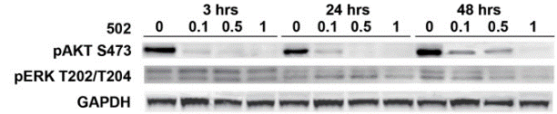
|
23826249 |
| Growth inhibition assay | Cell viability |
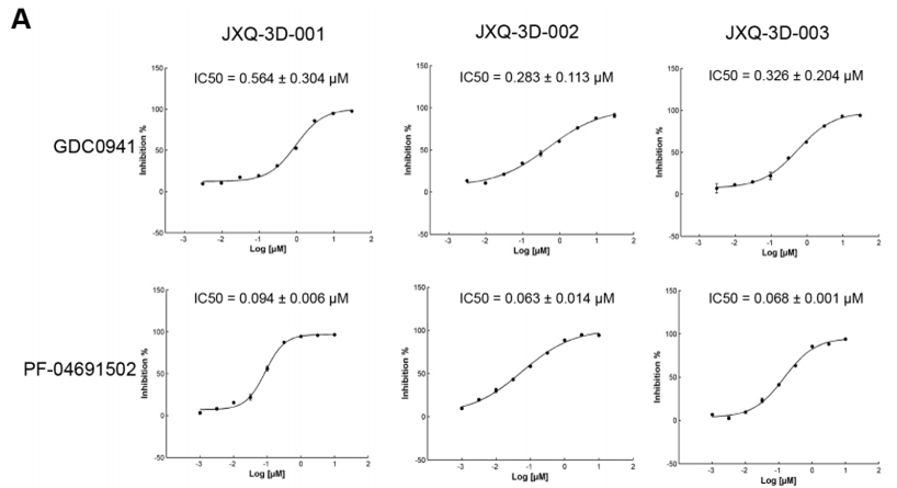
|
28029662 |
Clinical Trial Information
(data from https://clinicaltrials.gov, updated on 2024-05-22)
| NCT Number | Recruitment | Conditions | Sponsor/Collaborators | Start Date | Phases |
|---|---|---|---|---|---|
| NCT01658176 | Withdrawn | Breast Neoplasms |
Pfizer |
January 2013 | Phase 2 |
| NCT01420081 | Terminated | Endometrial Neoplasms |
Pfizer |
January 19 2012 | Phase 2 |
| NCT01347866 | Terminated | Advanced Cancer |
Pfizer |
October 2011 | Phase 1 |
Tech Support
Tel: +1-832-582-8158 Ext:3
If you have any other enquiries, please leave a message.
Signaling Pathway Map
Products are for research use only. Not for human use. We do not sell to patients.
©Copyright 2013 Selleck Chemicals. All Rights Reserved.