-
 Australia
-
 Austria
-
 Belgium
-
 Brazil
-
 Canada
-
 China
China
-
 Czech Republic
-
 Denmark
-
 Finland
-
 France
-
 Germany
-
 Greece
-
 Hong Kong
-
 Hungary
-
 Iceland
-
 India
India
-
 Ireland
-
 Israel
-
 Italy
Italy
-
 Japan
Japan
-
 Korea
Korea
-
 Luxembourg
-
 Malaysia
-
 Netherlands
-
 New Zealand
-
 Norway
-
 Poland
-
 Qatar
Qatar
-
 Romania
-
 Saudi Arabia
-
 Singapore
-
 Spain
Spain
-
 Sweden
-
 Switzerland
-
 Taiwan
-
 Turkey
-
 United Kingdom
-
 United States
research use only
Tubastatin A Hydrochloride HDAC6 Inhibitor
Cat.No.S2627
Chemical Structure
Molecular Weight: 371.86
Quality Control
Cell Culture, Treatment & Working Concentration
| Cell Lines | Assay Type | Concentration | Incubation Time | Formulation | Activity Description | PMID |
|---|---|---|---|---|---|---|
| neuron cultures | Kinase assay | 2.5 μM | DMSO | induces α-tubulin hyperacetylation | ||
| neuron cultures | Function assay | ~10 μM | DMSO | protects against glutathione depletion-induced oxidative stress | ||
| 134/04 | Function assay | 7.5 µM | impairs myotube formation | |||
| C2C12 | Function assay | 7.5 µM | impairs myotube formation | |||
| HaCaT keratinocytes | Function assay | 10 μM | blocks arsenite from inducing Nrf2 protein translation | |||
| JURL-MK1 | Function assay | 10 μM | enhances cell adhesivity to fibronectin | |||
| CML-T1 | Function assay | 10 μM | enhances cell adhesivity to fibronectin | |||
| K562 | Function assay | 10 μM | enhances cell adhesivity to fibronectin | |||
| HL-60 | Function assay | 10 μM | enhances cell adhesivity to fibronectin | |||
| KMCH | Growth inhibitory assay | ~10 μM | decreases proliferation and anchorage-independent growth | |||
| THP-1 | Function assay | ~10 μM | inhibits TNF-α and IL-6 secretion | |||
| RAW 264.7 | Function assay | ~10 μM | attenuates NO production | |||
| HT3 | Function assay | ~5 μM | DMSO | induces the differential α-tubulin acetylation | ||
| SiHa | Function assay | ~5 μM | DMSO | induces the differential α-tubulin acetylation | ||
| CaSki | Function assay | ~5 μM | DMSO | induces the differential α-tubulin acetylation | ||
| SiHa | Function assay | ~5 μM | DMSO | inhibits Thapsigargin- or EGF-induced SOCE activation | ||
| CaSki | Function assay | ~5 μM | DMSO | inhibits Thapsigargin- or EGF-induced SOCE activation | ||
| MCF-7 | Growth inhibitory assay | 30 μM | DMSO | IC50=15 μM | ||
| MCF-7 | Function assay | 30 μM | DMSO | increases the microtubule acetylation level. | ||
| MCF-7 | Function assay | 30 μM | DMSO | stabilizes microtubules against cold-induced depolymerization | ||
| MCF-7 | Function assay | 15 μM | DMSO | stabilizes microtubules against nocodazole-induced disassembly | ||
| MCF-7 | Function assay | 30 μM | DMSO | alteres the assembly dynamics of interphase microtubules | ||
| MCF-7 | Function assay | 30 μM | DMSO | increases the binding of HDAC6 with interphase microtubules | ||
| PC12 | Function assay | ~3 μM | DMSO | up-regulates anti-oxidative gene expression related to transcription factor XBP1s | ||
| PC12 | Growth inhibitory assay | ~3 μM | DMSO | reverse H2O2-induced growth inhibition | ||
| HEK293T | Function assay | ~3 μM | DMSO | up-regulated XBP1s protein level | ||
| HEK293T | Function assay | ~3 μM | DMSO | delays XBP1s protein degradation via acetylation-mediated proteasomal degradation | ||
| Huh7 | Function assay | ~5 μM | DMSO | suppresses proliferation of hepatitis C virus replicon with EC50 = 0.3 μM | ||
| SKMEL21 | Growth inhibitory assay | ~500 nM | DMSO | inhibits cell proliferation | ||
| SKMEL103 | Growth inhibitory assay | ~500 nM | DMSO | inhibits cell proliferation | ||
| SKMEL28 | Growth inhibitory assay | ~500 nM | DMSO | inhibits cell proliferation | ||
| WM164 | Growth inhibitory assay | ~500 nM | DMSO | inhibits cell proliferation | ||
| WM1361a | Growth inhibitory assay | ~500 nM | DMSO | inhibits cell proliferation | ||
| WM1366 | Growth inhibitory assay | ~500 nM | DMSO | inhibits cell proliferation | ||
| WM793 | Growth inhibitory assay | ~500 nM | DMSO | inhibits cell proliferation | ||
| WM35 | Growth inhibitory assay | ~500 nM | DMSO | inhibits cell proliferation | ||
| WM983a | Growth inhibitory assay | ~500 nM | DMSO | inhibits cell proliferation | ||
| WM793 | Function assay | ~6 μM | DMSO | induce G1 arrest | ||
| WM164 | Function assay | ~6 μM | DMSO | induce G1 arrest | ||
| WM983a | Function assay | ~6 μM | DMSO | induce G1 arrest | ||
| WM164 | Function assay | ~3 μM | DMSO | augments expression of MHC class I and melanoma associated antigens | ||
| WM983a | Function assay | ~3 μM | DMSO | augments expression of MHC class I and melanoma associated antigens | ||
| IPC298 | Function assay | ~3 μM | DMSO | augments expression of MHC class I and melanoma associated antigens | ||
| SKMEL30 | Function assay | ~3 μM | DMSO | augments expression of MHC class I and melanoma associated antigens | ||
| TCa83 | Function assay | induces PTEN expression and membrane translocation | ||||
| 293T | Function assay | ~2 μg/ml | induces PTEN expression and membrane translocation | |||
| SACC-83 | Function assay | ~2 μg/ml | induces PTEN expression and membrane translocation | |||
| 293T | Function assay | ~2 μg/ml | induces PTEN acetylation at K163 | |||
| U-87 MG | Function assay | ~2 μg/ml | inhibits the migration and invasion | |||
| U-87 MG | Function assay | ~10 μM | inhibits AKT phosphorylation | |||
| U-87 MG | Growth inhibitory assay | ~10 μM | inhibits cell growth | |||
| Click to View More Cell Line Experimental Data | ||||||
Solubility
|
In vitro |
DMSO
: 74 mg/mL
(198.99 mM)
Water : Insoluble Ethanol : Insoluble |
Molarity Calculator
|
In vivo |
|||||
In vivo Formulation Calculator (Clear solution)
Step 1: Enter information below (Recommended: An additional animal making an allowance for loss during the experiment)
Step 2: Enter the in vivo formulation (This is only the calculator, not formulation. Please contact us first if there is no in vivo formulation at the solubility Section.)
Calculation results:
Working concentration: mg/ml;
Method for preparing DMSO master liquid: mg drug pre-dissolved in μL DMSO ( Master liquid concentration mg/mL, Please contact us first if the concentration exceeds the DMSO solubility of the batch of drug. )
Method for preparing in vivo formulation: Take μL DMSO master liquid, next addμL PEG300, mix and clarify, next addμL Tween 80, mix and clarify, next add μL ddH2O, mix and clarify.
Method for preparing in vivo formulation: Take μL DMSO master liquid, next add μL Corn oil, mix and clarify.
Note: 1. Please make sure the liquid is clear before adding the next solvent.
2. Be sure to add the solvent(s) in order. You must ensure that the solution obtained, in the previous addition, is a clear solution before proceeding to add the next solvent. Physical methods such
as vortex, ultrasound or hot water bath can be used to aid dissolving.
Chemical Information, Storage & Stability
| Molecular Weight | 371.86 | Formula | C20H21N3O2.HCl |
Storage (From the date of receipt) | |
|---|---|---|---|---|---|
| CAS No. | 1310693-92-5 | Download SDF | Storage of Stock Solutions |
|
|
Mechanism of Action
| Targets/IC50/Ki |
HDAC6
(Cell-free assay) 15 nM
HDAC8
(Cell-free assay) 854 nM
|
|---|---|
| In vitro |
Tubastatin A is substantially selective for all 11 HDAC isoforms and maintains over 1000-fold selectivity against all isoforms excluding HDAC8, where it has approximately 57-fold selectivity. In homocysteic acid (HCA) induced neurodegeneration assays, Tubastatin A displays dose-dependent protection against HCA-induced neuronal cell death starting at 5 μM with near complete protection at 10 μM. At 100 ng/mL Tubastatin A increases Foxp3+ T-regulatory cells (Tregs) suppression of T cell proliferation in vitro. Tubastatin A treatment in C2C12 cells would lead to myotube formation impairment when alpha-tubulin is hyperacetylated early in the myogenic process; however, myotube elongation occurs when alpha-tubulin is hyeperacetylated in myotubes. A recent study indicates that Tubastatin A treatment increases cell elasticity as revealed by atomic force microscopy (AFM) tests without exerting drastic changes to the actin microfilament or microtubule networks in mouse ovarian cancer cell lines, MOSE-E and MOSE-L.
|
| Kinase Assay |
Enzyme Inhibition Assays
|
|
Enzyme inhibition assays are performed by the Reaction Biology Corporation, Malvern, PA, using the Reaction Biology HDAC Spectrum platform. (www.reactionbiology.com) The HDAC1, 2,
4, 5, 6, 7, 8, 9, 10, and 11 assays use isolated recombinant human protein; HDAC3/NcoR2 complex is used for the HDAC3 assay. Substrate for HDAC1, 2, 3, 6, 10, and 11 assays is a fluorogenic peptide from p53 residues 379-382 (RHKKAc); substrate for HDAC8 is fluorogenic diacyl peptide based on residues 379-382 of p53 (RHKAcKAc). Acetyl-Lys (trifluoroacetyl)-AMC substrate is used for HDAC4, 5, 7, and 9 assays. Tubastatin A is dissolved in DMSO and tested in 10-dose IC50 mode with 3-fold serial dilution starting at 30 μM. Control Compound Trichostatin A (TSA) is tested in a 10-dose IC50 with 3-fold serial dilution starting at 5 μM. IC50 values are extracted by curve-fitting the dose/response slopes.
|
|
| In vivo |
Daily treatment of Tubastatin A at 0.5mg/kg inhibits HDAC6 to promote Tregs suppressive activity in mouse models of inflammation and autoimmunity, including multiple forms of experimental colitis and fully major histocompatibility complex (MHC)-incompatible cardiac allograft rejection.
|
References |
|
Applications
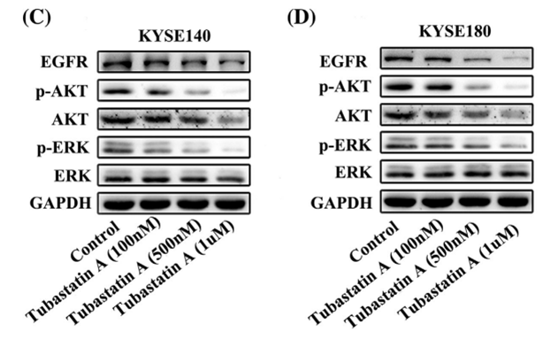
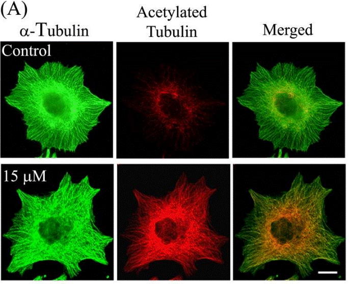
| Methods | Biomarkers | Images | PMID |
|---|---|---|---|
| Western blot | EGFR / p-AKT / AKT / p-ERK / ERK |
|
29665050 |
| Immunofluorescence | α-tubulin / Acetylated tubulin HDAC6 |
|
23798680 |
Tech Support
Tel: +1-832-582-8158 Ext:3
If you have any other enquiries, please leave a message.
Signaling Pathway Map
Products are for research use only. Not for human use. We do not sell to patients.
©Copyright 2013 Selleck Chemicals. All Rights Reserved.