-
 Australia
-
 Austria
-
 Belgium
-
 Brazil
-
 Canada
-
 China
China
-
 Czech Republic
-
 Denmark
-
 Finland
-
 France
-
 Germany
-
 Greece
-
 Hong Kong
-
 Hungary
-
 Iceland
-
 India
India
-
 Ireland
-
 Israel
-
 Italy
Italy
-
 Japan
Japan
-
 Korea
Korea
-
 Luxembourg
-
 Malaysia
-
 Netherlands
-
 New Zealand
-
 Norway
-
 Poland
-
 Qatar
Qatar
-
 Romania
-
 Saudi Arabia
-
 Singapore
-
 Spain
Spain
-
 Sweden
-
 Switzerland
-
 Taiwan
-
 Turkey
-
 United Kingdom
-
 United States
research use only
Scriptaid HDAC inhibitor
Cat.No.S8043
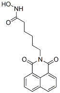
Chemical Structure
Molecular Weight: 326.35
Quality Control
Solubility
|
In vitro |
DMSO
: 65 mg/mL
(199.17 mM)
Water : Insoluble Ethanol : Insoluble |
Molarity Calculator
|
In vivo |
|||||
In vivo Formulation Calculator (Clear solution)
Step 1: Enter information below (Recommended: An additional animal making an allowance for loss during the experiment)
Step 2: Enter the in vivo formulation (This is only the calculator, not formulation. Please contact us first if there is no in vivo formulation at the solubility Section.)
Calculation results:
Working concentration: mg/ml;
Method for preparing DMSO master liquid: mg drug pre-dissolved in μL DMSO ( Master liquid concentration mg/mL, Please contact us first if the concentration exceeds the DMSO solubility of the batch of drug. )
Method for preparing in vivo formulation: Take μL DMSO master liquid, next addμL PEG300, mix and clarify, next addμL Tween 80, mix and clarify, next add μL ddH2O, mix and clarify.
Method for preparing in vivo formulation: Take μL DMSO master liquid, next add μL Corn oil, mix and clarify.
Note: 1. Please make sure the liquid is clear before adding the next solvent.
2. Be sure to add the solvent(s) in order. You must ensure that the solution obtained, in the previous addition, is a clear solution before proceeding to add the next solvent. Physical methods such
as vortex, ultrasound or hot water bath can be used to aid dissolving.
Chemical Information, Storage & Stability
| Molecular Weight | 326.35 | Formula | C18H18N2O4 |
Storage (From the date of receipt) | |
|---|---|---|---|---|---|
| CAS No. | 287383-59-9 | Download SDF | Storage of Stock Solutions |
|
|
| Synonyms | GCK 1026 | Smiles | C1=CC2=C3C(=C1)C(=O)N(C(=O)C3=CC=C2)CCCCCC(=O)NO | ||
Mechanism of Action
| Targets/IC50/Ki |
HDAC
|
|---|---|
| In vitro |
Scriptaid (6 μM) results in a >100-fold increase in histone acetylation in PANC-1 cell. This compound (8 μM) is not lethal to PANC-1 cell and has limited effects (80% survival) on MDAMB-468. It increases the transcription of pCMVb, p6SBE-luc and p6MBE-luc independent of a positive inducer of transcription. This chemical is capable of inducing high expression of p6MBE-luc, pCMVb, and pUB6/V5-LacZ, driven by viral (SV40 and CMV) or human (ubiquitin c, UB6) promoters, which do not depend on the specificity of the enhancer (SBE versus MBE), the type of promoter (viral versus cellular), the product of the reporter gene (luciferase versus b-gal), nor on the integration status of the reporter construct.
It induces high rates of somatic cell nuclear transfer (SCNT) oocytes development to the blastocyst stage and allowed full-term development (3.4, 4.2, 7.6, 6.8, and 4.1%) with all concentrations (50, 100, 250, 500, and 2000 nM respectively). This compound improves the full-term development of cloned B6D2F1embryos in a dose-dependent manner with the maximum effect at 250 nM. It enables the clone of all the important inbred mouse strains, such as C57BL/6, C3H/He, DBA/2, and 129/Sv. This chemical treatment enhances newly synthesized mRNA levels in cloned embryos. 250 nM of this compound treated for up to 48 h, does not inhibit the development of ICSI-fertilized embryos.
It inhibits T. gondii tachyzoite proliferation with IC50 of 39 nM. This compound (0.225 μM) completely protects the HS68 monolayers from T. gondii tachyzoite.
It inhibits growth of ER negative cell lines, MDA-MB-231, MDA-MB-435 and Hs578t with IC50 of 0.5-1.0 μg/mL after 48 h treatment. 1 μg/ml of this compound treated for 48 h induces an accumulation of both acetylated H3 and acetylated H4 histone tail proteins, and a maximal of 20,000-fold increase of ER mRNA transcript.
It inhibits the proliferation and viability of the Ishikawa endometrial cancer cell line, and the SK-OV-3 ovarian cancer cell line with IC50 of 9 μM and 55 μM, respectively, while the normal human endometrial epithelial cells shows little sensitivity. Endometrial cancer cells and ovarian cancer cells cultured for 2 days in the presence of this compound shows an accumulation in the G0/G1 phase (5 μM of this chemical) and G2/M phase (10 μM of this compound) of the cell cycle, with a concomitant decrease in the proportion of those in the S phase. 10 μM of this chemical induces a 56.1% of apoptotic Ishikawa cells with loss of mitochondrial membrane potential, and decreased levels of cyclin A and bcl-2 levels by 50% and 20%, respectively.
|
| Kinase Assay |
Immunoblotting assay of histone acetylation
|
|
PANC-1 cells are treated with 2 μg/mL of Scriptaid for 18 h in culture medium. Treated and untreated cells are harvested with trypsin-EDTA, washed with PBS, and resuspended in a protein sample buffer. Protein concentration is determined by BCA protein assay reagents. Fifty μg of proteins from each sample is loaded on a 12% denaturing polyacrylamide gel. Proteins are subsequently transferred to a nylon membrane using MilliblotGraphite Electroblotter I. The nylon membrane is incubated with rabbit antihuman acetyl-lysine antibody, followed by goat antirabbit antibody coupled to horseradish peroxidase, developed with SuperSignal substrates, and detected by film.
|
|
| In vivo |
Scriptaid elicits a dose-dependent decrease in lesion size (a maximal decrease of 45%) at 1.5 to 5.5 mg/kg and a concomitant attenuation in motor and cognitive deficits when delivered 30 minutes postinjury in a model of mode rate TBI. Comparable protection is achieved even when treatment is delayed to 12 h postinjury. The protection of motor and cognitive functions is long lasting, as similar improvements are detected 35 days postinjury. This compound induces an increase in surviving neurons (42%), as well as the number/length of their processes within the CA3 region of the hippocampus and the pericontusional cortex. This treatment prevents the decrease in phospho-AKT (p-AKT) and phosphorylated phosphatase and tensin homolog deleted on chromosome 10 ( p-PTEN) induced by TBI in cortical and CA3 hippocampal neurons.
This chemical (3.5 mg/kg) clearly inhibits tumor growth in a human breast cancer xenograft MDA-MB-231 model, reducing tumor volume by 75%.
|
References |
|
Tech Support
Tel: +1-832-582-8158 Ext:3
If you have any other enquiries, please leave a message.
Signaling Pathway Map
Products are for research use only. Not for human use. We do not sell to patients.
©Copyright 2013 Selleck Chemicals. All Rights Reserved.