-
 Australia
-
 Austria
-
 Belgium
-
 Brazil
-
 Canada
-
 China
China
-
 Czech Republic
-
 Denmark
-
 Finland
-
 France
-
 Germany
-
 Greece
-
 Hong Kong
-
 Hungary
-
 Iceland
-
 India
India
-
 Ireland
-
 Israel
-
 Italy
Italy
-
 Japan
Japan
-
 Korea
Korea
-
 Luxembourg
-
 Malaysia
-
 Netherlands
-
 New Zealand
-
 Norway
-
 Poland
-
 Qatar
Qatar
-
 Romania
-
 Saudi Arabia
-
 Singapore
-
 Spain
Spain
-
 Sweden
-
 Switzerland
-
 Taiwan
-
 Turkey
-
 United Kingdom
-
 United States
research use only
Roscovitine (Seliciclib) CDK inhibitor
Cat.No.S1153
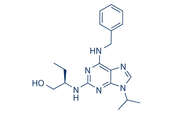
Chemical Structure
Molecular Weight: 354.45
Quality Control
Cell Culture, Treatment & Working Concentration
| Cell Lines | Assay Type | Concentration | Incubation Time | Formulation | Activity Description | PMID |
|---|---|---|---|---|---|---|
| LB771-HNC | Growth Inhibition Assay | IC50=48.9212 μM | SANGER | |||
| LB2241-RCC | Growth Inhibition Assay | IC50=48.6202 μM | SANGER | |||
| DU-4475 | Growth Inhibition Assay | IC50=48.4937 μM | SANGER | |||
| LB2518-MEL | Growth Inhibition Assay | IC50=47.0448 μM | SANGER | |||
| NCI-H209 | Growth Inhibition Assay | IC50=46.0115 μM | SANGER | |||
| CGTH-W-1 | Growth Inhibition Assay | IC50=44.9697 μM | SANGER | |||
| MS-1 | Growth Inhibition Assay | IC50=42.893 μM | SANGER | |||
| GI-ME-N | Growth Inhibition Assay | IC50=42.6671 μM | SANGER | |||
| DG-75 | Growth Inhibition Assay | IC50=42.6546 μM | SANGER | |||
| MLMA | Growth Inhibition Assay | IC50=42.2787 μM | SANGER | |||
| HT | Growth Inhibition Assay | IC50=42.0028 μM | SANGER | |||
| LC-1F | Growth Inhibition Assay | IC50=41.5705 μM | SANGER | |||
| NCI-H1882 | Growth Inhibition Assay | IC50=40.5998 μM | SANGER | |||
| NTERA-S-cl-D1 | Growth Inhibition Assay | IC50=39.5842 μM | SANGER | |||
| NCI-H345 | Growth Inhibition Assay | IC50=38.9106 μM | SANGER | |||
| MONO-MAC-6 | Growth Inhibition Assay | IC50=38.2477 μM | SANGER | |||
| RS4-11 | Growth Inhibition Assay | IC50=37.7069 μM | SANGER | |||
| ML-2 | Growth Inhibition Assay | IC50=37.6712 μM | SANGER | |||
| OPM-2 | Growth Inhibition Assay | IC50=37.2949 μM | SANGER | |||
| LU-139 | Growth Inhibition Assay | IC50=37.1856 μM | SANGER | |||
| COLO-684 | Growth Inhibition Assay | IC50=37.012 μM | SANGER | |||
| MOLT-4 | Growth Inhibition Assay | IC50=36.3276 μM | SANGER | |||
| TE-6 | Growth Inhibition Assay | IC50=36.3246 μM | SANGER | |||
| TE-441-T | Growth Inhibition Assay | IC50=36.1148 μM | SANGER | |||
| IMR-5 | Growth Inhibition Assay | IC50=35.3139 μM | SANGER | |||
| K5 | Growth Inhibition Assay | IC50=35.0861 μM | SANGER | |||
| TE-10 | Growth Inhibition Assay | IC50=34.9422 μM | SANGER | |||
| NCI-H2141 | Growth Inhibition Assay | IC50=34.6533 μM | SANGER | |||
| KGN | Growth Inhibition Assay | IC50=34.2524 μM | SANGER | |||
| LP-1 | Growth Inhibition Assay | IC50=33.8908 μM | SANGER | |||
| NCI-H64 | Growth Inhibition Assay | IC50=33.8597 μM | SANGER | |||
| RKO | Growth Inhibition Assay | IC50=33.5969 μM | SANGER | |||
| NCI-H526 | Growth Inhibition Assay | IC50=33.4936 μM | SANGER | |||
| GOTO | Growth Inhibition Assay | IC50=32.9129 μM | SANGER | |||
| Calu-6 | Growth Inhibition Assay | IC50=32.4745 μM | SANGER | |||
| LOUCY | Growth Inhibition Assay | IC50=32.1253 μM | SANGER | |||
| SK-N-FI | Growth Inhibition Assay | IC50=31.7535 μM | SANGER | |||
| SIG-M5 | Growth Inhibition Assay | IC50=31.6833 μM | SANGER | |||
| NKM-1 | Growth Inhibition Assay | IC50=31.1397 μM | SANGER | |||
| NCI-SNU-1 | Growth Inhibition Assay | IC50=31.1059 μM | SANGER | |||
| NCI-H82 | Growth Inhibition Assay | IC50=31.0135 μM | SANGER | |||
| NCI-H510A | Growth Inhibition Assay | IC50=30.0329 μM | SANGER | |||
| ES3 | Growth Inhibition Assay | IC50=29.9582 μM | SANGER | |||
| BB30-HNC | Growth Inhibition Assay | IC50=29.9483 μM | SANGER | |||
| KM12 | Growth Inhibition Assay | IC50=29.6239 μM | SANGER | |||
| GI-1 | Growth Inhibition Assay | IC50=29.0113 μM | SANGER | |||
| NOS-1 | Growth Inhibition Assay | IC50=28.9733 μM | SANGER | |||
| TE-8 | Growth Inhibition Assay | IC50=28.908 μM | SANGER | |||
| TE-9 | Growth Inhibition Assay | IC50=28.7969 μM | SANGER | |||
| HL-60 | Growth Inhibition Assay | IC50=27.9869 μM | SANGER | |||
| QIMR-WIL | Growth Inhibition Assay | IC50=27.9144 μM | SANGER | |||
| KARPAS-299 | Growth Inhibition Assay | IC50=26.8646 μM | SANGER | |||
| KURAMOCHI | Growth Inhibition Assay | IC50=26.8082 μM | SANGER | |||
| BL-41 | Growth Inhibition Assay | IC50=25.9597 μM | SANGER | |||
| NCI-H2126 | Growth Inhibition Assay | IC50=25.6529 μM | SANGER | |||
| HOP-62 | Growth Inhibition Assay | IC50=25.4425 μM | SANGER | |||
| IST-SL2 | Growth Inhibition Assay | IC50=24.5343 μM | SANGER | |||
| HH | Growth Inhibition Assay | IC50=24.3819 μM | SANGER | |||
| LS-513 | Growth Inhibition Assay | IC50=23.5179 μM | SANGER | |||
| EB-3 | Growth Inhibition Assay | IC50=23.1831 μM | SANGER | |||
| ACN | Growth Inhibition Assay | IC50=21.3389 μM | SANGER | |||
| NOMO-1 | Growth Inhibition Assay | IC50=21.2008 μM | SANGER | |||
| ES8 | Growth Inhibition Assay | IC50=21.06 μM | SANGER | |||
| CESS | Growth Inhibition Assay | IC50=20.8549 μM | SANGER | |||
| BL-70 | Growth Inhibition Assay | IC50=20.3274 μM | SANGER | |||
| MHH-PREB-1 | Growth Inhibition Assay | IC50=20.0356 μM | SANGER | |||
| BC-1 | Growth Inhibition Assay | IC50=19.1198 μM | SANGER | |||
| LC4-1 | Growth Inhibition Assay | IC50=18.8734 μM | SANGER | |||
| COLO-320-HSR | Growth Inhibition Assay | IC50=18.7688 μM | SANGER | |||
| A101D | Growth Inhibition Assay | IC50=18.3208 μM | SANGER | |||
| BC-3 | Growth Inhibition Assay | IC50=18.0305 μM | SANGER | |||
| TGW | Growth Inhibition Assay | IC50=17.8124 μM | SANGER | |||
| JAR | Growth Inhibition Assay | IC50=17.0152 μM | SANGER | |||
| HD-MY-Z | Growth Inhibition Assay | IC50=16.8246 μM | SANGER | |||
| NCI-H1304 | Growth Inhibition Assay | IC50=16.3601 μM | SANGER | |||
| OS-RC-2 | Growth Inhibition Assay | IC50=15.8382 μM | SANGER | |||
| OCI-AML2 | Growth Inhibition Assay | IC50=15.6482 μM | SANGER | |||
| HCC1599 | Growth Inhibition Assay | IC50=14.5975 μM | SANGER | |||
| SCC-3 | Growth Inhibition Assay | IC50=14.2956 μM | SANGER | |||
| RPMI-6666 | Growth Inhibition Assay | IC50=13.9121 μM | SANGER | |||
| MEG-01 | Growth Inhibition Assay | IC50=13.8379 μM | SANGER | |||
| Raji | Growth Inhibition Assay | IC50=13.7894 μM | SANGER | |||
| RPMI-8402 | Growth Inhibition Assay | IC50=13.6262 μM | SANGER | |||
| GCIY | Growth Inhibition Assay | IC50=12.8613 μM | SANGER | |||
| 697 | Growth Inhibition Assay | IC50=12.6007 μM | SANGER | |||
| D-247MG | Growth Inhibition Assay | IC50=12.3516 μM | SANGER | |||
| NB1 | Growth Inhibition Assay | IC50=12.3308 μM | SANGER | |||
| COR-L279 | Growth Inhibition Assay | IC50=12.2907 μM | SANGER | |||
| LB831-BLC | Growth Inhibition Assay | IC50=11.5624 μM | SANGER | |||
| ST486 | Growth Inhibition Assay | IC50=10.351 μM | SANGER | |||
| SK-UT-1 | Growth Inhibition Assay | IC50=10.35 μM | SANGER | |||
| BB65-RCC | Growth Inhibition Assay | IC50=9.97495 μM | SANGER | |||
| KARPAS-422 | Growth Inhibition Assay | IC50=9.96336 μM | SANGER | |||
| Becker | Growth Inhibition Assay | IC50=9.46082 μM | SANGER | |||
| KS-1 | Growth Inhibition Assay | IC50=9.45785 μM | SANGER | |||
| JiyoyeP-2003 | Growth Inhibition Assay | IC50=8.50264 μM | SANGER | |||
| NCCIT | Growth Inhibition Assay | IC50=7.55482 μM | SANGER | |||
| MRK-nu-1 | Growth Inhibition Assay | IC50=7.12969 μM | SANGER | |||
| A3-KAW | Growth Inhibition Assay | IC50=5.76116 μM | SANGER | |||
| SK-N-MC | qHTS assay | qHTS of pediatric cancer cell lines to identify multiple opportunities for drug repurposing: Primary screen for SK-N-MC cells | 15958589 | |||
| LP-1 | Apoptosis assay | 30 uM | 3 hrs | Induction of apoptosis in human LP-1 cells at 30 uM after 3 hrs using TUNEL staining by flow cytometry | 15958589 | |
| LP-1 | Cytotoxicity assay | 20 to 30 uM | 24 hrs | Cytotoxicity against human LP-1 cells assessed as reduction of cell viability at 20 to 30 uM treated for 24 hrs followed by washout measured after total 72 hrs growth period alamar blue assay relative to control | 15958589 | |
| LP-1 | Apoptosis assay | 30 uM | 1.5 hrs | Induction of apoptosis in human LP-1 cells assessed as reduction of RNA polymerase 2 phosphoserine 2 level at 30 uM after 1.5 hrs by immunoblotting | 15958589 | |
| LP-1 | Apoptosis assay | 30 uM | 3 hrs | Induction of apoptosis in human LP-1 cells assessed as reduction of Mcl-1 protein level at 30 uM after 3 hrs by immunoblotting | 15958589 | |
| LP-1 | Apoptosis assay | 30 uM | 3 to 5 hrs | Induction of apoptosis in human LP-1 cells assessed as increase in level of cleaved PARP at 30 uM after 3 to 5 hrs by immunoblotting | 15958589 | |
| NCI-H929 | Apoptosis assay | 30 uM | 5 hrs | Induction of apoptosis in human NCI-H929 cells assessed as increase in level of cleaved PARP at 30 uM after 5 hrs by immunoblotting | 15958589 | |
| NCI-H929 | Apoptosis assay | 30 uM | 1.5 hrs | Induction of apoptosis in human NCI-H929 cells assessed as fast slow migrating hyperphosphorylated RNA polymerase 2O form at 30 uM after 1.5 hrs by immunoblotting | 15958589 | |
| RPM18226 | Apoptosis assay | 30 uM | 1.5 hrs | Induction of apoptosis in human RPM18226 cells assessed as reduction of RNA polymerase 2 phosphoserine 2 level at 30 uM after 1.5 hrs by immunoblotting | 15958589 | |
| RPM18226 | Apoptosis assay | 30 uM | 3 hrs | Induction of apoptosis in human RPM18226 cells assessed as reduction of Mcl-1 protein level at 30 uM after 3 hrs by immunoblotting | 15958589 | |
| RPM18226 | Apoptosis assay | 30 uM | 3 to 5 hrs | Induction of apoptosis in human RPM18226 cells assessed as increase in level of cleaved PARP at 30 uM after 3 to 5 hrs by immunoblotting | 15958589 | |
| NCI-H929 | Apoptosis assay | 30 uM | 3 hrs | Induction of apoptosis in human NCI-H929 cells assessed as changes in XIAP protein level at 30 uM after 3 hrs by immunoblotting | 15958589 | |
| NCI-H929 | Apoptosis assay | 30 uM | 3 hrs | Induction of apoptosis in human NCI-H929 cells assessed as changes in survivin protein level at 30 uM after 3 hrs by immunoblotting | 15958589 | |
| RPM18226 | Apoptosis assay | 30 uM | 3 hrs | Induction of apoptosis in human RPM18226 cells at 30 uM after 3 hrs using TUNEL staining by flow cytometry | 15958589 | |
| NCI-H929 | Apoptosis assay | 30 uM | 1.5 hrs | Induction of apoptosis in human NCI-H929 cells assessed as reduction of RNA polymerase 2 phosphoserine 2 level at 30 uM after 1.5 hrs by immunoblotting | 15958589 | |
| NCI-H929 | Apoptosis assay | 30 uM | 1.5 hrs | Induction of apoptosis in human NCI-H929 cells assessed as dephosphorylation of pRb at S249/T252 at 30 uM after 1.5 hrs by immunoblotting | 15958589 | |
| NCI-H929 | Cytotoxicity assay | 20 to 30 uM | 16 hrs | Cytotoxicity against human NCI-H929 cells assessed as reduction of cell viability at 20 to 30 uM treated for 16 hrs followed by washout measured after total 72 hrs growth period alamar blue assay relative to control | 15958589 | |
| NCI-H929 | Apoptosis assay | 30 uM | 3 hrs | Induction of apoptosis in human NCI-H929 cells assessed as reduction of Mcl-1 protein level at 30 uM after 3 hrs by immunoblotting | 15958589 | |
| NCI-H929 | Apoptosis assay | 30 uM | 3 hrs | Induction of apoptosis in human NCI-H929 cells assessed as changes in Bcl-2 protein level at 30 uM after 3 hrs by immunoblotting | 15958589 | |
| NCI-H929 | Apoptosis assay | 30 uM | 3 hrs | Induction of apoptosis in human NCI-H929 cells at 30 uM after 3 hrs using TUNEL staining by flow cytometry | 15958589 | |
| NCI-H929 | Apoptosis assay | 30 uM | 1.5 hrs | Induction of apoptosis in human NCI-H929 cells assessed as reduction of RNA polymerase 2 phosphoserine 5 level at 30 uM after 1.5 hrs by immunoblotting | 15958589 | |
| NCI-H929 | Apoptosis assay | 30 uM | 1.5 hrs | Induction of apoptosis in human NCI-H929 cells assessed as reduction of Hdm2 level at 30 uM after 1.5 hrs by immunoblotting | 15958589 | |
| NCI-H929 | Apoptosis assay | 30 uM | 1.5 hrs | Induction of apoptosis in human NCI-H929 cells assessed as increase of p53 accumulation at 30 uM after 1.5 hrs by immunoblotting | 15958589 | |
| SK-N-MC | qHTS assay | qHTS of pediatric cancer cell lines to identify multiple opportunities for drug repurposing: Primary screen for SK-N-MC cells | 21080703 | |||
| HCT116 | Function assay | 30 to 40 umol/L | 24 hrs | Inhibition of cyclin A in human HCT116 cells assessed as decrease in protein level at 30 to 40 umol/L after 24 hrs by immunoblotting analysis | 21080703 | |
| HCT116 | Function assay | 30 to 40 umol/L | 24 hrs | Inhibition of cyclin B in human HCT116 cells assessed as decrease in protein level at 30 to 40 umol/L after 24 hrs by immunoblotting analysis | 21080703 | |
| HCT116 | Function assay | 30 to 40 umol/L | 24 hrs | Inhibition of cyclin D1 in human HCT116 cells assessed as decrease in protein level at 30 to 40 umol/L after 24 hrs by immunoblotting analysis | 21080703 | |
| HCT116 | Function assay | 30 to 40 umol/L | 24 hrs | Inhibition of CDK2 in human HCT116 cells assessed as decrease in protein level at 30 to 40 umol/L after 24 hrs by immunoblotting analysis | 21080703 | |
| HT-29 | Function assay | 2.5 to 40 uM | 24 hrs | Inhibition of retinoblastoma protein in human HT-29 cells assessed as reduction of cyclin A level at 2.5 to 40 uM after 24 hrs by immunoblotting | 21417417 | |
| MCF7 | Cell cycle assay | 24 hrs | Cell cycle arrest in human MCF7 cells assessed as accumulation at G2/M phase after 24 hrs using propidium iodide and BrdU staining by flow cytometry | 21417417 | ||
| RPMI8226 | Cell cycle assay | 24 hrs | Cell cycle arrest in human RPMI8226 cells assessed as accumulation at G2/M phase after 24 hrs using propidium iodide and BrdU staining by flow cytometry | 21417417 | ||
| MCF7 | Cell cycle assay | 24 hrs | Cell cycle arrest in human MCF7 cells assessed as decrease in S phase cell population after 24 hrs using propidium iodide and BrdU staining by flow cytometry | 21417417 | ||
| MCF7 | Cell cycle assay | 24 hrs | Cell cycle arrest in human MCF7 cells assessed as accumulation at sub-G1 phase after 24 hrs using propidium iodide and BrdU staining by flow cytometry | 21417417 | ||
| RPMI8226 | Cell cycle assay | 24 hrs | Cell cycle arrest in human RPMI8226 cells assessed as accumulation at sub-G1 phase after 24 hrs using propidium iodide and BrdU staining by flow cytometry | 21417417 | ||
| MCF7 | Cell cycle assay | 80 uM | 24 hrs | Cell cycle arrest in human MCF7 cells assessed as reduction of actively replicating DNA level at 80 uM after 24 hrs using propidium iodide and BrdU staining by flow cytometry | 21417417 | |
| MCF7 | Function assay | 20 uM | 24 hrs | Induction of p53-dependent transcriptional activity in human MCF7 cells assessed as increase of p21 WAF1 level at 20 uM after 24 hrs by immunofluorescence assay | 21417417 | |
| RPMI8226 | Cell cycle assay | 24 hrs | Cell cycle arrest in human RPMI8226 cells assessed as decrease in S phase cell population after 24 hrs using propidium iodide and BrdU staining by flow cytometry | 21417417 | ||
| RPMI8226 | Cell cycle assay | 80 uM | 24 hrs | Cell cycle arrest in human RPMI8226 cells assessed as reduction of actively replicating DNA level at 80 uM after 24 hrs using propidium iodide and BrdU staining by flow cytometry | 21417417 | |
| A549 | Apoptosis assay | 2 uM | 48 hrs | Induction of apoptosis in human A549 cells assessed as DNA fragmentation at 2 uM after 48 hrs by agarose gel electrophoresis | 23623491 | |
| Sf9 | Function assay | 10 mins | Inhibition of His-6-tagged recombinant human CDK2/cyclinE expressed in baculovirus-infected sf9 cells using histone H1 as substrate after 10 mins by liquid scintillation counting in presence of [gamma-32P]ATP, IC50 = 0.1 μM. | 24417566 | ||
| BJ | Function assay | 10 uM | 10 days | Suppression of senescence in human BJ cells assessed as increase in cell number at 10 uM after 10 days by senescence reversal assay | 24681986 | |
| BJ | Function assay | 10 uM | 10 days | Inhibition of ataxia telangiectasia-mutated in human BJ cells assessed as increase in cell number at 10 uM after 10 days by senescence reversal assay | 24681986 | |
| MCF7 | Function assay | 10 uM | 10 mins | Sensitization of infrared-induced DNA damage in human MCF7 cells assessed as reduction in colony formation at 10 uM pretreated for 10 mins followed by irradiation for 4 hrs measured after 10 days by crystal violet staining analysis | 26851505 | |
| Caco2 | Cell cycle assay | Cell cycle arrest in human Caco2 cells assessed as accumulation at G1/S phase by Hoechst staining based fluorescence assay | 28214231 | |||
| HaCaT | Cell cycle assay | Cell cycle arrest in human HaCaT cells assessed as accumulation at G1/S phase by Hoechst staining based fluorescence assay | 28214231 | |||
| HuH7 | Cell cycle assay | Cell cycle arrest in human HuH7 cells assessed as accumulation at G1/S phase by Hoechst staining based fluorescence assay | 28214231 | |||
| PC3 | Cell cycle assay | Cell cycle arrest in human PC3 cells assessed as accumulation at G2/M phase by Hoechst staining based fluorescence assay | 28214231 | |||
| MDA-MB-231 | Cell cycle assay | Cell cycle arrest in human MDA-MB-231 cells assessed as accumulation at G1/S phase by Hoechst staining based fluorescence assay | 28214231 | |||
| HCT116 | Cell cycle assay | Cell cycle arrest in human HCT116 cells assessed as accumulation at G1/S phase by Hoechst staining based fluorescence assay | 28214231 | |||
| SK-N-MC | qHTS assay | qHTS of pediatric cancer cell lines to identify multiple opportunities for drug repurposing: Primary screen for SK-N-MC cells | 28557430 | |||
| A673 | qHTS assay | qHTS of pediatric cancer cell lines to identify multiple opportunities for drug repurposing: Primary screen for A673 cells | 29435139 | |||
| DAOY | qHTS assay | qHTS of pediatric cancer cell lines to identify multiple opportunities for drug repurposing: Primary screen for DAOY cells | 29435139 | |||
| BT-37 | qHTS assay | qHTS of pediatric cancer cell lines to identify multiple opportunities for drug repurposing: Primary screen for BT-37 cells | 29435139 | |||
| SJ-GBM2 | qHTS assay | qHTS of pediatric cancer cell lines to identify multiple opportunities for drug repurposing: Primary screen for SJ-GBM2 cells | 29435139 | |||
| LAN-5 | qHTS assay | qHTS of pediatric cancer cell lines to identify multiple opportunities for drug repurposing: Primary screen for LAN-5 cells | 29435139 | |||
| SK-N-MC | qHTS assay | qHTS of pediatric cancer cell lines to identify multiple opportunities for drug repurposing: Primary screen for SK-N-MC cells | 30199702 | |||
| Click to View More Cell Line Experimental Data | ||||||
Solubility
|
In vitro |
DMSO
: 71 mg/mL
(200.31 mM)
Ethanol : 71 mg/mL Water : Insoluble |
Molarity Calculator
|
In vivo |
|||||
In vivo Formulation Calculator (Clear solution)
Step 1: Enter information below (Recommended: An additional animal making an allowance for loss during the experiment)
Step 2: Enter the in vivo formulation (This is only the calculator, not formulation. Please contact us first if there is no in vivo formulation at the solubility Section.)
Calculation results:
Working concentration: mg/ml;
Method for preparing DMSO master liquid: mg drug pre-dissolved in μL DMSO ( Master liquid concentration mg/mL, Please contact us first if the concentration exceeds the DMSO solubility of the batch of drug. )
Method for preparing in vivo formulation: Take μL DMSO master liquid, next addμL PEG300, mix and clarify, next addμL Tween 80, mix and clarify, next add μL ddH2O, mix and clarify.
Method for preparing in vivo formulation: Take μL DMSO master liquid, next add μL Corn oil, mix and clarify.
Note: 1. Please make sure the liquid is clear before adding the next solvent.
2. Be sure to add the solvent(s) in order. You must ensure that the solution obtained, in the previous addition, is a clear solution before proceeding to add the next solvent. Physical methods such
as vortex, ultrasound or hot water bath can be used to aid dissolving.
Chemical Information, Storage & Stability
| Molecular Weight | 354.45 | Formula | C19H26N6O |
Storage (From the date of receipt) | |
|---|---|---|---|---|---|
| CAS No. | 186692-46-6 | Download SDF | Storage of Stock Solutions |
|
|
| Synonyms | CYC202, Seliciclib, R-roscovitine | Smiles | CCC(CO)NC1=NC(=C2C(=N1)N(C=N2)C(C)C)NCC3=CC=CC=C3 | ||
Mechanism of Action
| Targets/IC50/Ki |
CDK5/p35
(Cell-free assay) 0.16 μM
Cdc2/CyclinB
(Cell-free assay) 0.65 μM
CDK2/CyclinA
(Cell-free assay) 0.7 μM
CDK2/CyclinE
(Cell-free assay) 0.7 μM
ERK2
(Cell-free assay) 14 μM
|
|---|---|
| In vitro |
Roscovitine displays high efficiency and high selectivity towards some cyclin-dependent kinases with IC50 of 0.65, 0.7, 0.7 and 0.16 μM for cdc2/cyclin B, cdk2/cyclin A, cdk2/cyclin E and cdk5/p53, respectively. This compound reversibly inhibits the prophaselmetaphase transition in the micromolar range of starfish oocytes and sea urchin embryos, inhibits in vitro M-phase-promoting factor activity and in vitro DNA synthesis in Xenopus egg extracts, and suppresses the proliferation of mammalian cell lines with an average IC50 of 16 μM. In mesangial cells, it results in a dose-dependent reduction of CDK2 activity that at concentrations of 7.5, 12.5 and 25 mM, this chemical causes a 25, 50% and 100% decrease in CDK2 activity, respectively. A recent study shows that this compound inhibits cdk5 kinase activity, cell proliferation, multicellular development, and cdk5 nuclear translocation in Dictyostelium discoideum, without affecting the expression of cdk5 protein during axenic growth. |
| Kinase Assay |
Enzymes
|
|
[API调用失败: invalid chat.event: ping, {'event': 'ping', 'data': '{"timestamp_ms":"1763706911366"}'}]
|
|
| In vivo |
Roscovitine, at a dose of 50 mg/kg, significantly inhibits growth of The Ewing's sarcoma family of tumors (ESFT) xenografts. This compound enhances the antitumor effect of doxorubicin without increased toxicity with a mechanism that involves cell cycle arrest rather than apoptosis in nude mice bearing established MCF7 xenografts. |
References |
|
Applications
| Methods | Biomarkers | Images | PMID |
|---|---|---|---|
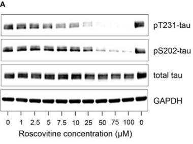
| Western blot | pT231-tau / pS202-tau / tau p-Rb / p-CDK2 / CDK2 / Cyclin D1 / Cyclin A2 / ERα / ERβ/ AIB1 / PELP1 |
|
30915013 |
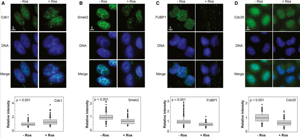
| Immunofluorescence | CDK1 / Smek2 / FUBP1 / Cdc20 E2F1 / FASN / Bmi1 / Cyclin D2 / CDK2 / CDK4 |
|
24534090 |
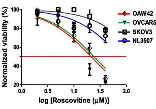
| Growth inhibition assay | Cell viability |
|
29996940 |
Clinical Trial Information
(data from https://clinicaltrials.gov, updated on 2024-05-22)
| NCT Number | Recruitment | Conditions | Sponsor/Collaborators | Start Date | Phases |
|---|---|---|---|---|---|
| NCT02649751 | Terminated | Cystic Fibrosis |
University Hospital Brest|ManRos Therapeutics|Cyclacel Pharmaceuticals Inc. |
February 22 2016 | Phase 2 |
Tech Support
Tel: +1-832-582-8158 Ext:3
If you have any other enquiries, please leave a message.
Frequently Asked Questions
Question 1:
How can I reconstitute it for in vivo studies?
Answer:
It in 1% DMSO+10% Tween 80+20% N-N-dimethylacetamide+PEG 400 is a clear solution which is okay for injection. And this compound in 1% DMSO+30% polyethylene glycol+1% Tween 80 at 30mg/ml is a suspension, which is fine for oral gavage.
Signaling Pathway Map
Products are for research use only. Not for human use. We do not sell to patients.
©Copyright 2013 Selleck Chemicals. All Rights Reserved.