-
 Australia
-
 Austria
-
 Belgium
-
 Brazil
-
 Canada
-
 China
China
-
 Czech Republic
-
 Denmark
-
 Finland
-
 France
-
 Germany
-
 Greece
-
 Hong Kong
-
 Hungary
-
 Iceland
-
 India
India
-
 Ireland
-
 Israel
-
 Italy
Italy
-
 Japan
Japan
-
 Korea
Korea
-
 Luxembourg
-
 Malaysia
-
 Netherlands
-
 New Zealand
-
 Norway
-
 Poland
-
 Qatar
Qatar
-
 Romania
-
 Saudi Arabia
-
 Singapore
-
 Spain
Spain
-
 Sweden
-
 Switzerland
-
 Taiwan
-
 Turkey
-
 United Kingdom
-
 United States
research use only
AT7519 HCl CDK inhibitor
Cat.No.S7808
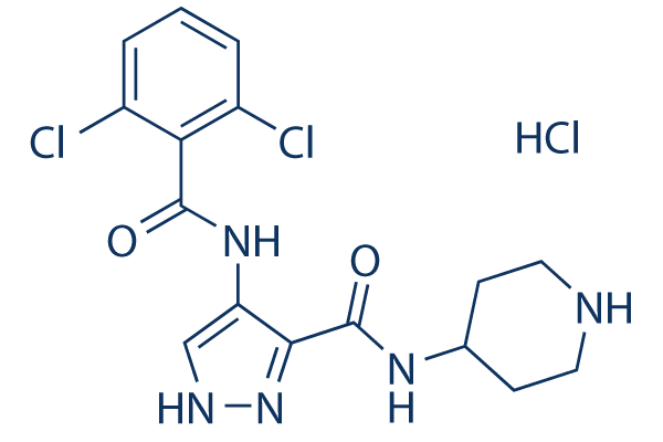
Chemical Structure
Molecular Weight: 418.71
Quality Control
Solubility
|
In vitro |
DMSO
: 52 mg/mL
(124.19 mM)
Water : 43 mg/mL Ethanol : 28 mg/mL |
Molarity Calculator
|
In vivo |
|||||
In vivo Formulation Calculator (Clear solution)
Step 1: Enter information below (Recommended: An additional animal making an allowance for loss during the experiment)
Step 2: Enter the in vivo formulation (This is only the calculator, not formulation. Please contact us first if there is no in vivo formulation at the solubility Section.)
Calculation results:
Working concentration: mg/ml;
Method for preparing DMSO master liquid: mg drug pre-dissolved in μL DMSO ( Master liquid concentration mg/mL, Please contact us first if the concentration exceeds the DMSO solubility of the batch of drug. )
Method for preparing in vivo formulation: Take μL DMSO master liquid, next addμL PEG300, mix and clarify, next addμL Tween 80, mix and clarify, next add μL ddH2O, mix and clarify.
Method for preparing in vivo formulation: Take μL DMSO master liquid, next add μL Corn oil, mix and clarify.
Note: 1. Please make sure the liquid is clear before adding the next solvent.
2. Be sure to add the solvent(s) in order. You must ensure that the solution obtained, in the previous addition, is a clear solution before proceeding to add the next solvent. Physical methods such
as vortex, ultrasound or hot water bath can be used to aid dissolving.
Chemical Information, Storage & Stability
| Molecular Weight | 418.71 | Formula | C16H18Cl3N5O2 |
Storage (From the date of receipt) | |
|---|---|---|---|---|---|
| CAS No. | 902135-91-5 | Download SDF | Storage of Stock Solutions |
|
|
| Synonyms | N/A | Smiles | C1CNCCC1NC(=O)C2=C(C=NN2)NC(=O)C3=C(C=CC=C3Cl)Cl.Cl | ||
Mechanism of Action
| Targets/IC50/Ki |
CDK9/CyclinT
(Cell-free assay) <10 nM
CDK5/p35
(Cell-free assay) 13 nM
CDK2/CyclinA
(Cell-free assay) 47 nM
GSK-3β
(Cell-free assay) 89 nM
CDK4/CyclinD1
(Cell-free assay) 100 nM
CDK6/CyclinD3
(Cell-free assay) 170 nM
CDK1/CyclinB
(Cell-free assay) 210 nM
CDK3/CyclinE
(Cell-free assay) 360 nM
|
|---|---|
| In vitro |
AT7519 is an ATP competitive CDK inhibitor with a Ki value of 38 nM for CDK1. AT7519 is inactive against all non-CDK kinases with the exception of GSK3β (IC50 = 89 nM). AT7519 shows potent antiproliferative activity in a variety of human tumor cell lines with IC50 values ranging from 40 nM for MCF-7 to 940 nM for SW620 consistent with the inhibition of CDK1 and CDK2. AT7519 induces dose-dependent cytotoxicity in multiple myeloma (MM) cell lines with IC50 values ranging from 0.5 to 2 μM at 48 hours, with the most sensitive cell lines being MM.1S (0.5 μM) and U266 (0.5 μM) and the most resistant MM.1R (>2 μM). It does not induce cytotoxicity in peripheral blood mononuclear cells (PBMNC). AT7519 partially overcomes the proliferative advantage conferred by IL6 and IGF-1 as well as the protective effect of bone marrow stromal cells (BMSCs). AT7519 induces rapid dephosphorylation of RNA pol II CTD at serine 2 and serine 5 sites, and leads to the inhibition of transcription, partially contributing to AT7519 induced cytotoxicity of MM cells. AT7519 induces activation of GSK-3β by down-regulating GSK-3β phosphorylation, which also contributes to AT7519 induced apoptosis independent of the inhibition of transcription.
|
| Kinase Assay |
In vitro Kinase Assays
|
|
Kinase assays for CDK1, CDK2 and GSK3-β are all carried out in a radiometric filter binding format. Assays for CDK5 are in DELFIA format and for CDKs 4 and 6 in ELISA format. For CDKs 1 and 2, the relevant CDK and 0.12 μg/mL Histone H1 are incubated in 20 mM MOPS, pH 7.2, 25 mM β-glycerophosphate, 5 mM EDTA, 15 mM MgCl2, 1 mM sodium orthovanadate, 1 mM DTT, 0.1 mg/mL BSA, 45 μM ATP (0.78 Ci/mmol) and different concentrations of AT7519 for 2 or 4 hours respectively. For GSK3-β, the relevant enzyme and 5 μM glycogen synthase peptide 2 along with 10 mM MOPS pH 7.0, 0.1 mg/mL BSA, 0.001% Brij-35, 0.5% glycerol, 0.2 mM EDTA, 10 mM MgCl2, 0.01% β-mercaptoethanol, 15 μM ATP (2.31 Ci/mmol) and different concentrations of AT7519 are incubated for 3 hours. Assay reactions are stopped by adding an excess of orthophosphoric acid and filtered using Millipore MAPH filter plates. The plates are then washed, scintillant added and radioactivity measured by scintillation counting on a Packard TopCount. For CDK5, CDK5/p35 and 1μM of a biotinylated Histone H1 peptide (Biotin-PKTPKKAKKL) are incubated in 25 mM Tris-HCl, pH 7.5, 2.5 mM MgCl2, 0.025% Brij-35, 0.1 mg/mL BSA, 1 mM DTT, 15 μM ATP and different concentrations of AT7519 for 30 minutes. Assay reactions are stopped using EDTA, transferred to Neutravidin-coated plates and phosphorylated peptide quantified by means of a rabbit phospho-cdk1 substrate polyclonal antibody and DELFIA europium-labelled anti-rabbit IgG secondary antibody using time-resolved fluorescence at λex=335nm, λem=620nm. For CDK 4 and 6 assays, plates are coated with GST- pRb769-921 and blocked with Superblock. CDK4 or 6 is incubated with 15 mM MgCl2, 50 mM HEPES, pH 7.4, 1 mM DTT, 1 mM EGTA, pH 8.0, 0.02% Triton X-100, 2.5% DMSO and different concentrations of AT7519; the reaction is initiated by addition of ATP. After 30 minutes, reactions are stopped by the addition of 0.5 M EDTA pH 8.0. Plates are then washed and incubated for one hour with the primary antibody (anti- p-Rb Serine 780) diluted in Superblock followed by secondary antibody (alkaline phosphatase linked anti-rabbit) for a further hour. Plates are developed using the Attophos system and fluorescence read on a Spectramax Gemini plate reader at excitation 450 nm and emission 580 nm. In all cases, IC50 values are calculated from replicate curves, using GraphPad Prism software.
|
|
| In vivo |
A twice daily dosing of AT7519 (9.1 mg/kg) causes tumor regression of both early-stage and advanced-stage s.c. tumors in the HCT116 and HT29 colon cancer xenograft models. AT7519 treatment (15 mg/kg) inhibits tumor growth and prolongs the median overall survival of mice in the human MM xenograft mouse model in association with increased caspase 3 activation.
|
References |
|
Clinical Trial Information
(data from https://clinicaltrials.gov, updated on 2024-05-22)
| NCT Number | Recruitment | Conditions | Sponsor/Collaborators | Start Date | Phases |
|---|---|---|---|---|---|
| NCT01183949 | Completed | Multiple Myeloma |
Astex Pharmaceuticals Inc.|Multiple Myeloma Research Consortium |
November 2010 | Phase 1|Phase 2 |
Tech Support
Tel: +1-832-582-8158 Ext:3
If you have any other enquiries, please leave a message.
Signaling Pathway Map
Products are for research use only. Not for human use. We do not sell to patients.
©Copyright 2013 Selleck Chemicals. All Rights Reserved.