-
 Australia
-
 Austria
-
 Belgium
-
 Brazil
-
 Canada
-
 China
China
-
 Czech Republic
-
 Denmark
-
 Finland
-
 France
-
 Germany
-
 Greece
-
 Hong Kong
-
 Hungary
-
 Iceland
-
 India
India
-
 Ireland
-
 Israel
-
 Italy
Italy
-
 Japan
Japan
-
 Korea
Korea
-
 Luxembourg
-
 Malaysia
-
 Netherlands
-
 New Zealand
-
 Norway
-
 Poland
-
 Qatar
Qatar
-
 Romania
-
 Saudi Arabia
-
 Singapore
-
 Spain
Spain
-
 Sweden
-
 Switzerland
-
 Taiwan
-
 Turkey
-
 United Kingdom
-
 United States
research use only
E7080 (Lenvatinib) VEGFR inhibitor
Cat.No.S1164
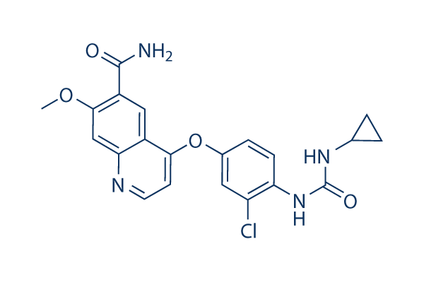
Chemical Structure
Molecular Weight: 426.85
Quality Control
Products Often Used Together with E7080 (Lenvatinib)
| Related Targets | EGFR PDGFR FGFR c-Met Src MEK CSF-1R FLT3 HER2 c-Kit |
|---|---|
| Other VEGFR Inhibitors | SAR131675 SU 5402 Cediranib (AZD2171) Vatalanib (PTK787) 2HCl Anlotinib (AL3818) Dihydrochloride Linifanib (ABT-869) Apatinib (YN968D1) Apatinib (YN968D1) mesylate Ki8751 Semaxanib (SU5416) |
Cell Culture, Treatment & Working Concentration
| Cell Lines | Assay Type | Concentration | Incubation Time | Formulation | Activity Description | PMID |
|---|---|---|---|---|---|---|
| TPC-1 and K1 cells | Function assay | 50 μM | 24 h | The inhibitory effects of lenvatinib on the viability of both cell lines were not influenced by the leptin treatment. | 30906321 | |
| ATC cells | Function assay | 1, 25 and 50 μM | 72 h | Phosphorylated/non-phosphorylated Akt or ERK1/2 proteins (evaluated by ELISA) in lenvatinib-treated samples were significantly reduced in ATC cell cultures. | 29517103 | |
| HCC cell lines Hep3B2.1-7, HuH-7, and JHH-7 | Proliferation assay | 6 days | Lenvatinib showed selective and potent antiproliferative activity against the HCC cell lines Hep3B2.1‐7, HuH‐7, and JHH‐7, with IC50 values of 0.23, 0.42, and 0.64 μmol/L, respectively. | 29733511 | ||
| HT29 cells | Cytotoxicity assay | 25, 50 nM | 72 h | cytotoxic dose: 50 nM and noncytotoxic dose: 25 nM | 24815456 | |
| DX3 and U2OS cells | Function assay | 1 μM and 10 μM | 16 hours | Lenvatinib inhibit tumor cells migration and invasion at concentrations that both inhibit its known targets and are achievable clinically. | 21781317 | |
| Click to View More Cell Line Experimental Data | ||||||
Solubility
|
In vitro |
DMSO
: 20 mg/mL
(46.85 mM)
Water : Insoluble Ethanol : Insoluble |
Molarity Calculator
|
In vivo |
|||||
In vivo Formulation Calculator (Clear solution)
Step 1: Enter information below (Recommended: An additional animal making an allowance for loss during the experiment)
Step 2: Enter the in vivo formulation (This is only the calculator, not formulation. Please contact us first if there is no in vivo formulation at the solubility Section.)
Calculation results:
Working concentration: mg/ml;
Method for preparing DMSO master liquid: mg drug pre-dissolved in μL DMSO ( Master liquid concentration mg/mL, Please contact us first if the concentration exceeds the DMSO solubility of the batch of drug. )
Method for preparing in vivo formulation: Take μL DMSO master liquid, next addμL PEG300, mix and clarify, next addμL Tween 80, mix and clarify, next add μL ddH2O, mix and clarify.
Method for preparing in vivo formulation: Take μL DMSO master liquid, next add μL Corn oil, mix and clarify.
Note: 1. Please make sure the liquid is clear before adding the next solvent.
2. Be sure to add the solvent(s) in order. You must ensure that the solution obtained, in the previous addition, is a clear solution before proceeding to add the next solvent. Physical methods such
as vortex, ultrasound or hot water bath can be used to aid dissolving.
Chemical Information, Storage & Stability
| Molecular Weight | 426.85 | Formula | C21H19ClN4O4 |
Storage (From the date of receipt) | |
|---|---|---|---|---|---|
| CAS No. | 417716-92-8 | Download SDF | Storage of Stock Solutions |
|
|
| Synonyms | E7080 | Smiles | COC1=CC2=NC=CC(=C2C=C1C(=O)N)OC3=CC(=C(C=C3)NC(=O)NC4CC4)Cl | ||
Mechanism of Action
| Targets/IC50/Ki |
RET
VEGFR2/KDR
(Cell-free assay) 4.0 nM
VEGFR3/FLT4
(Cell-free assay) 5.2 nM
VEGFR1/FLT1
(Cell-free assay) 22 nM
PDGFRβ
(Cell-free assay) 39 nM
FGFR1
(Cell-free assay) 46 nM
PDGFRα
(Cell-free assay) 51 nM
Kit
(Cell-free assay) 100 nM
|
|---|---|
| In vitro |
E7080, as a potent inhibitor of in vitro angiogenesis, shows a significantly inhibitory effect on VEGF/KDR and SCF/Kit signaling. According to the in vitro receptor tyrosine and serine/threonine kinase assays, this compound inhibits Flt-1, KDR, Flt-4 with IC50 of 22, 4.0 and 5.2 nM, respectively. In addition to these kinases, this compound also inhibits FGFR1 and PDGFR tyrosine kinases with IC50 value of 46, 51 and 100 nM for FGFR1, PDGFRα and PDGFRβ, respectively. This compound potently inhibits phosphorylation of VEGFR2 (IC50, 0.83 nM) and VEGFR3 (IC50, 0.36 nM) in HUVECs which is stimulated by VEGF and VEGF-C, respectively. A recent study shows that this compound treatment (both at 1 μM and 10 μM) results in a significant inhibition of cell migration and invasion by inhibiting FGFR and PDGFR signaling. |
| Kinase Assay |
In vitro kinase assay
|
|
Tyrosine kinase assays are performed by HTRF (KDR, VEGFR1, FGFR1, c-Met, EGFR) and ELISA (PDGFRβ), using the recombinant kinase domains of receptors. In both assays, 4 μL of serial dilutions of this compound are mixed in a 96-well round plate with 10 μL of enzyme, 16 μL of poly (GT) solution (250 ng) and 10 μL of ATP solution (1 μM ATP) (final concentration of DMSO is 0.1%). In wells for blanks, no enzyme is added. In control wells no test article is added. The kinase reaction is initiated by adding ATP solution to each well. After 30-minute incubation at 30°C, the reaction is stopped by adding 0.5 M EDTA (10 μL/well) to the reaction mixture in each well. Dilution buffer adequate to each kinase assay is added to the reaction mixture. In the HTRF assay, 50 μL of the reaction mixture is transferred to a 96-well 1/2 area black EIA/RIA plate, HTRF solution (50 μL/well) is added to the reaction mixture, and then kinase activity is determined by measurement of fluorescence with a time-resolved fluorescence detector at an excitation wavelength of 337 nm and an emission wavelengths of 620 and 665 nm. In the ELISA, 50 μL of the reaction mixture is incubated in avidin coated 96-well polystyrene plates at room temperature for 30 minutes. After washing with wash buffer, PY20-HRP solution (70 μL/well) is added and the reaction mixture is incubated at room temperature for 30 minutes. After washing with wash buffer, TMB reagent (100 μL/well) is added to each well. After several minutes (10–30 minutes), 1 M H3PO4 (100 μL/well) is added to each well. Kinase activity is determined by measurement of absorbance at 450 nm with a microplate reader.
|
|
| In vivo |
When orally administrated in a H146 xenograft model, Lenvatinib (E7080) inhibits the growth of H146 tumor at 30 and 100 mg/kg in a dose-dependent manner and leads to tumor regression at 100 mg/kg. Furthermore, this compound at 100 mg/kg decreases microvessel density more than anti-VEGF antibody and imatinib treatment. It significantly inhibits local tumor growth in a MDA-MB-231 mammary fat pad (m.f.p.) model with RTVs (calculated tumor volume on day 8/tumor volume on day 1) of 0.81, and reduces both angiogenesis and lymphangiogenesis of established metastatic nodules of MDA-MB-231 tumor in the lymph nodes. |
References |
|
Applications
| Methods | Biomarkers | Images | PMID |
|---|---|---|---|
| Western blot | Vimentin / E-cadherin / Snail / Zeb1 β-catenin Ki-67 / Cyclin D1 / CDK4 / p21 / p53 / Apaf-1 / p-NFκB / Bcl-2 / Cleaved-caspase 3 phospho-RET phospho-FGFR1 / FGFR1 /phospho-FRS2 / FRS2 / phospho-MEK / phospho-ERK |
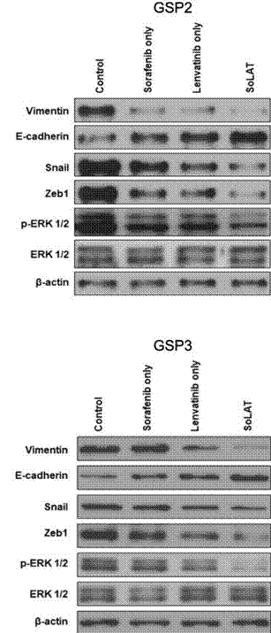
|
30286728 |
| Growth inhibition assay | Cell viability |
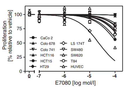
|
25425971 |
Clinical Trial Information
(data from https://clinicaltrials.gov, updated on 2024-05-22)
| NCT Number | Recruitment | Conditions | Sponsor/Collaborators | Start Date | Phases |
|---|---|---|---|---|---|
| NCT06161558 | Not yet recruiting | Neoplasms |
National Cancer Institute (NCI)|National Institutes of Health Clinical Center (CC) |
May 15 2024 | Phase 1 |
| NCT05846724 | Not yet recruiting | Kaposi Sarcoma|Classic Kaposi Sarcoma|Refractory Kaposi Sarcoma |
Fondazione IRCCS Ca'' Granda Ospedale Maggiore Policlinico |
February 1 2024 | Phase 2 |
| NCT05903833 | Not yet recruiting | Recurrent Vulvar Cancer|Persistent Vulvar Cancer|Metastatic Vulva Cancer|Locally Advanced Vulvar Cancer |
AGO Research GmbH |
January 1 2024 | Phase 2 |
| NCT05901194 | Not yet recruiting | Hepatocellular Carcinoma Non-resectable |
Assistance Publique - Hôpitaux de Paris|Laboratoire EISAI |
June 2023 | Phase 1|Phase 2 |
Tech Support
Tel: +1-832-582-8158 Ext:3
If you have any other enquiries, please leave a message.
Signaling Pathway Map
Products are for research use only. Not for human use. We do not sell to patients.
©Copyright 2013 Selleck Chemicals. All Rights Reserved.