- Inhibitors
- By product type
- Natural Products
- Inducing Agents
- Peptides
- Antibiotics
- Antibody-drug Conjugates(ADC)
- PROTAC
- Hydrotropic Agents
- Dyes
- By Signaling Pathways
- PI3K/Akt/mTOR
- Epigenetics
- Methylation
- Immunology & Inflammation
- Protein Tyrosine Kinase
- Angiogenesis
- Apoptosis
- Autophagy
By research - Antibodies
- Compound Libraries
- Popular Compound Libraries
- Customize Library
- Clinical and FDA-approved Related
- Bioactive Compound Libraries
- Inhibitor Related
- Natural Product Related
- Metabolism Related
- Cell Death Related
- By Signaling Pathway
- By Disease
- Anti-infection and Antiviral Related
- Neuronal and Immunology Related
- Fragment and Covalent Related
- FDA-approved Drug Library
- FDA-approved & Passed Phase I Drug Library
- Preclinical/Clinical Compound Library
- Bioactive Compound Library-I
- Bioactive Compound Library-Ⅱ
- Kinase Inhibitor Library
- Express-Pick Library
- Natural Product Library
- Human Endogenous Metabolite Compound Library
- Alkaloid Compound LibraryNew
- Angiogenesis Related compound Library
- Anti-Aging Compound Library
- Anti-alzheimer Disease Compound Library
- Antibiotics compound Library
- Anti-cancer Compound Library
- Anti-cancer Compound Library-Ⅱ
- Anti-cancer Metabolism Compound Library
- Anti-Cardiovascular Disease Compound Library
- Anti-diabetic Compound Library
- Anti-infection Compound Library
- Antioxidant Compound Library
- Anti-parasitic Compound Library
- Antiviral Compound Library
- Apoptosis Compound Library
- Autophagy Compound Library
- Calcium Channel Blocker LibraryNew
- Cambridge Cancer Compound Library
- Carbohydrate Metabolism Compound LibraryNew
- Cell Cycle compound library
- CNS-Penetrant Compound Library
- Covalent Inhibitor Library
- Cytokine Inhibitor LibraryNew
- Cytoskeletal Signaling Pathway Compound Library
- DNA Damage/DNA Repair compound Library
- Drug-like Compound Library
- Endoplasmic Reticulum Stress Compound Library
- Epigenetics Compound Library
- Exosome Secretion Related Compound LibraryNew
- FDA-approved Anticancer Drug LibraryNew
- Ferroptosis Compound Library
- Flavonoid Compound Library
- Fragment Library
- Glutamine Metabolism Compound Library
- Glycolysis Compound Library
- GPCR Compound Library
- Gut Microbial Metabolite Library
- HIF-1 Signaling Pathway Compound Library
- Highly Selective Inhibitor Library
- Histone modification compound library
- HTS Library for Drug Discovery
- Human Hormone Related Compound LibraryNew
- Human Transcription Factor Compound LibraryNew
- Immunology/Inflammation Compound Library
- Inhibitor Library
- Ion Channel Ligand Library
- JAK/STAT compound library
- Lipid Metabolism Compound LibraryNew
- Macrocyclic Compound Library
- MAPK Inhibitor Library
- Medicine Food Homology Compound Library
- Metabolism Compound Library
- Methylation Compound Library
- Mouse Metabolite Compound LibraryNew
- Natural Organic Compound Library
- Neuronal Signaling Compound Library
- NF-κB Signaling Compound Library
- Nucleoside Analogue Library
- Obesity Compound Library
- Oxidative Stress Compound LibraryNew
- Plant Extract Library
- Phenotypic Screening Library
- PI3K/Akt Inhibitor Library
- Protease Inhibitor Library
- Protein-protein Interaction Inhibitor Library
- Pyroptosis Compound Library
- Small Molecule Immuno-Oncology Compound Library
- Mitochondria-Targeted Compound LibraryNew
- Stem Cell Differentiation Compound LibraryNew
- Stem Cell Signaling Compound Library
- Natural Phenol Compound LibraryNew
- Natural Terpenoid Compound LibraryNew
- TGF-beta/Smad compound library
- Traditional Chinese Medicine Library
- Tyrosine Kinase Inhibitor Library
- Ubiquitination Compound Library
-
Cherry Picking
You can personalize your library with chemicals from within Selleck's inventory. Build the right library for your research endeavors by choosing from compounds in all of our available libraries.
Please contact us at info@selleckchem.com to customize your library.
You could select:
- Bioreagents
- qPCR
- 2x SYBR Green qPCR Master Mix
- 2x SYBR Green qPCR Master Mix(Low ROX)
- 2x SYBR Green qPCR Master Mix(High ROX)
- Protein Assay
- Protein A/G Magnetic Beads for IP
- Anti-Flag magnetic beads
- Anti-Flag Affinity Gel
- Anti-Myc magnetic beads
- Anti-HA magnetic beads
- Poly DYKDDDDK Tag Peptide lyophilized powder
- Protease Inhibitor Cocktail
- Protease Inhibitor Cocktail (EDTA-Free, 100X in DMSO)
- Phosphatase Inhibitor Cocktail (2 Tubes, 100X)
- Cell Biology
- Cell Counting Kit-8 (CCK-8)
- Animal Experiment
- Mouse Direct PCR Kit (For Genotyping)
- Featured Products
- MRTX1133
- Nab-Paclitaxel
- KP-457
- IAG933
- RMC-6236 (Daraxonrasib)
- RMC-7977
- Zoldonrasib (RMC-9805)
- GsMTx4
- Navitoclax (ABT-263)
- TSA (Trichostatin A)
- Y-27632 Dihydrochloride
- SB431542
- SB202190
- MK-2206 Dihydrochloride
- LY294002
- Alisertib (MLN8237)
- XAV-939
- CHIR-99021 (Laduviglusib)
- Bafilomycin A1 (Baf-A1)
- Thiazovivin (TZV)
- CP-673451
- Verteporfin
- DAPT
- Galunisertib (LY2157299)
- MG132
- SBE-β-CD
- Tween 80
- Bavdegalutamide (ARV-110)
- Z-VAD-FMK
- Wnt-C59 (C59)
- IWR-1-endo
- (+)-JQ1
- 3-Deazaneplanocin A (DZNep) Hydrochloride
- RepSox (E-616452)
- Erastin
- Q-VD-Oph
- Puromycin Dihydrochloride
- Cycloheximide
- Telaglenastat (CB-839)
- A-83-01
- Ceralasertib (AZD6738)
- Liproxstatin-1
- Emricasan (IDN-6556)
- PMA (Phorbol 12-myristate 13-acetate)
- Dibutyryl cAMP (Bucladesine) sodium
- Nedisertib (M3814)
- PLX5622
- IKE (Imidazole Ketone Erastin)
- STM2457
- Saruparib (AZD5305)
- New Products
- Contact Us
research use only
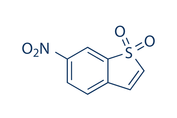
Stattic STAT3 Inhibitor
Cat.No.S7024

Chemical Structure
Molecular Weight: 211.19
Quality Control
Batch:
Purity:
99.88%
99.88
Cell Culture, Treatment & Working Concentration
| Cell Lines | Assay Type | Concentration | Incubation Time | Formulation | Activity Description | PMID |
|---|---|---|---|---|---|---|
| H9c2 | Function Assay | 10 μM | 4 h | reverses the effects of IL-27 | 26339633 | |
| NPC | Function Assay | 0-7.5 µM | abolishes EMT-like molecular alterations, and cell migration and invasion induced by RKIP knockdown | 25915430 | ||
| HASMC | Function Assay | 1.25-5 μM | 20 min | DMSO | inhibits p-(Y)-STAT-1,3,5 signals | 25849622 |
| H9c2 | Function Assay | 2/10 μM | 2 h | DMSO | abrogates the cytoprotective effects of IL-27 against SH | 25820907 |
| A431 | Growth Inhibition Assay | 2 μM | 2 h | blocks EGF-reversed decreases in cell viability | 25720435 | |
| A431 | Growth Inhibition Assay | 2 μM | 2 h | increases in apoptosis induced by shikonin | 25720435 | |
| SiHa | Cell Viability Assay | 5-75 nM | 24 h | shows morphology of a typical apoptotic cell and dose-dependent loss of cell viability | 25539644 | |
| SiHa | Function Assay | 5-75 nM | 24 h | reduces the phosphorylation at the tyrosine residue 705 | 25539644 | |
| ECA109 | Growth Inhibition Assay | 0-20 μM | 24 h | IC50=5.50 μM | 25492480 | |
| TE13 | Growth Inhibition Assay | 0-20 μM | 24 h | IC50=6.15 μM | 25492480 | |
| KYSE150 | Growth Inhibition Assay | 0-20 μM | 24 h | IC50=12.64 μM | 25492480 | |
| ECA109 | Clonogenic Survival Assay | 0.5 μM | 24 h | suppresses the clonogenic formation | 25492480 | |
| TE13 | Clonogenic Survival Assay | 0.5 μM | 24 h | suppresses the clonogenic formation | 25492480 | |
| KYSE150 | Clonogenic Survival Assay | 0.5 μM | 24 h | suppresses the clonogenic formation | 25492480 | |
| ECA109 | Function Assay | 0.5 μM | 24 h | enhances IR-induced generation of DSBs | 25492480 | |
| PC3M-1E8 | Function Assay | 2.5/5/10 μM | 0-4 h | inhibits the STAT3 activation in a dose- and time-dependent manner | 25261365 | |
| PC3M-1E8 | Function Assay | 10 μM | 24 h | downregulates Bcl-xL, survivin and c-Myc | 25261365 | |
| PC3M-1E8 | Function Assay | 10 μM | 24 h | inhibits IL-6 induced STAT3 activation and the IL-6-induced STAT3 activation | 25261365 | |
| PC3M-1E8 | Clonogenic Survival Assay | 2.5/5/10 μM | inhibits the colony formation significantly | 25261365 | ||
| MDA-MB-231 | Function Assay | 20 μM | 2 h | exhibits Snail and E-cadherin expression | 25153349 | |
| H9c2 | Function Assay | 20 µM | 30 min | DMSO | abolishes propofol-induced AKT phosphorylation at both ser473 and thr308 | 25105067 |
| HaCaT | Growth Inhibition Assay | 10 µM | 20 min | DMSO | enhances sorafenib- and sunitinib-induced growth inhibition | 25013907 |
| Caki-1 | Growth Inhibition Assay | 10 µM | 20 min | DMSO | enhances sorafenib- and sunitinib-induced growth inhibition | 25013907 |
| HaCaT | Apoptosis Assay | 10 µM | 20 min | DMSO | increases proportions of apoptotic cells due to treatment with sorafenib or sunitinib | 25013907 |
| FHL-primed hNSCs | Cell Viability Assay | 0.02-5 μM | 72 h | leads to the loss of cell viability at high concentration | 24945434 | |
| ELL-primed hNSCs | Cell Viability Assay | 0.02-5 μM | 72 h | leads to the loss of cell viability at high concentration | 24945434 | |
| SS | Cell Viability Assay | 1-10 μM | 72 h | DMSO | causes a dose-dependent inhibition of the viability | 24756111 |
| SeAx | Cell Viability Assay | 1-10 μM | 72 h | DMSO | causes a dose-dependent inhibition of the viability | 24756111 |
| HuT-78 | Cell Viability Assay | 1-10 μM | 72 h | DMSO | causes a dose-dependent inhibition of the viability | 24756111 |
| CD4+ | Apoptosis Assay | 10 μm | 24 h | DMSO | induces apoptosis strongly | 24756111 |
| MCF-7 | Growth Inhibition Assay | 0.469-3.75 μM | 5 d | reduces cell number significantly | 24728078 | |
| MCF-7/LCC1 | Growth Inhibition Assay | 0.469-3.75 μM | 5 d | reduces cell number significantly | 24728078 | |
| MCF-7/LCC9 | Growth Inhibition Assay | 0.469-3.75 μM | 5 d | reduces cell number significantly | 24728078 | |
| HaCaT | Growth Inhibition Assay | 10 µM | 20 min | DMSO | enhances everolimus-induced cell growth inhibition | 24423131 |
| HaCaT | Apoptosis Assay | 10 µM | 20 min | DMSO | enhances the apoptotic effects of everolimus | 24423131 |
| MDA-MB-231 | Function Assay | 10 µM | 24 h | DMSO | reduces P-STAT3 expression | 24376586 |
| SUM-159 | Function Assay | 10 µM | 24 h | DMSO | reduces P-STAT3 expression | 24376586 |
| SK-BR-3 | Function Assay | 10 µM | 24 h | DMSO | reduces P-STAT3 expression | 24376586 |
| MCF7-HER2 | Growth Inhibition Assay | 0-10 μM | 48 h | DMSO | induces cell death dose dependently | 24297508 |
| MCF7-HER2 | Function Assay | 5 μM | 24 h | DMSO | diminishes Sox-2, Oct-4, and slug expression | 24297508 |
| MCF7-HER2 | Function Assay | 5 μM | 24 h | DMSO | decreases the expression levels of EMT markers, vimentin and slug | 24297508 |
| MCF7-HER2 | Growth Inhibition Assay | 5 μM | 24 h | DMSO | enhances cell growth inhibition combined with Herceptin | 24297508 |
| HMECs | Function Assay | 10 μM | 2 h | inhibits IFNα mediated phosphorylation of STAT1, STAT2 and STAT3 | 24211327 | |
| HTR8/SVneo | Function Assay | 1 μM | 1 h | suppressed OSM-induced STAT3 phosphorylation | 24060241 | |
| HTR8/SVneo | Function Assay | 0.5/1 μM | 48 h | restores the expression of E-cadherin suppressed by OSM | 24060241 | |
| HTR8/SVneo | Function Assay | 1 μM | 48 h | significantly increases migration by OSM | 24060241 | |
| C13* | Apoptosis Assay | 0-10 μM | 24/48 h | induces apoptosis in a dose and time dependent manner | 23962558 | |
| OV2008 | Apoptosis Assay | 0-10 μM | 24/48 h | induces apoptosis in a dose and time dependent manner | 23962558 | |
| C13* | Apoptosis Assay | 24/48 h | enhances cisplatin-induced apoptosis | 23962558 | ||
| OV2008 | Apoptosis Assay | 24/48 h | enhances cisplatin-induced apoptosis | 23962558 | ||
| W480 | Function Assay | 2.5/10 μM | 30 min | DMSO | sensitizes cells to chemoradiotherapy in a dose-dependent manner | 23934972 |
| SW837 | Function Assay | 2.5/10 μM | 30 min | DMSO | sensitizes cells to chemoradiotherapy in a dose-dependent manner | 23934972 |
| T24 | Function Assay | 2/10/20 μM | 24 h | causes dose-dependent inhibition of the CXCL12-induced increase of invading cells | 23526079 | |
| CNE1 | Function Assay | 20 µM | 48 h | blocks the IL-6 increased phosphorylation of Stat3 | 23382914 | |
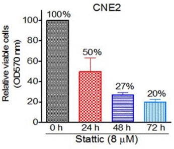
| CNE2 | Function Assay | 20 µM | 48 h | blocks the IL-6 increased phosphorylation of Stat3 | 23382914 | |
| HONE1 | Function Assay | 20 µM | 48 h | blocks the IL-6 increased phosphorylation of Stat3 | 23382914 | |
| CNE1 | Growth Inhibition Assay | 4 μM | significantly reduces cell viability | 23382914 | ||
| CNE1 | Function Assay | 0-20 μM | 0-4 h | inhibits Stat3 activation in a dose- and time-dependent manner | 23382914 | |
| CNE2 | Function Assay | 0-20 μM | 0-4 h | inhibits Stat3 activation in a dose- and time-dependent manner | 23382914 | |
| HONE1 | Function Assay | 0-20 μM | 0-4 h | inhibits Stat3 activation in a dose- and time-dependent manner | 23382914 | |
| CNE1 | Cell Viability Assay | 0.5-64 μM | 48 h | suppresses cell viability in a dose- and time-dependent manner | 23382914 | |
| CNE2 | Cell Viability Assay | 0.5-64 μM | 48 h | suppresses cell viability in a dose- and time-dependent manner | 23382914 | |
| HONE1 | Cell Viability Assay | 0.5-64 μM | 48 h | suppresses cell viability in a dose- and time-dependent manner | 23382914 | |
| C666-1 | Cell Viability Assay | 0.5-64 μM | 48 h | suppresses cell viability in a dose- and time-dependent manner | 23382914 | |
| CNE1 | Apoptosis Assay | 10 µM | 48 h | induces apoptosis | 23382914 | |
| CNE2 | Apoptosis Assay | 10 µM | 48 h | induces apoptosis | 23382914 | |
| HONE1 | Apoptosis Assay | 10 µM | 48 h | induces apoptosis | 23382914 | |
| CNE2 | Cell Viability Assay | 1/2 μM | 48 h | sensitize cells to radiotherapy | 23382914 | |
| HONE1 | Cell Viability Assay | 1/2 μM | 48 h | sensitize cells to radiotherapy | 23382914 | |
| C666-1 | Cell Viability Assay | 1/2 μM | 48 h | sensitize cells to radiotherapy | 23382914 | |
| HEC-1A | Function Assay | 1 μM | 24 h | DMSO | blocks the MUC20-enhanced invasion triggered by 10% FBS | 23262208 |
| RL95-2 | Function Assay | 1 μM | 24 h | DMSO | blocks the MUC20-enhanced invasion triggered by 10% FBS | 23262208 |
| HEC-1A | Function Assay | 1 μM | 24 h | DMSO | blocks the MUC20-enhanced invasion triggered by EGF | 23262208 |
| RL95-2 | Function Assay | 1 μM | 24 h | DMSO | blocks the MUC20-enhanced invasion triggered by EGF | 23262208 |
| CT26 | Function Assay | 20 mM | 1 h | suppresses HGF-induced VEGF expression | 23233163 | |
| UM-SCC-17B | Growth Inhibition Assay | IC50=2.562 ± 0.409 μM, GI50=1.279 ± 0.194 μM | 22770899 | |||
| OSC-19 | Growth Inhibition Assay | IC50=3.481 ± 0.953 μM, GI50=1.366 ± 0.770 μM | 22770899 | |||
| Cal33 | Growth Inhibition Assay | IC50=2.282 ± 0.423 μM, GI50=1.349 ± 0.363 μM | 22770899 | |||
| UM-SCC-22B | Growth Inhibition Assay | IC50=2.648 ± 0.542 μM, GI50=1.320 ± 0.204 μM | 22770899 | |||
| UM-SCC-17B | Function Assay | 0-30 μM | 0-24 h | inhibits STAT3 activation dose and time dependently | 22770899 | |
| OSC-19 | Function Assay | 0-30 μM | 0-24 h | inhibits STAT3 activation dose and time dependently | 22770899 | |
| Cal33 | Function Assay | 0-30 μM | 0-24 h | inhibits STAT3 activation dose and time dependently | 22770899 | |
| UM-SCC-22B | Function Assay | 0-30 μM | 0-24 h | inhibits STAT3 activation dose and time dependently | 22770899 | |
| U-87MG | Cell Viability Assay | 0-10 μM | 72 h | DMSO | inhibits cell viability dose dependently | 25436682 |
| U-373MG | Cell Viability Assay | 0-10 μM | 72 h | DMSO | inhibits cell viability dose dependently | 25436682 |
| SH-SY5Y | Cell Viability Assay | 0-10 μM | 72 h | DMSO | inhibits cell viability dose dependently | 25436682 |
| Tu-9648 | Cell Viability Assay | 0-10 μM | 72 h | DMSO | inhibits cell viability dose dependently | 25436682 |
| Neuro-2a | Cell Viability Assay | 0-10 μM | 72 h | DMSO | inhibits cell viability dose dependently | 25436682 |
| PCNs | Cell Viability Assay | 0-10 μM | 72 h | DMSO | inhibits cell viability dose dependently | 25436682 |
| PGCs | Cell Viability Assay | 0-10 μM | 72 h | DMSO | inhibits cell viability dose dependently | 25436682 |
| RAW264.7 | Function Assay | 10 μM | 12 h | abrogates the mRNA expressions of JAK2, STAT1, STAT2, and STAT3 induced by DON and T-2 toxin | 22454431 | |
| RAW264.7 | Apoptosis Assay | 5/10 μM | 45 min | enhances toxins induced apoptosis and MMP loss | 22454431 | |
| SW480 | Cell Viability Assay | 5/10/20 μM | 72 h | inhibits cell viability of the ALDH+/CD133+ cells | 21900397 | |
| HCT116 | Cell Viability Assay | 5/10/20 μM | 72 h | inhibits cell viability of the ALDH+/CD133+ cells | 21900397 | |
| DLD-1 | Cell Viability Assay | 5/10/20 μM | 72 h | inhibits cell viability of the ALDH+/CD133+ cells | 21900397 | |
| SNU387 | Cell Viability Assay | 20 μM | 24 h | reduces cell viability | 21311975 | |
| SNU398 | Cell Viability Assay | 20 μM | 24 h | reduces cell viability | 21311975 | |
| HepG2 | Cell Viability Assay | 20 μM | 24 h | reduces cell viability | 21311975 | |
| Huh-7 | Cell Viability Assay | 20 μM | 24 h | reduces cell viability | 21311975 | |
| VSMC | Growth Inhibition Assay | 3/5/10 μM | 30 min | DMSO | prevents PDGF- and thrombin-mediated VSMC proliferation in a dose-dependent manner | 20847306 |
| MDA-MB-231 | Apoptosis Assay | 10 μM | 24 h | DMSO | induces apoptosis | 17114005 |
| MDA-MB-435S | Apoptosis Assay | 10 μM | 24 h | DMSO | induces apoptosis | 17114005 |
| AsPC1 | Antiproliferative assay | 72 hrs | Antiproliferative activity against human AsPC1 cells assessed as inhibition of cell proliferation after 72 hrs by MTS assay, IC50 = 1.32 μM. | 24904966 | ||
| MDA-MB-231 | Antiproliferative assay | 72 hrs | Antiproliferative activity against ER-negative and triple-negative human MDA-MB-231 cells assessed as inhibition of cell proliferation after 72 hrs by MTS assay, IC50 = 2.89 μM. | 24904966 | ||
| MCF7 | Antiproliferative assay | 72 hrs | Antiproliferative activity against ER-positive human MCF7 cells assessed as inhibition of cell proliferation after 72 hrs by MTS assay, IC50 = 3.6 μM. | 24904966 | ||
| PANC1 | Antiproliferative assay | 72 hrs | Antiproliferative activity against human PANC1 cells assessed as inhibition of cell proliferation after 72 hrs by MTS assay, IC50 = 3.77 μM. | 24904966 | ||
| MDA-MB-231 | Cytotoxicity assay | 48 hrs | Cytotoxicity against human MDA-MB-231 cells assessed as growth inhibition after 48 hrs by MTT assay, IC50 = 1.56 μM. | 26396689 | ||
| MDA-MB-435S | Cytotoxicity assay | 48 hrs | Cytotoxicity against human MDA-MB-435S cells assessed as growth inhibition after 48 hrs by MTT assay, IC50 = 1.87 μM. | 26396689 | ||
| MCF7 | Cytotoxicity assay | 48 hrs | Cytotoxicity against human MCF7 cells assessed as growth inhibition after 48 hrs by MTT assay, IC50 = 2.16 μM. | 26396689 | ||
| A549 | Cytotoxicity assay | 48 hrs | Cytotoxicity against human A549 cells assessed as growth inhibition after 48 hrs by MTT assay, IC50 = 2.5 μM. | 26396689 | ||
| DU145 | Cytotoxicity assay | 48 hrs | Cytotoxicity against human DU145 cells assessed as growth inhibition after 48 hrs by MTT assay, IC50 = 2.5 μM. | 26396689 | ||
| PANC1 | Cytotoxicity assay | 48 hrs | Cytotoxicity against human PANC1 cells assessed as growth inhibition after 48 hrs by MTT assay, IC50 = 2.9 μM. | 26396689 | ||
| HCT116 | Antiproliferative assay | 72 hrs | Antiproliferative activity against human HCT116 cells after 72 hrs by MTT assay, IC50 = 1.08 μM. | 27718470 | ||
| MDA-MB-231 | Antiproliferative assay | 72 hrs | Antiproliferative activity against human MDA-MB-231 cells after 72 hrs by MTT assay, IC50 = 1.68 μM. | 27718470 | ||
| MCF7 | Antiproliferative assay | 72 hrs | Antiproliferative activity against human MCF7 cells after 72 hrs by MTT assay, IC50 = 2.36 μM. | 27718470 | ||
| A549 | Antiproliferative assay | 72 hrs | Antiproliferative activity against human A549 cells after 72 hrs by MTT assay, IC50 = 4.4 μM. | 27718470 | ||
| AD293 | Function assay | 6 hrs | Inhibition of IFNgamma-stimulated GFP/FLAG-tagged STAT3 dimerization in human AD293 cells incubated for 6 hrs by Western blot analysis, IC50 = 5.1 μM. | 30228000 | ||
| MDA-MB-231 | Function assay | 1 to 10 uM | 12 hrs | Inhibition of STAT3 phosphorylation at Tyr705 in human MDA-MB-231 cells at 1 to 10 uM after 12 hrs by western blot analysis | 24904966 | |
| MDA-MB-231 | Anticancer assay | 1 to 10 uM | 48 hrs | Anticancer activity against human MDA-MB-231 cells assessed as cell growth inhibition, apoptosis and cellular morphological changes at 1 to 10 uM after 48 hrs by light microscopy | 24904966 | |
| MDA-MB-231 | Function assay | 1 to 10 uM | 12 hrs | Decrease in STAT3 protein expression in human MDA-MB-231 cells at 1 to 10 uM after 12 hrs by western blot analysis | 24904966 | |
| MCF7 | Function assay | 12 hrs | Inhibition of STAT3 phosphorylation at Y705 in human MCF7 cells after 12 hrs by Western blot analysis | 26396689 | ||
| MDA-MB-435S | Function assay | 12 hrs | Inhibition of STAT3 phosphorylation at Y705 in human MDA-MB-435S cells after 12 hrs by Western blot analysis | 26396689 | ||
| MDA-MB-231 | Function assay | 12 hrs | Inhibition of STAT3 phosphorylation at Y705 in human MDA-MB-231 cells after 12 hrs by Western blot analysis | 26396689 | ||
| Click to View More Cell Line Experimental Data | ||||||
Chemical Information, Storage & Stability
| Molecular Weight | 211.19 | Formula | C8H5NO4S |
Storage (From the date of receipt) | |
|---|---|---|---|---|---|
| CAS No. | 19983-44-9 | Download SDF | Storage of Stock Solutions |
|
|
| Synonyms | N/A | Smiles | C1=CC(=CC2=C1C=CS2(=O)=O)[N+](=O)[O-] | ||
Solubility
|
In vitro |
DMSO
: 42 mg/mL
(198.87 mM)
Water : Insoluble Ethanol : Insoluble |
Molarity Calculator
|
In vivo |
|||||
In vivo Formulation Calculator (Clear solution)
Step 1: Enter information below (Recommended: An additional animal making an allowance for loss during the experiment)
mg/kg
g
μL
Step 2: Enter the in vivo formulation (This is only the calculator, not formulation. Please contact us first if there is no in vivo formulation at the solubility Section.)
% DMSO
%
% Tween 80
% ddH2O
%DMSO
%
Calculation results:
Working concentration: mg/ml;
Method for preparing DMSO master liquid: mg drug pre-dissolved in μL DMSO ( Master liquid concentration mg/mL, Please contact us first if the concentration exceeds the DMSO solubility of the batch of drug. )
Method for preparing in vivo formulation: Take μL DMSO master liquid, next addμL PEG300, mix and clarify, next addμL Tween 80, mix and clarify, next add μL ddH2O, mix and clarify.
Method for preparing in vivo formulation: Take μL DMSO master liquid, next add μL Corn oil, mix and clarify.
Note: 1. Please make sure the liquid is clear before adding the next solvent.
2. Be sure to add the solvent(s) in order. You must ensure that the solution obtained, in the previous addition, is a clear solution before proceeding to add the next solvent. Physical methods such
as vortex, ultrasound or hot water bath can be used to aid dissolving.
Mechanism of Action
| Features |
Stattic is the first non-peptide small molecule with inhibitory activity against STAT3 SH2 domain regardless of the STAT3 phosphorylation state in vitro.
|
|---|---|
| Targets/IC50/Ki |
STAT3 [1]
(Cell-free assay) 5.1 μM
|
| In vitro |
Stattic inhibits binding of a phosphotyrosine-containing peptide derived from the gp130 receptor to the STAT3 SH2 domain in a strongly temperature-dependent manner. This compound only has a very weak effect on binding of a tyrosinephosphorylated peptide to the SH2-domain of the tyrosine kinase Lck. And it doesn’t inhibit dimerization of two other dimeric transcription factors (c-Myc/Max and Jun/Jun). It also inhibits fluorescein-labeled phosphopeptides to the SH2 domains of STAT1 and STAT5b. This chemical selectively inhibits DNA binding of STAT3 homodimers at a concentration of 10 μM. Shown to inhibit cellular phosphorylation of STAT3 at Tyr705 with little effect towards STAT1 phosphorylation at Tyr701 (in HepG2 cells) or the phosphorylation of JAK1, JAK2, and c-Src (in MDA-MB-231 and MDA-MB-235S cells). This compound increases the apoptotic rate of STAT3-dependent breast cancer cell lines. [1] |
| Kinase Assay |
High-Throughput Screening and FluorescencePolarization Assays
|
|
Screening is performed at approximately 30 °C. The specificity of screening hits is validated in analogous assays for binding of the test compounds to the SH2 domains of STAT1, STAT5, and Lck. The final concentration of buffer components used for all FP assays is 10 mM HEPES (pH 7.5), 1 mM EDTA, 0.1% Nonidet P-40, 50 mM NaCl, and 10% DMSO. The absence of dithiothreitol is essential for inhibitory activity. The sequences of the peptides are: STAT3, 5-carboxyfluorescein-GY(PO3H2)LPQTV-NH2; STAT1, 5-carboxyfluorescein-GY(PO3H2)DKPHVL; STAT5, 5-carboxyfluorescein-GY(PO3H2)LVLDKW; and Lck, 5-carboxyfluorescein-GY(PO3H2)EEIP. For specificity analysis at 30 °C, proteins are used at 150 nM (STAT1, STAT3, and STAT5). For specificity analysis at 37 °C, proteins are used at 370 nM (STAT3) or 100 nM (Lck). Proteins are incubated with test compounds in Eppendorf tubes at the indicated temperatures for 60 min prior addition of the respective 5-carboxyfluorescein labeled peptides (final concentration: 10 nM). Before measurement at room temperature, the mixtures are allowed to equilibrate for at least 30 min. Test compounds are used at the indicated concentrations diluted from a 20× stock in DMSO. Binding curves and inhibition curves are fitted with SigmaPlot. All competition curves are repeated three times in independent experiment
|
References |
Applications
| Methods | Biomarkers | Images | PMID |
|---|---|---|---|
| Western blot | PARP / C-PARP / Caspase-3 / C-Caspse-3 Survivin / c-Myc / Bcl-xl p-STAT3 / STAT3 |

|
23382914 |
| Immunofluorescence | p-STAT3 / STAT3 / Survivin |

|
25261365 |
| Growth inhibition assay | Cell viability |

|
23382914 |
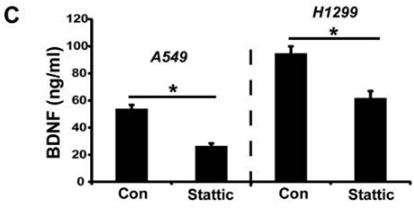
| ELISA | BDNF |

|
27456333 |
Tech Support
Tel: +1-832-582-8158 Ext:3
If you have any other enquiries, please leave a message.


