-
 Australia
-
 Austria
-
 Belgium
-
 Brazil
-
 Canada
-
 China
China
-
 Czech Republic
-
 Denmark
-
 Finland
-
 France
-
 Germany
-
 Greece
-
 Hong Kong
-
 Hungary
-
 Iceland
-
 India
India
-
 Ireland
-
 Israel
-
 Italy
Italy
-
 Japan
Japan
-
 Korea
Korea
-
 Luxembourg
-
 Malaysia
-
 Netherlands
-
 New Zealand
-
 Norway
-
 Poland
-
 Qatar
Qatar
-
 Romania
-
 Saudi Arabia
-
 Singapore
-
 Spain
Spain
-
 Sweden
-
 Switzerland
-
 Taiwan
-
 Turkey
-
 United Kingdom
-
 United States
research use only
NSC 74859 (S3I-201) STAT3 Inhibitor
Cat.No.S1155
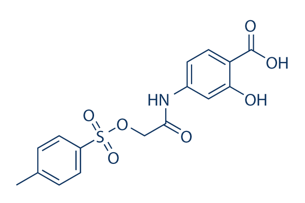
Chemical Structure
Molecular Weight: 365.36
Quality Control
| Related Targets | EGFR JAK Pim |
|---|---|
| Other STAT Inhibitors | Napabucasin (BBI608) Stattic Cryptotanshinone (Tanshinone C) C188-9 (TTI-101) SH-4-54 BP-1-102 AS1517499 HO-3867 Nifuroxazide Homoharringtonine (HHT) |
Cell Culture, Treatment & Working Concentration
| Cell Lines | Assay Type | Concentration | Incubation Time | Formulation | Activity Description | PMID |
|---|---|---|---|---|---|---|
| U87 | Growth Inhibition Assay | 72 h | IC50=55.1 μM | 20072652 | ||
| U373 | Growth Inhibition Assay | 72 h | IC50=52.5 μM | 20072652 | ||
| HPAC | Growth Inhibition Assay | 72 h | IC50>100 μM | 20072652 | ||
| PANC-1 | Growth Inhibition Assay | 72 h | IC50>100 μM | 20072652 | ||
| SK-BR-3 | Growth Inhibition Assay | 72 h | IC50>100 μM | 20072652 | ||
| U-373 MG | Cytotoxicity Assay | 3/10 μM | 24 h | reduces FN-γ-induced cell neurotoxicity | 20888416 | |
| MDA-MB-231 | Growth Inhibition Assay | 72 h | IC50>100 μM | 20072652 | ||
| HUVEC | Function Assay | 0.5-20 μM | 24 h | DMSO | suppresses the hypoxia-induced accumulation of HIF-1α | 21523559 |
| Huh7 | Growth Inhibition Assay | 100 nM | 48 h | DMSO | inhibits the IL-6 stimulation promoted cell proliferation | 23364389 |
| PLC/PRF/5 | Growth Inhibition Assay | 100 nM | 48 h | DMSO | inhibits the IL-6 stimulation promoted cell proliferation | 23364389 |
| H460 | Function Assay | 50/100 μM | 48 h | inhibits the Stat3C increased miR-92a expression | 23820254 | |
| H1299 | Function Assay | 50/100 μM | 48 h | suppresses miR-92a expression dose-dependently | 23820254 | |
| T-cell | Growth Inhibition Assay | IC50=50 μM | 24068731 | |||
| U373 | Growth Inhibition Assay | 125 μM | 24 h | DMSO | disrupts STAT3 signaling and proliferation | 24070820 |
| HUT-102 | Apoptosis Assay | 75-300 μM | 24/48 h | suppresses cell proliferation in a dose-dependent manner and induces cell apoptosis | 24090995 | |
| MT-2 | Apoptosis Assay | 75-300 μM | 24/48 h | suppresses cell proliferation in a dose-dependent manner and induces cell apoptosis | 24090995 | |
| H460 | Apoptosis Assay | 100 nM | 24 h | enhances cell death co-treated with LY294002 | 24472538 | |
| A459 | Apoptosis Assay | 100 nM | 24 h | induces cell apoptosis co-treated with BEZ235 | 24472538 | |
| H460 | Apoptosis Assay | 100 nM | 24 h | induces cell apoptosis co-treated with BEZ235 | 24472538 | |
| GC | Growth Inhibition Assay | 50-125 μM | 72 h | attenuates the cell growth in a dose-dependent manner | 25774503 | |
| GH3 | Growth Inhibition Assay | 50-125 μM | 72 h | attenuates the cell growth in a dose-dependent manner | 25774503 | |
| BT474R | Function Assay | 50 μM | 10-60 d | inhibits STAT3 activity | 25327561 | |
| NCI-N87R | Function Assay | 50 μM | 10-60 d | inhibits STAT3 activity | 25327561 | |
| MDA-MB-468 | Function assay | 100 uM | 24 hrs | Inhibition of Stat3 activation in human MDA-MB-468 cells at 100 uM after 24 hrs | 17463090 | |
| MDA-MB-435 | Function assay | 100 uM | 24 hrs | Inhibition of Stat3 activation in human MDA-MB-435 cells at 100 uM after 24 hrs | 17463090 | |
| MDA-MB-231 | Function assay | 100 uM | 24 hrs | Inhibition of Stat3 activation in human MDA-MB-231 cells at 100 uM after 24 hrs | 17463090 | |
| NIH3T3 | Function assay | 100 uM | 24 hrs | Reduction of pTyr-705 Stat3 level in v-Src expressing mouse NIH3T3 cells at 100 uM after 24 hrs | 17463090 | |
| NIH3T3 | Growth inhibition assay | 100 uM | 4 days | Growth inhibition of mouse NIH3T3 cells expressing v-Src at 100 uM after 4 days by trypan blue exclusion assay | 17463090 | |
| MDA-MB-435 | Growth inhibition assay | 100 uM | 4 days | Growth inhibition of human MDA-MB-435 cells expressing v-Src at 100 uM after 4 days by trypan blue exclusion assay | 17463090 | |
| MDA-MB-231 | Growth inhibition assay | 100 uM | 4 days | Growth inhibition of human MDA-MB-231 cells expressing v-Src at 100 uM after 4 days by trypan blue exclusion assay | 17463090 | |
| MDA-MB-468 | Growth inhibition assay | 100 uM | 4 days | Growth inhibition of human MDA-MB-468 cells expressing v-Src at 100 uM after 4 days by trypan blue exclusion assay | 17463090 | |
| NIH3T3 | Growth inhibition assay | 100 uM | Growth inhibition of mouse NIH3T3 cells expressing v-Ras at 100 uM for every 3 days by soft-agar colony-formation assay | 17463090 | ||
| MDA-MB-435 | Apoptosis assay | 30 to 100 uM | 48 hrs | Induction of apoptosis in human MDA-MB-435 cells expressing active Stat3 at 30 to 100 uM after 48 hrs | 17463090 | |
| MDA-MB-231 | Apoptosis assay | 100 uM | 24 hrs | Reduction of apoptosis in Stat3 transfected human MDA-MB-231 cells at 100 uM after 24 hrs | 17463090 | |
| MDA-MB-231 | Function assay | 100 uM | 48 hrs | Reduction of cyclin D1 gene expression in human MDA-MB-231 cells at 100 uM after 48 hrs | 17463090 | |
| MDA-MB-231 | Apoptosis assay | 100 uM | 24 hrs | Induction of apoptosis in Stat3 SH2 domain transfected human MDA-MB-231 cells at 100 uM after 24 hrs | 17463090 | |
| MDA-MB-231 | Apoptosis assay | 100 uM | 24 hrs | Induction of apoptosis in Stat3C transfected human MDA-MB-231 cells at 100 uM after 24 hrs | 17463090 | |
| NIH3T3 | Function assay | 100 uM | 48 hrs | Reduction of cyclin D1 gene expression in v-Src transfected mouse NIH3T3 cells at 100 uM after 48 hrs | 17463090 | |
| NIH3T3 | Function assay | 100 uM | 48 hrs | Reduction of Bcl-xL gene expression in v-Src transfected mouse NIH3T3 cells at 100 uM after 48 hrs | 17463090 | |
| NIH3T3 | Function assay | 100 uM | 48 hrs | Reduction of survivin gene expression in v-Src transfected mouse NIH3T3 cells at 100 uM after 48 hrs | 17463090 | |
| MDA-MB-231 | Function assay | 100 uM | 48 hrs | Reduction of Bcl-xL gene expression in human MDA-MB-231 cells at 100 uM after 48 hrs | 17463090 | |
| MDA-MB-231 | Function assay | 100 uM | 48 hrs | Reduction of survivin gene expression in human MDA-MB-231 cells at 100 uM after 48 hrs | 17463090 | |
| MDA-MB-231 | Antitumor assay | 5 mg/kg | 2 weeks | Antitumor activity against human MDA-MB-231 cells expressing active Stat3 xenografted in mouse at 5 mg/kg, iv for every 3 days for 2 weeks | 17463090 | |
| NIH3T3 | Growth inhibition assay | 100 uM | Growth inhibition of mouse NIH3T3 cells expressing v-Src at 100 uM for every 3 days by soft-agar colony-formation assay | 17463090 | ||
| A673 | qHTS assay | qHTS of pediatric cancer cell lines to identify multiple opportunities for drug repurposing: Primary screen for A673 cells | 29435139 | |||
| SK-N-MC | qHTS assay | qHTS of pediatric cancer cell lines to identify multiple opportunities for drug repurposing: Primary screen for SK-N-MC cells | 29435139 | |||
| NB1643 | qHTS assay | qHTS of pediatric cancer cell lines to identify multiple opportunities for drug repurposing: Primary screen for NB1643 cells | 29435139 | |||
| LAN-5 | qHTS assay | qHTS of pediatric cancer cell lines to identify multiple opportunities for drug repurposing: Primary screen for LAN-5 cells | 29435139 | |||
| Click to View More Cell Line Experimental Data | ||||||
Solubility
|
In vitro |
DMSO
: 73 mg/mL
(199.8 mM)
Water : Insoluble Ethanol : Insoluble |
Molarity Calculator
|
In vivo |
|||||
In vivo Formulation Calculator (Clear solution)
Step 1: Enter information below (Recommended: An additional animal making an allowance for loss during the experiment)
Step 2: Enter the in vivo formulation (This is only the calculator, not formulation. Please contact us first if there is no in vivo formulation at the solubility Section.)
Calculation results:
Working concentration: mg/ml;
Method for preparing DMSO master liquid: mg drug pre-dissolved in μL DMSO ( Master liquid concentration mg/mL, Please contact us first if the concentration exceeds the DMSO solubility of the batch of drug. )
Method for preparing in vivo formulation: Take μL DMSO master liquid, next addμL PEG300, mix and clarify, next addμL Tween 80, mix and clarify, next add μL ddH2O, mix and clarify.
Method for preparing in vivo formulation: Take μL DMSO master liquid, next add μL Corn oil, mix and clarify.
Note: 1. Please make sure the liquid is clear before adding the next solvent.
2. Be sure to add the solvent(s) in order. You must ensure that the solution obtained, in the previous addition, is a clear solution before proceeding to add the next solvent. Physical methods such
as vortex, ultrasound or hot water bath can be used to aid dissolving.
Chemical Information, Storage & Stability
| Molecular Weight | 365.36 | Formula | C16H15NO7S |
Storage (From the date of receipt) | |
|---|---|---|---|---|---|
| CAS No. | 501919-59-1 | Download SDF | Storage of Stock Solutions |
|
|
Mechanism of Action
| Features |
A chemical probe inhibitor of Stat3 activity.
|
|---|---|
| Targets/IC50/Ki |
STAT3
(Cell-free assay) 86 μM
|
| In vitro |
NSC 74859 (S3I-201) inhibits growth and induces apoptosis preferentially in tumor cells that contain persistently activated Stat3 by inhibiting Stat3·Stat3 complex formation and Stat3 DNA-binding and transcriptional activities. Moreover, it also inhibits the expression of the Stat3-regulated genes encoding cyclin D1, Bcl-xL, and survivin.This compound inhibits breast carcinoma MDA-MB-435, MDA-MB-453 and MDA-MB-231 cell lines with IC50 of 100 μM. In addition, the cells with impaired TGF-β signaling are four times as sensitive to S3I-201.A recent study shows that it potentiates the antiproliferative effect in HepG2 and Huh-7 cells via the STAT3 signalling pathway. |
| Kinase Assay |
In vitro Stat3 DNA-binding assay and EMSA analysis
|
|
Briefly, 100 mL of biotinyl-e-Ac-EPQpYEEIEL-OH (in 50 mM Tris/150 mM NaCl, pH 7.5) is added to each well of streptavidin-coated 96-well microtiter plates and incubated with shaking at 4 °C overnight. Then plates are rinsed with PBS/Tween 20 and then two times with 200 mL of BSA-T-PBS (0.2% BSA/0.1% Tween 20/PBS). Then 50 mL of Lck-SH2-GST fusion protein (6.4 ng/ml in BSA-T-PBS) is added to each well of the 96-well plate in the presence and absence of 50 mL of NSC 74859 (S3I-201) (for 30 and 100 mM final concentrations), and the plate is shaken at room temperature for 4 hours. After solutions are removed, each well is rinsed four times with BSA-T-PBS (200 mL), and 100 mL of polyclonal rabbit anti-GST antibody (100 ng/mL in BSA-T-PBS) is added to each well and incubated at 4 °C overnight. After washing with BSA-T-PBS, 100 mL of 200 ng/mL BSA-T-PBS horseradish peroxidase-conjugated mouse anti-rabbit antibody is added to each well and incubated for 45 minutes at room temperature. After four washing steps with BSA-T-PBS and three washing steps with PBS-T, 100 mL of peroxidase substrate is added to each well and incubated for 5-15 minutes. The peroxidase reaction is stopped by adding 100 mL of 1 M sulfuric acid solution, and absorbance is read at 450 nm with an ELISA plate rea
|
|
| In vivo |
NSC 74859 (S3I-201) (5 mg/kg, i.v. every 2 or every 3 days) shows the antitumor efficacy in mouse models with human breast tumor xenografts that harbor constitutively active Stat3.This compound treatment reduces Varicella-zoster virus (VZV) replication on the basis of the bioluminescence signal and the number of positive skin xenografts compared with DMSO-treated mice by inhibiting STAT3 phosphorylation. |
References |
|
Applications
| Methods | Biomarkers | Images | PMID |
|---|---|---|---|
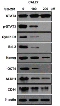
| Western blot | STAT3 / p-STAT3 / Cyclin D1 / Bcl-2 / Nanog / OCT4 / ALDH1 / CD44 PD-L1 |
|
26556875 |
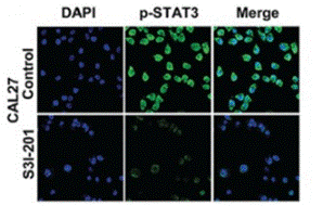
| Immunofluorescence | p-STAT3 Oct4 / Twist |
|
26556875 |
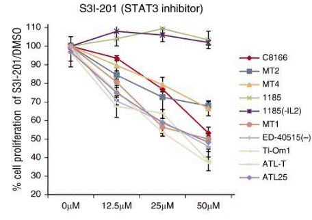
| Growth inhibition assay | Cell viability |
|
26813676 |
Tech Support
Tel: +1-832-582-8158 Ext:3
If you have any other enquiries, please leave a message.
Signaling Pathway Map
Products are for research use only. Not for human use. We do not sell to patients.
©Copyright 2013 Selleck Chemicals. All Rights Reserved.