-
 Australia
-
 Austria
-
 Belgium
-
 Brazil
-
 Canada
-
 China
China
-
 Czech Republic
-
 Denmark
-
 Finland
-
 France
-
 Germany
-
 Greece
-
 Hong Kong
-
 Hungary
-
 Iceland
-
 India
India
-
 Ireland
-
 Israel
-
 Italy
Italy
-
 Japan
Japan
-
 Korea
Korea
-
 Luxembourg
-
 Malaysia
-
 Netherlands
-
 New Zealand
-
 Norway
-
 Poland
-
 Qatar
Qatar
-
 Romania
-
 Saudi Arabia
-
 Singapore
-
 Spain
Spain
-
 Sweden
-
 Switzerland
-
 Taiwan
-
 Turkey
-
 United Kingdom
-
 United States
research use only
SNX-2112 HSP inhibitor
Cat.No.S2639
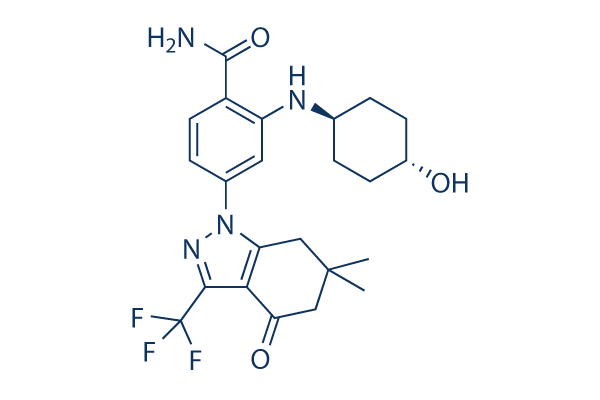
Chemical Structure
Molecular Weight: 464.48
Quality Control
Cell Culture, Treatment & Working Concentration
| Cell Lines | Assay Type | Concentration | Incubation Time | Formulation | Activity Description | PMID |
|---|---|---|---|---|---|---|
| A375 cells | Function assay | 24 h | Inhibition of Hsp90 in human A375 cells assessed as pS6 degradation after 24 hrs by high content screening, IC50=0.001 μM | |||
| LNCAP cells | Proliferation assay | 72-144 h | Antiproliferative activity against human LNCAP cells after 72 to 144 hrs by cyquant DNA dye method, IC50=0.003 μM | |||
| human SW620 cells | Proliferation assay | 72-144 h | Antiproliferative activity against human SW620 cells after 72 to 144 hrs by cyquant DNA dye method, IC50=0.003 μM | |||
| HT-29 cells | Proliferation assay | 72-144 h | Antiproliferative activity against human HT-29 cells after 72 to 144 hrs by cyquant DNA dye method, IC50=0.003 μM | |||
| human SK-MEL-5 cells | Proliferation assay | 72-144 h | Antiproliferative activity against human SK-MEL-5 cells after 72 to 144 hrs by cyquant DNA dye method, IC50=0.006 μM | |||
| sf9 cells | Function assay | 3 h | Binding affinity to human N-terminal polyHis-tagged HSP90beta (D9 to E236) expressed in insect sf9 cells after 3 hrs by fluorescence polarization assay, Ki=0.006 μM | |||
| K562 cells | Proliferation assay | 72-144 h | Antiproliferative activity against human K562 cells after 72 to 144 hrs by cyquant DNA dye method, IC50=0.006 μM | |||
| MDA-MB-231 cells | Proliferation assay | 72-144 h | Antiproliferative activity against human MDA-MB-231 cells after 72 to 144 hrs by cyquant DNA dye method, IC50=0.006 μM | |||
| PC3 cells | Proliferation assay | 72-144 h | Antiproliferative activity against human PC3 cells after 72 to 144 hrs by cyquant DNA dye method, IC50=0.017 μM | |||
| NCI-H460 cells | Proliferation assay | 72-144 h | Antiproliferative activity against human NCI-H460 cells after 72 to 144 hrs by cyquant DNA dye method, IC50=0.018 μM | |||
| MCF7 cells | Function assay | Inhibition of HSP90alpha in human MCF7 cells assessed as degradation of Her-2, EC50=0.019 μM | ||||
| AU565 cells | Function assay | 24 h | Inhibition of Hsp90 in human AU565 cells assessed as pERK degradation after 24 hrs by high content screening, IC50=0.041 μM | |||
| HCT15 cells | Proliferation assay | 72-144 h | Antiproliferative activity against human HCT15 cells after 72 to 144 hrs by cyquant DNA dye method, IC50=0.052 μM | |||
| MRC5 cells | Cytotoxicity assay | 48 h | Cytotoxicity against human MRC5 cells after 48 hrs by MTT assay, CC50=21.34 μM | |||
| Click to View More Cell Line Experimental Data | ||||||
Solubility
|
In vitro |
DMSO
: 93 mg/mL
(200.22 mM)
Ethanol : 1 mg/mL Water : Insoluble |
Molarity Calculator
|
In vivo |
|||||
In vivo Formulation Calculator (Clear solution)
Step 1: Enter information below (Recommended: An additional animal making an allowance for loss during the experiment)
Step 2: Enter the in vivo formulation (This is only the calculator, not formulation. Please contact us first if there is no in vivo formulation at the solubility Section.)
Calculation results:
Working concentration: mg/ml;
Method for preparing DMSO master liquid: mg drug pre-dissolved in μL DMSO ( Master liquid concentration mg/mL, Please contact us first if the concentration exceeds the DMSO solubility of the batch of drug. )
Method for preparing in vivo formulation: Take μL DMSO master liquid, next addμL PEG300, mix and clarify, next addμL Tween 80, mix and clarify, next add μL ddH2O, mix and clarify.
Method for preparing in vivo formulation: Take μL DMSO master liquid, next add μL Corn oil, mix and clarify.
Note: 1. Please make sure the liquid is clear before adding the next solvent.
2. Be sure to add the solvent(s) in order. You must ensure that the solution obtained, in the previous addition, is a clear solution before proceeding to add the next solvent. Physical methods such
as vortex, ultrasound or hot water bath can be used to aid dissolving.
Chemical Information, Storage & Stability
| Molecular Weight | 464.48 | Formula | C23H27F3N4O3 |
Storage (From the date of receipt) | |
|---|---|---|---|---|---|
| CAS No. | 908112-43-6 | Download SDF | Storage of Stock Solutions |
|
|
| Synonyms | PF-04928473 | Smiles | CC1(CC2=C(C(=O)C1)C(=NN2C3=CC(=C(C=C3)C(=O)N)NC4CCC(CC4)O)C(F)(F)F)C | ||
Mechanism of Action
| Targets/IC50/Ki |
HSP90α
(cell-free assay) 30 nM(Ka)
HSP90β
(cell-free assay) 30 nM(Ka)
|
|---|---|
| In vitro |
Treatment of BT-474 cells with 1 μM SNX-2112 results in down-regulation of HER2 expression within 3 to 6 hours of drug exposure with near-complete loss of HER2 expression by 10 hours. Treatment with this compound also results in a decline in total Akt expression. It inhibits cell proliferation with IC50 values ranging from 10 to 50 nM, in BT474, SKBR-3, SKOV-3, MDA-468, MCF-7 and H1650 cancer cells. And these antiproliferative effects are associated with hypophosphorylation of Rb, arrest of G1 and modest levels of apotosis. This chemical competitively binds to the N-terminal adenosine triphosphate binding site of Hsp90. It induces apoptosis via caspase-8, -9, -3, and poly (ADPribose) polymerase cleavage. The compound inhibits cytokine-inducedAkt and extracellular signal-related kinase (ERK) activation and also overcomes the growth advantages conferred by interleukin-6, insulin-like growth factor-1, and bone marrow stromal cells. It inhibits tube formation by human umbilical vein endothelial cells via abrogation of eNOS/Akt pathway and markedly inhibits osteoclast formation via down-regulation of ERK/c-fos and PU.1. Cell lines (eight cell lines from osteosarcoma, neuroblastoma, hepatoblastoma, and ymphoma) studied demonstrates sensitivity to this agent with IC50 values ranging from 10-100 nM. A higher dose (70 nM) exhibits a more prolonged inhibition and larger sub-G1 accumulation. Observed levels of Akt1 and C-Raf are markedly reduced over time along with an increase in PARP cleavage. A recent research indicates this compound induces autophagy in a time- and dose-dependent manner via Akt/mTOR/p70S6K inhibition. It induces significant apoptosis and utophagy in human melanoma A-375 cells, and may be an effective targeted therapy agent. |
| Kinase Assay |
ATP Displacement Assay
|
|
For the protein affinity-displacement assay, a purine-based affinity resin is generated by incubating ATP-linked Sepharose with Jurkat cell lysate (flash frozen and homogenized in saline) at 4 °C. This is then incubated with SNX-2112 for 90 minutes. Proteins eluted by this compound are then resolved by SDS-PAGE, visualized with silver staining, and excised from the gel for MS-based identification. Briefly, after destaining and trypsin digestion, peptides are extracted with μC18 ZipTips and then eluted and spotted directly to a conventional stainless steel matrix-assisted laser desorption/ionization target with a saturated solution of α-cyano-4- hydroxycinnamic acid in 50% acetonitrile, 0.15% formic acid. Mass spectra are then acquired using a MALDI-TOF/TOF 4700 Proteomics Analyzer. MS spectra are acquired (1,000 shots per spectrum), and the three peaks from each with the greatest signal-to-noise ratio are automatically submitted for tandem MS analysis (3000 shots per spectrum). The collision energy is 1keV. Air is used as the collision gas. Protein identification is done from the MS and tandem MS data using GPS Explorer software with the integrated Mascot database search engine.
|
|
| In vivo |
SNX-2112 is an active metabolite of the compounds SNX-5542, which inhibits multiple myeloma (MM) cell growth and prolongs survival in a xenograft murine model and blockade of Hsp90 by this compound not only inhibits MM cell growth but also acts in the bone marrow microenvironment to block angiogenesis and osteoclastogenesis. |
References |
|
Tech Support
Tel: +1-832-582-8158 Ext:3
If you have any other enquiries, please leave a message.
Signaling Pathway Map
Products are for research use only. Not for human use. We do not sell to patients.
©Copyright 2013 Selleck Chemicals. All Rights Reserved.