-
 Australia
-
 Austria
-
 Belgium
-
 Brazil
-
 Canada
-
 China
China
-
 Czech Republic
-
 Denmark
-
 Finland
-
 France
-
 Germany
-
 Greece
-
 Hong Kong
-
 Hungary
-
 Iceland
-
 India
India
-
 Ireland
-
 Israel
-
 Italy
Italy
-
 Japan
Japan
-
 Korea
Korea
-
 Luxembourg
-
 Malaysia
-
 Netherlands
-
 New Zealand
-
 Norway
-
 Poland
-
 Qatar
Qatar
-
 Romania
-
 Saudi Arabia
-
 Singapore
-
 Spain
Spain
-
 Sweden
-
 Switzerland
-
 Taiwan
-
 Turkey
-
 United Kingdom
-
 United States
research use only
GW9662 PPARγ Antagonist
Cat.No.S2915
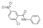
Chemical Structure
Molecular Weight: 276.68
Quality Control
| Related Targets | HDAC PARP ATM/ATR DNA-PK WRN DNA/RNA Synthesis Topoisomerase Sirtuin Casein Kinase eIF |
|---|---|
| Other PPAR Inhibitors | T0070907 GW6471 WY-14643 (Pirinixic Acid) GSK3787 GW0742 AZ6102 Harmine Palmitoylethanolamide Astaxanthin Eupatilin |
Cell Culture, Treatment & Working Concentration
| Cell Lines | Assay Type | Concentration | Incubation Time | Formulation | Activity Description | PMID |
|---|---|---|---|---|---|---|
| 293H | Function assay | 30 mins | Antagonist activity at human PPARgamma expressed in 293H cells assessed as reduction in transcriptional response preincubated for 30 mins followed by addition and measured after 16 hrs by reporter gene-based FRET assay, EC50=0.002μM | 31294974 | ||
| Click to View More Cell Line Experimental Data | ||||||
Solubility
|
In vitro |
DMSO
: 55 mg/mL
(198.78 mM)
Water : Insoluble Ethanol : Insoluble |
Molarity Calculator
|
In vivo |
|||||
In vivo Formulation Calculator (Clear solution)
Step 1: Enter information below (Recommended: An additional animal making an allowance for loss during the experiment)
Step 2: Enter the in vivo formulation (This is only the calculator, not formulation. Please contact us first if there is no in vivo formulation at the solubility Section.)
Calculation results:
Working concentration: mg/ml;
Method for preparing DMSO master liquid: mg drug pre-dissolved in μL DMSO ( Master liquid concentration mg/mL, Please contact us first if the concentration exceeds the DMSO solubility of the batch of drug. )
Method for preparing in vivo formulation: Take μL DMSO master liquid, next addμL PEG300, mix and clarify, next addμL Tween 80, mix and clarify, next add μL ddH2O, mix and clarify.
Method for preparing in vivo formulation: Take μL DMSO master liquid, next add μL Corn oil, mix and clarify.
Note: 1. Please make sure the liquid is clear before adding the next solvent.
2. Be sure to add the solvent(s) in order. You must ensure that the solution obtained, in the previous addition, is a clear solution before proceeding to add the next solvent. Physical methods such
as vortex, ultrasound or hot water bath can be used to aid dissolving.
Chemical Information, Storage & Stability
| Molecular Weight | 276.68 | Formula | C13H9ClN2O3 |
Storage (From the date of receipt) | |
|---|---|---|---|---|---|
| CAS No. | 22978-25-2 | Download SDF | Storage of Stock Solutions |
|
|
| Synonyms | N/A | Smiles | C1=CC=C(C=C1)NC(=O)C2=C(C=CC(=C2)[N+](=O)[O-])Cl | ||
Mechanism of Action
| Targets/IC50/Ki |
PPARγ
(Cell-free assay) 3.3 nM
PPARα
(Cell-free assay) 32 nM
|
|---|---|
| In vitro |
GW9662 binds to Cys(285) on PPARgamma which is conserved among all three PPARs. This compound acts as an antagonist of PPARgamma which is confirmed in an assay of adipocyte differentiation inhibition. It prevents activation of PPARγ and inhibits growth of human mammary tumour cell lines (MCF7, MDA-MB-468, MDA-MB-231) with IC50 of 20 μM-30 μM, suggesting either the existence of PPARγ agonistic properties of this chemical or growth-inhibitory mechanisms independent of PPARγ. Co-treatment with this compound (10 μM) results in statistically lower viable cell numbers after 7 days in MDA-MB-231 cells. PPARγ1 ligands could suppress RANKL-induced osteoclast formation in primary murine myeloid (BMs) and RAW264.7 cells. Importantly, suppression by these ligands is reversed in a concentration-dependent fashion with this chemical (2 μM). It (2 μM) blocks IL-4 suppression of osteoclast formation in BMs. This compound (1 μM) blocks RANKL activation of NF-κB in RAW264.7 cells. GW9662 (10 μM) inhibits hormone- and agonist-induced adipogenesis of primary preadipocytes from patients with thyroid eye disease. |
| Kinase Assay |
Binding assay
|
|
The human PPARα, PPARγ, and PPARδ ligand binding domains (LBDs) are expressed in E. coli as polyhistidine-tagged fusion proteins. Receptors are immobilized on SPA beads by addition of the desired receptor (15 nM) to a slurry of streptavidin-modifed SPA beads (0.5 mg/mL) in assay buffer. The mixture is allowed to equilibrate for at least 1 hour at room temperature, and the beads are pelleted by centrifugation at 1×10³ g. The supernate is discarded, and the beads are resuspended in the original volume of fresh assay buffer with gentle mixing. The centrifugation/resuspension procedure is repeated, and the resulting slurry of receptor-coated beads is used immediately or stored at 4 ℃ for up to 1 week before use. [3H]GW2443 are used as radioligands for determination of competition binding to PPARα, PPARγ, and PPARδ, respectively. Unless otherwise indicated, the buffer used for all assays is 50 mM HEPES (pH 7), 50 mM NaCl, 5 mM CHAPS, 0.1 mg/mL BSA, and 10 mM DTT. For some experiments, the HEPES (pH 7) is replaced with 50 mM Tris (pH 8).
|
|
| In vivo |
Pretreatment with LPS (1 mg/kg i.p.) significantly attenuates all markers of renal injury and dysfunction caused by ischemia/reperfusion (I/R) injury in rats. Most notably, this compound (1 mg/kg i.p.) abolishes the protective effects of LPS. |
References |
|
Applications
| Methods | Biomarkers | Images | PMID |
|---|---|---|---|
| Western blot | Vimentin / Slug / MMP9 / MMP2 |
|
30912275 |
| Growth inhibition assay | Cell proliferation |
|
30912275 |
Tech Support
Tel: +1-832-582-8158 Ext:3
If you have any other enquiries, please leave a message.
Signaling Pathway Map
Products are for research use only. Not for human use. We do not sell to patients.
©Copyright 2013 Selleck Chemicals. All Rights Reserved.