-
 Australia
-
 Austria
-
 Belgium
-
 Brazil
-
 Canada
-
 China
China
-
 Czech Republic
-
 Denmark
-
 Finland
-
 France
-
 Germany
-
 Greece
-
 Hong Kong
-
 Hungary
-
 Iceland
-
 India
India
-
 Ireland
-
 Israel
-
 Italy
Italy
-
 Japan
Japan
-
 Korea
Korea
-
 Luxembourg
-
 Malaysia
-
 Netherlands
-
 New Zealand
-
 Norway
-
 Poland
-
 Qatar
Qatar
-
 Romania
-
 Saudi Arabia
-
 Singapore
-
 Spain
Spain
-
 Sweden
-
 Switzerland
-
 Taiwan
-
 Turkey
-
 United Kingdom
-
 United States
research use only
TPCA-1 IKK-2 Inhibitor
Cat.No.S2824
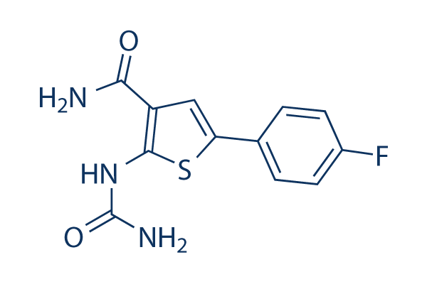
Chemical Structure
Molecular Weight: 279.29
Quality Control
| Related Targets | NF-κB HDAC Antioxidant ROS Nrf2 AP-1 MALT NOD |
|---|---|
| Other IκB/IKK Inhibitors | TBK1/IKKε-IN-5 Wedelolactone IKK-16 BMS-345541 Bay 11-7085 IMD 0354 MRT67307 HCl SC-514 Mesalazine (5-ASA) LY2409881 |
Cell Culture, Treatment & Working Concentration
| Cell Lines | Assay Type | Concentration | Incubation Time | Formulation | Activity Description | PMID |
|---|---|---|---|---|---|---|
| C57BL/6 mouse BMDM cells | Cytotoxicity assay | 24 h | Cytotoxicity against C57BL/6 mouse BMDM cells assessed as LDH release after 24 hrs | 22533790 | ||
| C57BL/6 mouse BMDM cells | Function assay | 0.5 μM | 1 h | Antiinflammatory activity in C57BL/6 mouse BMDM cells assessed as inhibition of LPS-stimulated TNFalpha production at 0.5 uM pretreated for 1 hr before LPS challenge after 8 to 24 hrs by immunoassay | 22533790 | |
| MDA-MB-231 | Cytotoxicity assay | 3 days | Cytotoxicity against human MDA-MB-231 cells assessed as cell growth inhibition after 3 days by presto blue dye based plate reader method | 27077228 | ||
| TC32 | qHTS assay | qHTS of pediatric cancer cell lines to identify multiple opportunities for drug repurposing: Primary screen for TC32 cells | 29435139 | |||
| DAOY | qHTS assay | qHTS of pediatric cancer cell lines to identify multiple opportunities for drug repurposing: Primary screen for DAOY cells | 29435139 | |||
| SJ-GBM2 | qHTS assay | qHTS of pediatric cancer cell lines to identify multiple opportunities for drug repurposing: Primary screen for SJ-GBM2 cells | 29435139 | |||
| A673 | qHTS assay | qHTS of pediatric cancer cell lines to identify multiple opportunities for drug repurposing: Primary screen for A673 cells | 29435139 | |||
| SK-N-MC | qHTS assay | qHTS of pediatric cancer cell lines to identify multiple opportunities for drug repurposing: Primary screen for SK-N-MC cells | 29435139 | |||
| BT-37 | qHTS assay | qHTS of pediatric cancer cell lines to identify multiple opportunities for drug repurposing: Primary screen for BT-37 cells | 29435139 | |||
| NB-EBc1 | qHTS assay | qHTS of pediatric cancer cell lines to identify multiple opportunities for drug repurposing: Primary screen for NB-EBc1 cells | 29435139 | |||
| U-2 OS | qHTS assay | qHTS of pediatric cancer cell lines to identify multiple opportunities for drug repurposing: Primary screen for U-2 OS cells | 29435139 | |||
| Saos-2 | qHTS assay | qHTS of pediatric cancer cell lines to identify multiple opportunities for drug repurposing: Primary screen for Saos-2 cells | 29435139 | |||
| SK-N-SH | qHTS assay | qHTS of pediatric cancer cell lines to identify multiple opportunities for drug repurposing: Primary screen for SK-N-SH cells | 29435139 | |||
| NB1643 | qHTS assay | qHTS of pediatric cancer cell lines to identify multiple opportunities for drug repurposing: Primary screen for NB1643 cells | 29435139 | |||
| LAN-5 | qHTS assay | qHTS of pediatric cancer cell lines to identify multiple opportunities for drug repurposing: Primary screen for LAN-5 cells | 29435139 | |||
| Rh18 | qHTS assay | qHTS of pediatric cancer cell lines to identify multiple opportunities for drug repurposing: Primary screen for Rh18 cells | 29435139 | |||
| OHS-50 | qHTS assay | qHTS of pediatric cancer cell lines to identify multiple opportunities for drug repurposing: Primary screen for OHS-50 cells | 29435139 | |||
| RD | qHTS assay | qHTS of pediatric cancer cell lines to identify multiple opportunities for drug repurposing: Primary screen for RD cells | 29435139 | |||
| MG 63 (6-TG R) | qHTS assay | qHTS of pediatric cancer cell lines to identify multiple opportunities for drug repurposing: Primary screen for MG 63 (6-TG R) cells | 29435139 | |||
| Rh30 | qHTS assay | qHTS of pediatric cancer cell lines to identify multiple opportunities for drug repurposing: Primary screen for Rh30 cells | 29435139 | |||
| Rh41 | qHTS assay | qHTS of pediatric cancer cell lines to identify multiple opportunities for drug repurposing: Primary screen for Rh41 cells | 29435139 | |||
| A673 | qHTS assay | qHTS of pediatric cancer cell lines to identify multiple opportunities for drug repurposing: Confirmatory screen for A673 cells) | 29435139 | |||
| SK-N-MC | qHTS assay | qHTS of pediatric cancer cell lines to identify multiple opportunities for drug repurposing: Confirmatory screen for SK-N-MC cells | 29435139 | |||
| TC32 | qHTS assay | qHTS of pediatric cancer cell lines to identify multiple opportunities for drug repurposing: Confirmatory screen for TC32 cells | 29435139 | |||
| MG 63 (6-TG R) | qHTS assay | qHTS of pediatric cancer cell lines to identify multiple opportunities for drug repurposing: Confirmatory screen for MG 63 (6-TG R) cells | 29435139 | |||
| U-2 OS | qHTS assay | qHTS of pediatric cancer cell lines to identify multiple opportunities for drug repurposing: Confirmatory screen for U-2 OS cells | 29435139 | |||
| SK-N-SH | qHTS assay | qHTS of pediatric cancer cell lines to identify multiple opportunities for drug repurposing: Confirmatory screen for SK-N-SH cells | 29435139 | |||
| Click to View More Cell Line Experimental Data | ||||||
Solubility
|
In vitro |
DMSO
: 56 mg/mL
(200.5 mM)
Water : Insoluble Ethanol : Insoluble |
Molarity Calculator
|
In vivo |
|||||
In vivo Formulation Calculator (Clear solution)
Step 1: Enter information below (Recommended: An additional animal making an allowance for loss during the experiment)
Step 2: Enter the in vivo formulation (This is only the calculator, not formulation. Please contact us first if there is no in vivo formulation at the solubility Section.)
Calculation results:
Working concentration: mg/ml;
Method for preparing DMSO master liquid: mg drug pre-dissolved in μL DMSO ( Master liquid concentration mg/mL, Please contact us first if the concentration exceeds the DMSO solubility of the batch of drug. )
Method for preparing in vivo formulation: Take μL DMSO master liquid, next addμL PEG300, mix and clarify, next addμL Tween 80, mix and clarify, next add μL ddH2O, mix and clarify.
Method for preparing in vivo formulation: Take μL DMSO master liquid, next add μL Corn oil, mix and clarify.
Note: 1. Please make sure the liquid is clear before adding the next solvent.
2. Be sure to add the solvent(s) in order. You must ensure that the solution obtained, in the previous addition, is a clear solution before proceeding to add the next solvent. Physical methods such
as vortex, ultrasound or hot water bath can be used to aid dissolving.
Chemical Information, Storage & Stability
| Molecular Weight | 279.29 | Formula | C12H10FN3O2S |
Storage (From the date of receipt) | |
|---|---|---|---|---|---|
| CAS No. | 507475-17-4 | Download SDF | Storage of Stock Solutions |
|
|
| Synonyms | GW683965 | Smiles | C1=CC(=CC=C1C2=CC(=C(S2)NC(=O)N)C(=O)N)F | ||
Mechanism of Action
| Targets/IC50/Ki |
NF-κB
STAT3
IKK2
(Cell-free assay) 17.9 nM
|
|---|---|
| In vitro |
In a time-resolved fluorescence resonance energy transfer assay, TPCA-1 inhibits human IKK-2 activity with an IC50 of 17.9 nM. In addition, this compound is demonstrated to be ATP-competitive. Besides, it exhibits IC50 values of 400 nM and 3600 nM against IKK-1 and JNK3, respectively. This chemical inhibits the production of TNF-α, IL-6, and IL-8 in a concentration-dependent manner, exhibiting IC50 values of 170, 290, and 320 nM, respectively. It inhibits glioma cell proliferation, as well as TNF-induced RelA (p65) nuclear translocation and NFκB-dependent IL8 gene expression. Importantly, this compound inhibits IFN-induced gene expression, completely suppressing MX1 and GBP1 gene expression, while having only a minor effect on ISG15 expression. |
| Kinase Assay |
IKK-2 Assay
|
|
Recombinant human IKK-2 (residues 1-756) is expressed in baculovirus as an N-terminal GST-tagged fusion protein, and its activity is assessed using a time-resolved fluorescence resonance energy transfer assay. In brief, IKK-2 (5 nM final) diluted in assay buffer (50 mM HEPES, 10 mM MgCl2, 1 mM CHAPS, pH 7.4, with 1 mM DTT and 0.01% w/v BSA) is added to wells containing various concentrations of this compound or DMSO vehicle (3% final). The reaction is initiated by the addition of GST-IκBα substrate (25 nM final)/ATP (1 μM final), in a total volume of 30 μL. The reaction is incubated for 30 min at room temperature, then terminated by the addition of 15 μL of 50 mM EDTA. Detection reagent (15 μL) in buffer (100 mM HEPES, pH 7.4, 150 mM NaCl, and 0.1% w/v BSA) containing antiphosphoserine- IκBα-32/36 monoclonal antibody 12C2, labeled with W-1024 europium chelate, and an allophycocyanin-labeled anti-GST antibody is added, and the reaction is further incubated for 60 min at room temperature. The degree of phosphorylation of GST- IκBαis measured as a ratio of specific 665-nm energy transfer signal to reference europium 620-nm signal, using a Packard Discovery plate reader.
|
|
| In vivo |
Prophylactic administration of TPCA-1 at 3, 10, or 20 mg/kg, i.p., b.i.d., results in a dose-dependent reduction in the severity of murine collagen-induced arthritis (CIA). The significantly reduced disease severity and delay of disease onset resulting from administration of this compound at 10 mg/kg, i.p., b.i.d. are comparable to the effects of the antirheumatic drug, i.p., every other day. Nuclear localization of p65, as well as levels of IL-1beta, IL-6, TNF-alpha, and interferon-gamma, is significantly reduced in the paw tissue of this compound-treated mice. In addition, administration of this chemical in vivo results in significantly decreased collagen-induced T cell proliferation ex vivo. Therapeutic administration of this compound at 20 mg/kg, but not at 3 or 10 mg/kg, i.p., b.i.d., significantly reduces the severity of CIA. |
References |
|
Tech Support
Tel: +1-832-582-8158 Ext:3
If you have any other enquiries, please leave a message.
Signaling Pathway Map
Products are for research use only. Not for human use. We do not sell to patients.
©Copyright 2013 Selleck Chemicals. All Rights Reserved.