-
 Australia
-
 Austria
-
 Belgium
-
 Brazil
-
 Canada
-
 China
China
-
 Czech Republic
-
 Denmark
-
 Finland
-
 France
-
 Germany
-
 Greece
-
 Hong Kong
-
 Hungary
-
 Iceland
-
 India
India
-
 Ireland
-
 Israel
-
 Italy
Italy
-
 Japan
Japan
-
 Korea
Korea
-
 Luxembourg
-
 Malaysia
-
 Netherlands
-
 New Zealand
-
 Norway
-
 Poland
-
 Qatar
Qatar
-
 Romania
-
 Saudi Arabia
-
 Singapore
-
 Spain
Spain
-
 Sweden
-
 Switzerland
-
 Taiwan
-
 Turkey
-
 United Kingdom
-
 United States
research use only
BAY 11-7082 (BAY 11-7821) NF-κB Inhibitor
Cat.No.S2913
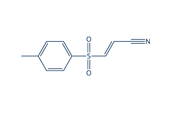
Chemical Structure
Molecular Weight: 207.25
Quality Control
| Related Targets | HDAC Antioxidant ROS IκB/IKK Nrf2 AP-1 MALT NOD |
|---|---|
| Other NF-κB Inhibitors | DCZ0415 Omaveloxolone (RTA-408) JSH-23 QNZ (EVP4593) Caffeic Acid Phenethyl Ester SC75741 DHA (Dihydroartemisinin) Andrographolide CBL0137 Withaferin A (WFA) |
Cell Culture, Treatment & Working Concentration
| Cell Lines | Assay Type | Concentration | Incubation Time | Formulation | Activity Description | PMID |
|---|---|---|---|---|---|---|
| HeLa | Function Assay | 10 μM | 1.5 h | abolishes BPA induced up regulation of FN and MMP-9 | 25797437 | |
| SiHa | Function Assay | 10 μM | 1.5 h | abolishes BPA induced up regulation of FN and MMP-9 | 25797437 | |
| ARPE-19 | Function Assay | 1 μM | 0.5 h | suppresses TG-induced IL-8 promoter activation | 25593029 | |
| HCT116 | Function Assay | 5 μM | 2 h | DMSO | attenuates silymarin-induced downregulation of cyclin D1 | 25479723 |
| HMECs | Function Assay | 5 μM | 2 h | abolishes TNF-α-induced VCAM-1 expression | 25193116 | |
| A549 | Function Assay | 10 µM | 12 h | suppresses Dvl-3 induced activation of p65 | 25156800 | |
| RAW 264.7 | Function Assay | 5 μM | 1 h | inhibits TNF-α and IL-12 p40 production | 25019567 | |
| macrophages | Function Assay | 5 µM | 3 h | partially blocks YPFS-induced expression of iNOS and COX-2 | 24967898 | |
| HUVECs | Function Assay | 3-30 μM | 1 h | reduces the expression of miR-146a in a dose-dependent manner | 24863965 | |
| HeLa | Function Assay | 5 μM | 24 h | DMSO | reduces the activity of TNF-α promoter | 24657783 |
| A549 | Function Assay | 10 μM | 1 h | inhibits the increase of phospho-IκBα in PA103-infected cultures | 24612488 | |
| HUVEC | Function Assay | 20 µM | 0.5 h | DMSO | prevents the induction of EAM expression | 24551209 |
| A549RT-eto | Apoptosis Assay | 10 μM | 24 h | DMSO | accelerates FERO-mediated apoptosis | 24535083 |
| THP-1 | Function Assay | 0.1/1 μM | 0.5 h | abrogates TNF-α secretion as well as the increased secretion of IL-6 and IL-1β | 24378536 | |
| SKCXCR2 | Growth Inhibition Assay | 2 µM | 48 h | decreases cell proliferation significantly | 24376747 | |
| SKCXCR2 | Function Assay | 2 µM | 48 h | blocks the CXCL1-induced cell invasion | 24376747 | |
| OVCXCR2 | Function Assay | 2 µM | 48 h | blocks the CXCL1-induced cell invasion | 24376747 | |
| DSCs | Function Assay | 2.5 μM | 0.5 h | reverses the enhancement of CCL2/CCR2 expression of DSCs induced by IL-33 | 24344240 | |
| WPs | Function Assay | 25 μM | 5 min | suppresses ATP and vWF secretion | 24331207 | |
| A549RT-eto | Apoptosis Assay | 10 μM | 24 h | accelerates F14 extract-mediated apoptosis combined treatment with F14 | 24220725 | |
| A549RT-eto | Function Assay | 10 μM | 24 h | decreases the expression levels of NF-κB and P-gp | 24220725 | |
| FaDu | Function Assay | 2 h | inhibits p65 expression and blocks TNFα-induced TWIST expression | 24220622 | ||
| IVD | Function Assay | 10 μM | 3 d | reverses TNF-α–mediated suppression of the disc matrix macromolecules aggrecan and collagen II | 24176808 | |
| IVD | Function Assay | 10 μM | 3 d | abrogates TNF-α–induced up-regulation of ADAMTS-4 and ADAMTS-5 | 24176808 | |
| iNKT | Function Assay | 10/100 μM | 0.5 h | inhibits the induction of A2AR mRNA and other factor | 24124453 | |
| PC-3 | Function Assay | 2.5/5/10 μM | 0.5 h | blocks IGF-II-induced STS mRNA expression | 24055520 | |
| THP-1 | Function Assay | 10 μM | 1 h | abolishes the effect of rHSP27 on SR-A mRNA | 23939398 | |
| A549 | Function Assay | 1 μM | 48 h | enhances the up-regulation of IκB and subsequent decrease in Bax expression induced by combined stimulation | 23900080 | |
| A549 | Apoptosis Assay | 1 μM | 48 h | reduces the cell death induced by combined stimulation | 23900080 | |
| NCI-N87 | Growth Inhibition Assay | 10/20/30 μM | 6/24 h | suppresses cell viability significantly | 23846545 | |
| AGS | Growth Inhibition Assay | 10/20/30 μM | 6/24 h | suppresses cell viability significantly | 23846545 | |
| MGC80-3 | Growth Inhibition Assay | 10/20/30 μM | 6/24 h | suppresses cell viability significantly | 23846545 | |
| HGC-27 | Function Assay | 7.5/15/30 μM | 6 h | induces the dephosphorylation and up-regulation of IκBα | 23846545 | |
| MGC80-3 | Function Assay | 7.5/15/30 μM | 6 h | induces the dephosphorylation and up-regulation of IκBα | 23846545 | |
| HGC-27 | Apoptosis Assay | 7.5/15/30 μM | 6 h | induces apoptosis in a time- and dose-dependent manner | 23846545 | |
| HBE | Function Assay | 10μM | 3h | abolishes the increases of IL-6 expression induced by CSE | 23824089 | |
| HepG2 | Function Assay | 0.3/1/3 μM | 48 h | reduces IL6-induced PON1 expression | 23791833 | |
| THP-1 | Function Assay | 5 µM | 1 h | DMSO | inhibits MTB-induced NFκB activation | 23634218 |
| THP-1 | Growth Inhibition Assay | 5 µM | 4/8 d | DMSO | reduces the viability of intracellular MTB | 23634218 |
| MDM | Growth Inhibition Assay | 5 µM | 4/8 d | DMSO | reduces the viability of intracellular MTB | 23634218 |
| AM | Growth Inhibition Assay | 5 µM | 4/8 d | DMSO | reduces the viability of intracellular MTB | 23634218 |
| RAW 264 | Function Assay | 0.2-5 µM | 30/60/90 min | inhibits the phosphatase activity of PTP1B | 23578302 | |
| HUVEC | Function Assay | 10 μM | 0.5 h | DMSO | counteractes the loss of Tie2 mRNA | 23563632 |
| HT29 | Function Assay | 10/30/100 μM | 1 h | inhibites both TWEAK-induced p100 processing | 23527154 | |
| HT29 | Function Assay | 10/30/100 μM | 1 h | inhibits TNF-induced phosphorylation and degradation of IκBα | 23527154 | |
| MM.1S | Apoptosis Assay | 30 µM | 3 h | induces MM cell death involves necrosis | 23527154 | |
| KMS-12-BM | Apoptosis Assay | 30 µM | 3 h | induces MM cell death involves necrosis | 23527154 | |
| BAFs | Function Assay | 0.5/1 μM | 24 h | inhibits TNFα/DEX induced CYP19A1 transcripts | 23485457 | |
| SP6.5 | Function Assay | 5 μM | 2 h | decreases translocation of p65 in the nucleus | 23443086 | |
| VUP | Function Assay | 5 μM | 2 h | decreases translocation of p65 in the nucleus | 23443086 | |
| OCM1 | Function Assay | 5 μM | 2 h | decreases translocation of p65 in the nucleus | 23443086 | |
| OM431 | Function Assay | 5 μM | 2 h | decreases translocation of p65 in the nucleus | 23443086 | |
| SP6.5 | Growth Inhibition Assay | 2.5-20 μM | 24 h | IC50=5 μM, exhibits strong anti-proliferative effects in a dose-dependent manner | 23443086 | |
| VUP | Growth Inhibition Assay | 2.5-20 μM | 24 h | IC50=5 μM, exhibits strong anti-proliferative effects in a dose-dependent manner | 23443086 | |
| OCM1 | Growth Inhibition Assay | 2.5-20 μM | 24 h | IC50=5 μM, exhibits strong anti-proliferative effects in a dose-dependent manner | 23443086 | |
| OM431 | Growth Inhibition Assay | 2.5-20 μM | 24 h | IC50=5 μM, exhibits strong anti-proliferative effects in a dose-dependent manner | 23443086 | |
| SP6.5 | Apoptosis Assay | 5 μM | 24 h | induces apoptosis | 23443086 | |
| VUP | Apoptosis Assay | 5 μM | 24 h | induces apoptosis | 23443086 | |
| OCM1 | Apoptosis Assay | 5 μM | 24 h | induces apoptosis | 23443086 | |
| OM431 | Apoptosis Assay | 5 μM | 24 h | induces apoptosis | 23443086 | |
| SP6.5 | Function Assay | 5 μM | 12 h | reduces the migration | 23443086 | |
| VUP | Function Assay | 5 μM | 12 h | reduces the migration | 23443086 | |
| OCM1 | Function Assay | 5 μM | 12 h | reduces the migration | 23443086 | |
| OM431 | Function Assay | 5 μM | 12 h | reduces the migration | 23443086 | |
| HBL-1 | Growth Inhibition Assay | 3 μM | 24/48/72 h | DMSO | slows cell growth modestly | 23441730 |
| RAW 264.7 | Function Assay | 2-15 μM | 1 h | DMSO | suppresses the activation of IKK family members | 23441730 |
| IL-1R | Function Assay | 2-15 μM | 1 h | DMSO | suppresses the activation of IKK family members | 23441730 |
| RAW 264.7 | Function Assay | 15 μM | 1 h | DMSO | suppresses the activation of and JNK | 23441730 |
| IL-1R | Function Assay | 15 μM | 1 h | DMSO | suppresses the activation of and JNK | 23441730 |
| U2OS | Function Assay | 15 μM | 1 h | DMSO | prevents the LPS- or IL-1-stimulated formation of K63-pUb chains | 23441730 |
| MT‐1 | Function Assay | 8 µm | 3 h | decreases the levels of p‐STAT3 and p‐4EBP1 | 23278479 | |
| MT‐2 | Function Assay | 8 µm | 3 h | decreases the levels of p‐STAT3 and p‐4EBP1 | 23278479 | |
| MT‐1 | Function Assay | 8 µm | 3 h | decreases the levels of the p65 subunit of NF‐κB | 23278479 | |
| MT‐2 | Function Assay | 8 µm | 3 h | decreases the levels of the p65 subunit of NF‐κB | 23278479 | |
| MCF-7 | Function Assay | 2.5-15 μM | 0.5 h | DMSO | causes the gradual loss of cell adhesion | 23093227 |
| HaCaT | Function Assay | 5.0 μM | 1 h | attenuates the TCOH-induced production of IL-6 | 23041168 | |
| A549 | Function Assay | 1 h | inhibits LTA-induced SP-A mRNA production significantly | 23031213 | ||
| OA chondrocytes | Function Assay | 10 μM | 1 h | blocks the AGE-BSA-induced gene/protein expression of GRP78 or COX-2 (p<0.05) | 22982228 | |
| RAW264.7 | Function Assay | 15 μM | 15-120 min | blocks the production of NO, PGE2, and TNF-α | 22745523 | |
| RAW264.7 | Growth Inhibition Assay | 5-30 μM | 24 h | inhibits cell growth in a dose-dependent manner | 22745523 | |
| HBL6 | Apoptosis Assay | 0.5/5/25 μM | 6/24 h | decreases cell viability and leeads to apoptosis in a dose-dependent manner | 22074820 | |
| HT29 | Function Assay | 1-10 μM | 10 h | increases HO-1 mRNA and protein expression | 21620964 | |
| Ca9–22 | Apoptosis Assay | 10 μM | 1 h | completely inhibits ALA-PDT-induced apoptosis | 21138480 | |
| Ca9–22 | Function Assay | 10 μM | 1 h | completely abrogates the ALA-PDT-induced JNK activation | 21138480 | |
| A-549 | Growth Inhibition Assay | 10 μM | 24/48 h | inhibits cell growth in a time-dependent manner | 20866043 | |
| AP | Function Assay | 5/10 μM | 48 h | downregulates the BAD protein level a dose-dependent manner | 20596645 | |
| AQ1 | Function Assay | 5/10 μM | 48 h | downregulates the BAD protein level a dose-dependent manner | 20596645 | |
| AP | Function Assay | 20 μM | 4/8 h | downregulates the BAD protein level a time-dependent manner | 20596645 | |
| AQ1 | Function Assay | 20 μM | 4/8 h | downregulates the BAD protein level a time-dependent manner | 20596645 | |
| THP-1 | Function Assay | 5 μM | 0.5 h | attenuates the LPS-induced p-IκBα protein by 72% | 20309718 | |
| K562 | Growth Inhibition Assay | 2-30 μM | 24 h | IC50=8 μM,inhibits cell growth in a dose-dependent manner | 19646807 | |
| Jurket | Growth Inhibition Assay | 2-30 μM | 24 h | IC50=7.1 μM, inhibits cell growth in a dose-dependent manner | 19646807 | |
| U937 | Growth Inhibition Assay | 2-30 μM | 24 h | IC50=10.5 μM, inhibits cell growth in a dose-dependent manner | 19646807 | |
| PBMC | Growth Inhibition Assay | 2-30 μM | 24 h | IC50=40.2 μM, inhibits cell growth in a dose-dependent manner | 19646807 | |
| K562 | Apoptosis Assay | 2-20 μM | 24 h | induces a dose-dependent apoptosis | 19646807 | |
| THP1 | Cytotoxicity assay | 72 hrs | Cytotoxicity against human THP1 cells assessed as reduction in cell viability after 72 hrs by MTT assay, TC50 = 1.5 μM. | 28410442 | ||
| RAW264.7 | Function assay | 6 hrs | Inhibition of LPS-induced NF-kappaB activation in mouse RAW264.7 cells treated 30 mins before LPS challenge measured after 6 hrs by luciferase reporter gene assay, IC50 = 1.72 μM. | 24315191 | ||
| HEK293 | Function assay | 6 hrs | Inhibition of TNF-alpha-induced NF-kappaB activity in HEK293 cells after 6 hrs by luciferase reporter gene assay, IC50 = 2 μM. | 24533857 | ||
| HEK293 | Function assay | 6 hrs | Inhibition of TNF-alpha-induced NF-kappaB activity in HEK293 cells after 6 hrs by luciferase reporter gene assay, IC50 = 2 μM. | 24992702 | ||
| HEK293 | Function assay | 6 hrs | Inhibition of TNFalpha-induced NF-kappaB activity (unknown origin) transfected in HEK293 cells after 6 hrs by luciferase reporter gene assay, IC50 = 2 μM. | 26343828 | ||
| HEK293 | Function assay | 6 hrs | Inhibition of TNFalpha-induced NF-kappaB activity expressed in human HEK 293 cells after 6 hrs by luciferase reporter gene assay, IC50 = 2 μM. | 22850207 | ||
| HEK293 | Function assay | 6 hrs | Inhibition of TNFalpha-induced human NFkappaB activity in HEK293 cells incubated for 6 hrs followed by compound wash out measured after 5 mins by by luciferase assay, IC50 = 2.01 μM. | 22712432 | ||
| HEK293 | Cytotoxicity assay | Cytotoxicity against HEK293 cells, IC50 = 3.8 μM. | 24533857 | |||
| HEK293 | Function assay | 6 hrs | Inhibition of TNFalpha-induced NFkappaB (unknown origin) activation expressed in HEK293 cells after 6 hrs by luciferase reporter gene assay, IC50 = 5 μM. | 23316950 | ||
| HEK293 | Function assay | Effect on Cdc2 expressed in HEK293 cells assessed as effect on Cdc2:Cdc25C interaction complexes in presence of camptothecin by EYFP and/or YFP Venus fragment based reporter gene assay | 16680159 | |||
| HEK293 | Function assay | 20 uM | 24 hrs | Inhibition of TNF-alpha stimulated NFkappaB transactivation in HEK293 cells at 20 uM measured after 24 hrs by dual luciferase reporter gene assay | 27736063 | |
| RAW264.7 | Function assay | 20 uM | 1 hr | Inhibition of LPS-induced NFkB activation in mouse RAW264.7 cells assessed as reduction in nuclear translocation of p65 at 20 uM preincubated for 1 hr followed by LPS stimulation measured after 3 hrs by Western blot method | 28667873 | |
| RAW264.7 | Function assay | 20 uM | 6 hrs | Inhibition of LPS-induced NF-kappaB activation in mouse RAW264.7 cells at 20 uM treated 30 mins before LPS challenge measured after 6 hrs by luciferase reporter gene assay | 24315191 | |
| RAW264.7 | Antinflammatory assay | 20 uM | 18 hrs | Antinflammatory activity in mouse RAW264.7 cells assessed as inhibition of LPS-induced nitric oxide production at 20 uM treated 30 mins before LPS challenge measured after 18 hrs by Griess assay | 24315191 | |
| THP1 | Antinflammatory assay | 5 uM | 24 hrs | Antiinflammatory activity in human THP1 cells assessed as inhibition of TPA/ionomycin-induced extracellular IL-1beta level at 5 uM incubated 1 hr prior to TPA/ionomycin challenge measured after 24 hrs by ELISA | 24400858 | |
| THP1 | Antinflammatory assay | 5 uM | 24 hrs | Antiinflammatory activity in human THP1 cells assessed as inhibition of TPA/ionomycin-induced extracellular TNF-alpha production at 5 uM incubated 1 hr prior to TPA/ionomycin challenge measured after 24 hrs by ELISA | 24400858 | |
| THP1 | Antinflammatory assay | 5 uM | 24 hrs | Antiinflammatory activity in human THP1 cells assessed as inhibition of TPA/ionomycin-induced intracellular proIL-1beta level at 5 uM incubated 1 hr prior to TPA/ionomycin challenge measured after 24 hrs by ELISA | 24400858 | |
| THP1 | Antinflammatory assay | 5 uM | 24 hrs | Antiinflammatory activity in human THP1 cells assessed as inhibition of TPA/ionomycin-induced intracellular IL-1beta level at 5 uM incubated 1 hr prior to TPA/ionomycin challenge measured after 24 hrs by ELISA | 24400858 | |
| RAW264.7 | Function assay | 0.3 ug/ml | 12 hrs | Inhibition of LPS-induced NF-kB p65 phosphorylation in mouse RAW264.7 cells at 0.3 ug/ml preincubated for 12 hrs followed by LPS stimulation for 3 hrs by Western blot method | 28284806 | |
| RAW264.7 | Function assay | 0.3 ug/ml | 12 hrs | Inhibition of LPS-induced NF-kB p65 activation in mouse RAW264.7 cells at 0.3 ug/ml preincubated for 12 hrs followed by LPS stimulation for 3 hrs by DAPI staining based inverted fluorescence microscopic method | 28284806 | |
| RAW264.7 | Function assay | 0.3 ug/ml | 12 hrs | Inhibition of NF-kB p65 in mouse RAW264.7 cells assessed as reduction in LPS-induced iNOS expression at 0.3 ug/ml preincubated for 12 hrs followed by LPS stimulation for 3 hrs by Western blot method | 28284806 | |
| RAW264.7 | Function assay | 0.3 ug/ml | 12 hrs | Inhibition of NF-kB p65 in mouse RAW264.7 cells assessed as reduction in LPS-induced COX2 expression at 0.3 ug/ml preincubated for 12 hrs followed by LPS stimulation for 3 hrs by Western blot method | 28284806 | |
| HEK293 | Function assay | 20 uM | 24 hrs | Inhibition of TNFalpha-induced NFkappaB activation in HEK293 cells at 20 uM after 24 hrs by dual luciferase reporter gene assay | 28873303 | |
| RAW264.7 | Function assay | 20 uM | 2 hrs | Inhibition of NFkappaB nuclear translocation in LPS-stimulated mouse RAW264.7 cells at 20 uM pretreated for 2 hrs followed by LPS-induction by DAPI-staining based immunofluorescence microscopic method | 29759725 | |
| BGC823 | Function assay | 5 uM | 12 hrs | Inhibition of colony formation in human BGC823 cells at 5 uM treated for 12 hrs followed by incubation in drug free medium for 14 days by crystal violet staining based assay | 28881286 | |
| SGC7901 | Function assay | 5 uM | 12 hrs | Inhibition of colony formation in human SGC7901 cells at 5 uM treated for 12 hrs followed by incubation in drug free medium for 14 days by crystal violet staining based assay | 28881286 | |
| RAW264.7 | Function assay | 10 uM | 2 hrs | Inhibition of LPS-induced IL-6 mRNA expression in mouse RAW264.7 cells at 10 uM pre-incubated for 2 hrs before LPS stimulation for 24 hrs by qRT-PCR method | 27038497 | |
| RAW264.7 | Function assay | 10 uM | 2 hrs | Inhibition of LPS-induced IL-1beta mRNA expression in mouse RAW264.7 cells at 10 uM pre-incubated for 2 hrs before LPS stimulation for 24 hrs by qRT-PCR method | 27038497 | |
| RAW264.7 | Function assay | 10 uM | 2 hrs | Inhibition of LPS-induced iNOS mRNA expression in mouse RAW264.7 cells at 10 uM pre-incubated for 2 hrs before LPS stimulation for 24 hrs by qRT-PCR method | 27038497 | |
| Click to View More Cell Line Experimental Data | ||||||
Solubility
|
In vitro |
DMSO
: 41 mg/mL
(197.82 mM)
Ethanol : 10 mg/mL Water : Insoluble |
Molarity Calculator
|
In vivo |
|||||
In vivo Formulation Calculator (Clear solution)
Step 1: Enter information below (Recommended: An additional animal making an allowance for loss during the experiment)
Step 2: Enter the in vivo formulation (This is only the calculator, not formulation. Please contact us first if there is no in vivo formulation at the solubility Section.)
Calculation results:
Working concentration: mg/ml;
Method for preparing DMSO master liquid: mg drug pre-dissolved in μL DMSO ( Master liquid concentration mg/mL, Please contact us first if the concentration exceeds the DMSO solubility of the batch of drug. )
Method for preparing in vivo formulation: Take μL DMSO master liquid, next addμL PEG300, mix and clarify, next addμL Tween 80, mix and clarify, next add μL ddH2O, mix and clarify.
Method for preparing in vivo formulation: Take μL DMSO master liquid, next add μL Corn oil, mix and clarify.
Note: 1. Please make sure the liquid is clear before adding the next solvent.
2. Be sure to add the solvent(s) in order. You must ensure that the solution obtained, in the previous addition, is a clear solution before proceeding to add the next solvent. Physical methods such
as vortex, ultrasound or hot water bath can be used to aid dissolving.
Chemical Information, Storage & Stability
| Molecular Weight | 207.25 | Formula | C10H9NO2S |
Storage (From the date of receipt) | |
|---|---|---|---|---|---|
| CAS No. | 19542-67-7 | Download SDF | Storage of Stock Solutions |
|
|
| Synonyms | BAY 11-7821 | Smiles | CC1=CC=C(C=C1)S(=O)(=O)C=CC#N | ||
Mechanism of Action
| Targets/IC50/Ki |
E2-conjugating enzymes
(Cell-free assay) USP7
(Cell-free assay) 0.19 μM
USP21
(Cell-free assay) 0.96 μM
USP6
(Cell-free assay) 1.7 μM
IκBα phosphorylation
(Tumor cells) 10 μM
|
|---|---|
| In vitro |
BAY 11-7082 completely and specifically abrogates NF-κB DNA binding, downregulating the NF-κB-inducible cytokine IL-6 and inducing apoptosis. This compound (< 8 μM) is able to effectively inhibit both basal and TNFα stimulated NFκB luciferase activity in a dose dependent manner. It (8 μM) strongly inhibits the rate of proliferation in NCI-H1703 cells. This compound (5 μM) rapidly and efficiently reduces the DNA binding of NF-kappaB in HTLV-I-infected T-cell lines and down-regulates the expression of the antiapoptotic gene, Bcl-x(L), whereas it has little effect on the DNA binding of another transcription factor, AP-1. This chemical-induced apoptosis of primary ATL cells is more prominent than that of normal peripheral blood mononuclear cells, and apoptosis of these cells is also associated with down-regulation of NF-kappaB activity. It (5 μM) selectively induces apoptosis of HTLV-I–infected T-cell lines associated with down-regulation of the expression of cyclin D1, cyclin D2, and Bcl-xL. This compound (100 μM) prevents the nuclear translocation of p65 elicited by NMDA and the NMDA-induced increase of NF-κB binding in mouse hippocampal slices. It prevents NMDA toxicity occurring in CA1 region of hippocampal slices with 40% neuroprotection at 20 μM and 70% neuroprotection at 100 μM. This chemical at all concentrations tested significantly inhibits NF-κB p65 DNA-binding activity in adipose tissue, whereas in skeletal muscle, it at 50 μM and 100 μM significantly inhibits NF-κB p65 DNA-binding activity. It (100 μM) reduces IKK-β protein in human adipose tissue and skeletal muscle. This compound (100 μM) significantly decreases the release of TNF-α from adipose tissue, whereas the release of IL-6 and IL-8 is significantly inhibited at all concentrations of this chemical tested. It (50 μM) significantly decreases the release of TNF-α, IL-6, and IL-8 in skeletal muscle. This compound is also found to inactivate the E2-conjugating enzymes Ubc (ubiquitin conjugating) 13 and UbcH7 and the E3 ligase LUBAC (linear ubiquitin assembly complex), and thus induces B-cell lymphoma and leukaemic T-cell death. |
| In vivo |
BAY 11-7082, an NF-κB inhibitor, induces apoptosis and S phase arrest in gastric cancer cells. |
References |
|
Applications
| Methods | Biomarkers | Images | PMID |
|---|---|---|---|
| Western blot | NF-κB p-IKKβ/ IκBα p-IRAK4 / IRAK4 NF-κB p-IKKβ/ IκBα p-IRAK4 / IRAK4 |
|
31332209 |
| Growth inhibition assay | Cell viability Cell viability |
|
31332209 |
Tech Support
Tel: +1-832-582-8158 Ext:3
If you have any other enquiries, please leave a message.
Signaling Pathway Map
Products are for research use only. Not for human use. We do not sell to patients.
©Copyright 2013 Selleck Chemicals. All Rights Reserved.