research use only
PAC-1 Caspase activator
Cat.No.S2738

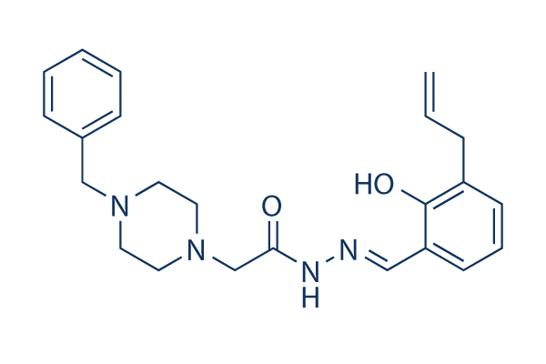
Chemical Structure
Molecular Weight: 392.49
Quality Control
| Related Targets | Bcl-2 PD-1/PD-L1 Ferroptosis p53 Apoptosis related Synthetic Lethality STAT TNF-alpha Ras KRas |
|---|---|
| Other Caspase Inhibitors | Z-VAD-FMK Z-DEVD-FMK Q-VD-Oph Emricasan (IDN-6556) Belnacasan (VX-765) Z-IETD-FMK Ac-DEVD-CHO Z-LEHD-FMK TFA Z-VAD(OH)-FMK Z-YVAD-FMK |
Solubility
|
In vitro |
DMSO
: 78 mg/mL
(198.73 mM)
Ethanol : 78 mg/mL Water : Insoluble |
Molarity Calculator
|
In vivo |
|||||
In vivo Formulation Calculator (Clear solution)
Step 1: Enter information below (Recommended: An additional animal making an allowance for loss during the experiment)
Step 2: Enter the in vivo formulation (This is only the calculator, not formulation. Please contact us first if there is no in vivo formulation at the solubility Section.)
Calculation results:
Working concentration: mg/ml;
Method for preparing DMSO master liquid: mg drug pre-dissolved in μL DMSO ( Master liquid concentration mg/mL, Please contact us first if the concentration exceeds the DMSO solubility of the batch of drug. )
Method for preparing in vivo formulation: Take μL DMSO master liquid, next addμL PEG300, mix and clarify, next addμL Tween 80, mix and clarify, next add μL ddH2O, mix and clarify.
Method for preparing in vivo formulation: Take μL DMSO master liquid, next add μL Corn oil, mix and clarify.
Note: 1. Please make sure the liquid is clear before adding the next solvent.
2. Be sure to add the solvent(s) in order. You must ensure that the solution obtained, in the previous addition, is a clear solution before proceeding to add the next solvent. Physical methods such
as vortex, ultrasound or hot water bath can be used to aid dissolving.
Chemical Information, Storage & Stability
| Molecular Weight | 392.49 | Formula | C23H28N4O2 |
Storage (From the date of receipt) | |
|---|---|---|---|---|---|
| CAS No. | 315183-21-2 | Download SDF | Storage of Stock Solutions |
|
|
| Synonyms | N/A | Smiles | C=CCC1=C(C(=CC=C1)C=NNC(=O)CN2CCN(CC2)CC3=CC=CC=C3)O | ||
Mechanism of Action
| Features |
The first small molecule known to directly activate procaspase-3 to caspase-3.
|
|---|---|
| Targets/IC50/Ki |
Procaspase-3
0.22 μM(EC50)
|
| In vitro |
PAC-1 activates procaspase-7 in a less efficient manner with EC50 of 4.5 μM. Elevated caspase 3 level in cancer cell lines allows this compound to selectively induce apoptosis in a manner proportional to procaspase-3 concentration with IC50 of 0.35 μM for NCI-H226 cells to ~3.5 μM for UACC-62 cells. It induces apoptosis in the primary cancerous cells with IC50 values of 3 nM to 1.41 μM, more potently than in the adjacent noncancerous cells with IC50 of 5.02 μM to 9.98 μM, which is also directly related to the distinct procaspase-3 concentration. This chemical activates procaspase-3 by chelating zinc ions, thus relieving the zinc-mediated inhibition and allowing procaspase-3 to auto-activate itself to caspase-3. It is capable to induce cell death in Bax/Bak double-knockout cells and Bcl-2 and Bcl-xL-overexpressing cells with the same efficacy as its wild-type counterpart in a delayed manner. This compound induces cytochrome c release in a caspase-3 independent manner, which subsequently triggers downstream caspase-3 activation and cell death. It can not induce cell death and caspase-3 activation in Apaf-1 knockout cells, suggesting that apoptosome formation is essential for caspase-3 activation by PAC-1-mediated cell death.
|
| Kinase Assay |
In vitro procaspase-3 activation
|
|
Procaspase-3 is expressed and purified in Escherichia coli. Various concentrations of PAC-1 are added to 90 μL of a 50 ng/mL of procaspase-3 in caspase assay buffer in a 96-well plate, The plate is incubated for 12 hours at 37 °C. A 10 μL volume of a 2 mM solution of caspase-3 peptidic substrate acetyl Asp-Glu-Val-Asp-p-nitroanilide (Ac-DEVD-pNa) in caspase assay buffer is then added to each well. The plate is read every 2 minutes at 405 nm for 2 hours in a Spectra Max Plus 384 well plate reader. The slope of the linear portion for each well is determined, and the relative increase in activation from untreated control wells is calculated.
|
|
| In vivo |
Administration of PAC-1 at 5 mg with low and steady releasing significantly inhibits the growth of ACHN renal cancer xenograft in mice. Oral administration of this compound (50 or 100 mg/kg) significantly retards tumor growth of NCI-H226 lung cancer xenograft in a dose-dependent manner, and markedly prevents the cancer cells from infiltrating the lung tissue. The in vivo anti-tumor effect of this chemical is ascribed to procaspase-3 activation and subsequently apoptosis induction consistent with the activity in vitro.
|
References |
|
Applications
| Methods | Biomarkers | Images | PMID |
|---|---|---|---|
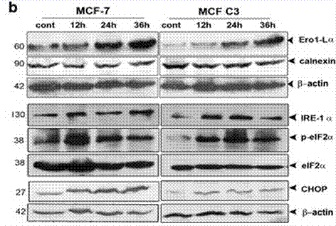
| Western blot | Ero1-Lα / Calnexin / IRE-1α / p-eIF2α / eIF2α / CHOP |

|
24357799 |
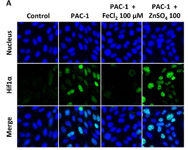
| Immunofluorescence | Hif1α p-H2AX / Rad51 |

|
30287840 |
Clinical Trial Information
(data from https://clinicaltrials.gov, updated on 2024-05-22)
| NCT Number | Recruitment | Conditions | Sponsor/Collaborators | Start Date | Phases |
|---|---|---|---|---|---|
| NCT05967741 | Recruiting | Platelet Aggregation Spontaneous|Vascular Thrombosis |
University of California Davis |
July 20 2023 | Not Applicable |
| NCT03927248 | Withdrawn | Metastatic Renal Cell Carcinoma |
HealthPartners Institute |
September 2020 | Phase 1|Phase 2 |
| NCT03441412 | Completed | Healthy Volunteers |
Vastra Gotaland Region|Gothia Forum - Center for Clinical Trial|Uppsala University |
February 28 2018 | Phase 1 |
Tech Support
Tel: +1-832-582-8158 Ext:3
If you have any other enquiries, please leave a message.


