-
 Australia
-
 Austria
-
 Belgium
-
 Brazil
-
 Canada
-
 China
China
-
 Czech Republic
-
 Denmark
-
 Finland
-
 France
-
 Germany
-
 Greece
-
 Hong Kong
-
 Hungary
-
 Iceland
-
 India
India
-
 Ireland
-
 Israel
-
 Italy
Italy
-
 Japan
Japan
-
 Korea
Korea
-
 Luxembourg
-
 Malaysia
-
 Netherlands
-
 New Zealand
-
 Norway
-
 Poland
-
 Qatar
Qatar
-
 Romania
-
 Saudi Arabia
-
 Singapore
-
 Spain
Spain
-
 Sweden
-
 Switzerland
-
 Taiwan
-
 Turkey
-
 United Kingdom
-
 United States
research use only
ICG-001 Wnt/β-catenin Inhibitor
Cat.No.S2662
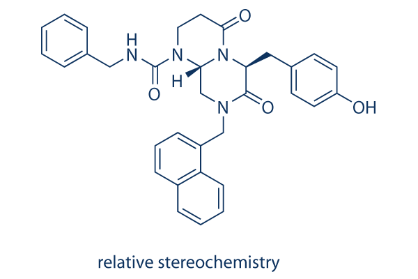
Chemical Structure
Molecular Weight: 548.63
Quality Control
Products Often Used Together with ICG-001
| Related Targets | JAK TGF-beta/Smad ERK GSK-3 ROCK Hedgehog/Smoothened PKA Secretase STAT Casein Kinase |
|---|---|
| Other Wnt/beta-catenin Inhibitors | IWR-1-endo PRI-724 (Foscenvivint) IWP-2 Tegatrabetan (BC-2059) Isoquercitrin SKL2001 BML-284 Hydrochloride (Wnt agonist 1, AMBMP) PNU-74654 LF3 Salinomycin (Procoxacin) |
Cell Culture, Treatment & Working Concentration
| Cell Lines | Assay Type | Concentration | Incubation Time | Formulation | Activity Description | PMID |
|---|---|---|---|---|---|---|
| SH-SY5Y | Apoptosis Assay | 50 μm | 24 h | DMSO | blocks the protective effect of melatonin against PrP (106–126)-induced apoptotic signals | 25251028 |
| AsPC-1 | Growth Inhibition Assay | 1-20 μM | 2/4/6 d | inhibits the cell growth in a dose-dependent manner | 25082960 | |
| MiaPaCa-2 | Growth Inhibition Assay | 1-20 μM | 2/4/6 d | inhibits the cell growth in a dose-dependent manner | 25082960 | |
| PANC-1 | Growth Inhibition Assay | 1-20 μM | 2/4/6 d | inhibits the cell growth in a dose-dependent manner | 25082960 | |
| L3.6pl | Growth Inhibition Assay | 1-20 μM | 2/4/6 d | inhibits the cell growth in a dose-dependent manner | 25082960 | |
| SH-SY5Y | Apoptosis Assay | 10 μM | 24 h | inhibits the neuroprotective effects of hypoxia against PrP (106-126)-mediated neuronal cell death | 23900566 | |
| HKC-8 | Function Assay | 10 µM | 24 h | abolishes β-catenin–mediated RAS induction | 25012166 | |
| HK-2 | Function Assay | 10 µM | 3 h | reduced the expression of TGF-β1, α-SMA, and CTGF after treatment with HHE | 23690997 | |
| HepT1 | Apoptosis Assay | 0-100 μM | 24 h | IC50=34 μM | 23266718 | |
| HuH6 | Apoptosis Assay | 0-100 μM | 24 h | IC50=39 μM | 23266718 | |
| MCF7 | Function Assay | 5 μm | inhibits leptin-mediated increased expression of Snail, Slug, and Zeb2 | 22270359 | ||
| RLE-6TN | Function Assay | 2.5/5/7.5 μM | 48 h | inhibits TGF-β1-induced α-SMA induction and EMT | 22241478 | |
| HKC-8 | Function Assay | 5/10/20 μM | 48 h | blocks β-catenin-driven gene expression | 21816937 | |
| SW480 | Growth Inhibition Assay | 2-100 μM | IC50=5.8±0.68 μM | 15782138 | ||
| SW480 | Function assay | Inhibition of CBP binding to beta-casein in human SW480 cells by immunoblot analysis, IC50 = 1.3 μM. | 23232060 | |||
| A549 | Antiproliferative assay | 72 hrs | Antiproliferative activity against human A549 cells after 72 hrs by MTT assay, GI50 = 6.1 μM. | 24950489 | ||
| HepG2 | Antiproliferative assay | 72 hrs | Antiproliferative activity against human HepG2 cells after 72 hrs by MTT assay, GI50 = 12.7 μM. | 24950489 | ||
| LoVo | Antiproliferative assay | 72 hrs | Antiproliferative activity against human LoVo cells after 72 hrs by MTT assay, GI50 = 15.6 μM. | 24950489 | ||
| HT-29 | Antiproliferative assay | 72 hrs | Antiproliferative activity against human HT-29 cells after 72 hrs by MTT assay, GI50 = 17.2 μM. | 24950489 | ||
| HT29 | Function assay | 24 hrs | Inhibition of Wnt signaling in human HT29 cells assessed as inhibition of beta-catenin-mediated Tcf/Lef transcriptional activity after 24 hrs by dual luciferase reporter gene assay relative to control, IC50 = 18.7 μM. | 24950489 | ||
| TC32 | qHTS assay | qHTS of pediatric cancer cell lines to identify multiple opportunities for drug repurposing: Primary screen for TC32 cells | 29435139 | |||
| A673 | qHTS assay | qHTS of pediatric cancer cell lines to identify multiple opportunities for drug repurposing: Primary screen for A673 cells | 29435139 | |||
| DAOY | qHTS assay | qHTS of pediatric cancer cell lines to identify multiple opportunities for drug repurposing: Primary screen for DAOY cells | 29435139 | |||
| BT-37 | qHTS assay | qHTS of pediatric cancer cell lines to identify multiple opportunities for drug repurposing: Primary screen for BT-37 cells | 29435139 | |||
| RD | qHTS assay | qHTS of pediatric cancer cell lines to identify multiple opportunities for drug repurposing: Primary screen for RD cells | 29435139 | |||
| MG 63 (6-TG R) | qHTS assay | qHTS of pediatric cancer cell lines to identify multiple opportunities for drug repurposing: Primary screen for MG 63 (6-TG R) cells | 29435139 | |||
| NB1643 | qHTS assay | qHTS of pediatric cancer cell lines to identify multiple opportunities for drug repurposing: Primary screen for NB1643 cells | 29435139 | |||
| OHS-50 | qHTS assay | qHTS of pediatric cancer cell lines to identify multiple opportunities for drug repurposing: Primary screen for OHS-50 cells | 29435139 | |||
| SJ-GBM2 | qHTS assay | qHTS of pediatric cancer cell lines to identify multiple opportunities for drug repurposing: Primary screen for SJ-GBM2 cells | 29435139 | |||
| SK-N-MC | qHTS assay | qHTS of pediatric cancer cell lines to identify multiple opportunities for drug repurposing: Primary screen for SK-N-MC cells | 29435139 | |||
| NB-EBc1 | qHTS assay | qHTS of pediatric cancer cell lines to identify multiple opportunities for drug repurposing: Primary screen for NB-EBc1 cells | 29435139 | |||
| LAN-5 | qHTS assay | qHTS of pediatric cancer cell lines to identify multiple opportunities for drug repurposing: Primary screen for LAN-5 cells | 29435139 | |||
| LoVo | Cytotoxicity assay | 10 uM | 72 hrs | Cytotoxicity against Wnt/beta-catenin signalling dependent human LoVo cells assessed as cell viability at 10 uM after 72 hrs by ATPlite assay | ChEMBL | |
| NCI-H1703 | Function assay | 10 uM | 24 hrs | Inhibition of TNIK in human NCI-H1703 cells transfected with lentiviral vector 7TFP assessed as reduction of GSK3 inhibitor X activated TNIK-mediated Wnt/TCF/beta-catenin-dependent transcription at 10 uM after 24 hrs by luciferase reporter assay | ChEMBL | |
| HCT116 | Cytotoxicity assay | 10 uM | 72 hrs | Cytotoxicity against Wnt/beta-catenin signalling dependent human HCT116 cells assessed as cell viability at 10 uM after 72 hrs by ATPlite assay | ChEMBL | |
| Click to View More Cell Line Experimental Data | ||||||
Solubility
|
In vitro |
DMSO
: 30 mg/mL
(54.68 mM)
Water : Insoluble Ethanol : Insoluble |
Molarity Calculator
|
In vivo |
|||||
In vivo Formulation Calculator (Clear solution)
Step 1: Enter information below (Recommended: An additional animal making an allowance for loss during the experiment)
Step 2: Enter the in vivo formulation (This is only the calculator, not formulation. Please contact us first if there is no in vivo formulation at the solubility Section.)
Calculation results:
Working concentration: mg/ml;
Method for preparing DMSO master liquid: mg drug pre-dissolved in μL DMSO ( Master liquid concentration mg/mL, Please contact us first if the concentration exceeds the DMSO solubility of the batch of drug. )
Method for preparing in vivo formulation: Take μL DMSO master liquid, next addμL PEG300, mix and clarify, next addμL Tween 80, mix and clarify, next add μL ddH2O, mix and clarify.
Method for preparing in vivo formulation: Take μL DMSO master liquid, next add μL Corn oil, mix and clarify.
Note: 1. Please make sure the liquid is clear before adding the next solvent.
2. Be sure to add the solvent(s) in order. You must ensure that the solution obtained, in the previous addition, is a clear solution before proceeding to add the next solvent. Physical methods such
as vortex, ultrasound or hot water bath can be used to aid dissolving.
Chemical Information, Storage & Stability
| Molecular Weight | 548.63 | Formula | C33H32N4O4 |
Storage (From the date of receipt) | |
|---|---|---|---|---|---|
| CAS No. | 780757-88-2 (relative stereochemistry); 847591-62-2 (absolute stereochemistry) | Download SDF | Storage of Stock Solutions |
|
|
Mechanism of Action
| Targets/IC50/Ki |
CBP
(Cell-free assay) 3 μM
|
|---|---|
| In vitro |
ICG-001 has no effect on the related reporter construct, FOPFLASH, which contains mutated TCF sites. After treatment with 25μM of this compound for 8 hours, SW480 cell reduces the steady-state levels of Survivin and Cyclin D1 RNA and protein, both of which can be up-regulated by β-catenin. This compound selectively induces apoptosis in transformed cells but not in normal colon cells, reduces in vitro growth of colon carcinoma cells. It can phenotypically rescue normal nerve growth factor (NGF) -induced neuronal differentiation and neurite outgrowth in the presenilin-1 mutant cells, emphasizing the importance of the TCF/β-catenin signaling pathway on neurite outgrowth and neuronal differentiation. A recent study demonstrates that 5μM of this chemical inhibits leptin-induced EMT, invasion and tumorsphere formation in MCF7 cells. |
| Kinase Assay |
DUAL-Luciferase Reporter Assay
|
|
The Dual-Luciferase Reporter (DLR) Assay System provides an efficient means of performing dual reporter assays. In the DLRTM Assay, the activities of firefly (Photinus pyralis) and Renilla (Renilla reniformis, also known as sea pansy) luciferases are measured sequentially from a single sample. The firefly luciferase reporter is measured first by adding Luciferase Assay Reagent II (LAR II) to generate a “glow-type” luminescent signal. After quantifying the firefly luminescence, this reaction is quenched, and the Renilla luciferase reaction is initiated by simultaneously adding Stop & Glo® Reagent to the same tube. The Stop & Glo® Reagent also produces a “glow-type” signal from the Renilla luciferase, which decays slowly over the course of the measurement. In the DLRTM Assay System, both reporters yield linear assays with subattomole (<10-18) sensitivities and no endogenous activity of either reporter in the experimental host cells. Furthermore, the integrated format of the DLRTM Assay provides rapid quantitation of both reporters either in transfected cells or in cell-free transcription/translation reactions.
|
|
| In vivo |
Administration of a water-soluble analog of ICG-001 for 9 weeks reduces the formation of colon and small intestinal polyps by 42% as effectively as the nonsteroidal antiinflammatory agent, which has consistently demonstrated efficacy in this model. No overt toxicity is detected throughout the course of treatment. In the SW620 nude mouse xenograft model of tumor regression, 150 mg/kg, i.v. of this compound demonstrates a dramatic reduction in tumor volume over the 19-day course of treatment, with no mortality or weight loss. This chemical (5 mg/kg per day) significantly inhibits beta-catenin signaling and attenuates bleomycin-induced lung fibrosis in mice, while concurrently preserving the epithelium. |
References |
|
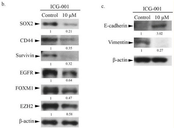
Applications
| Methods | Biomarkers | Images | PMID |
|---|---|---|---|
| Western blot | SOX-2 / CD44 / Survivin / EGFR / FOXM1 / EZH2 / Vimentin pβ-catenin beta-catenin / F-actin ABC / Beta-catenin / E-cadherin / N-cadherin / MMP-9 / c-MYC / Actin / Histone H3 CCNB1 / Cyclin D1 |
|
25897700 |
Tech Support
Tel: +1-832-582-8158 Ext:3
If you have any other enquiries, please leave a message.
Frequently Asked Questions
Question 1:
If the compound is stored in DMSO at -80, how long would it be stable? For cell culture, how long should I change for the fresh medium with it?
Answer:
It in DMSO solution can be stored at 4 degree for 1 week and -20 degree for 1 month. The best storage condition is solid powder, even at -80 the solution is not stable enough for long term storage. For cell culture, you need change medium every 48h.
Signaling Pathway Map
Products are for research use only. Not for human use. We do not sell to patients.
©Copyright 2013 Selleck Chemicals. All Rights Reserved.