-
 Australia
-
 Austria
-
 Belgium
-
 Brazil
-
 Canada
-
 China
China
-
 Czech Republic
-
 Denmark
-
 Finland
-
 France
-
 Germany
-
 Greece
-
 Hong Kong
-
 Hungary
-
 Iceland
-
 India
India
-
 Ireland
-
 Israel
-
 Italy
Italy
-
 Japan
Japan
-
 Korea
Korea
-
 Luxembourg
-
 Malaysia
-
 Netherlands
-
 New Zealand
-
 Norway
-
 Poland
-
 Qatar
Qatar
-
 Romania
-
 Saudi Arabia
-
 Singapore
-
 Spain
Spain
-
 Sweden
-
 Switzerland
-
 Taiwan
-
 Turkey
-
 United Kingdom
-
 United States
research use only
AR-A014418 GSK-3 inhibitor
Cat.No.S7435
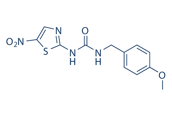
Chemical Structure
Molecular Weight: 308.31
Quality Control
| Related Targets | PI3K Akt mTOR ATM/ATR DNA-PK AMPK PDPK1 PTEN PP2A PDK |
|---|---|
| Other GSK-3 Inhibitors | CHIR-99021 (Laduviglusib) Laduviglusib (CHIR-99021) Hydrochloride SB216763 CHIR-98014 TWS119 GSK-3 Inhibitor IX (BIO) LY2090314 Tideglusib SB415286 1-Azakenpaullone |
Cell Culture, Treatment & Working Concentration
| Cell Lines | Assay Type | Concentration | Incubation Time | Formulation | Activity Description | PMID |
|---|---|---|---|---|---|---|
| human BxPC3 cells | Growth inhibition assay | 72 h | Growth inhibition of human BxPC3 cells after 72 hrs by MTS assay, IC50=14 μM | |||
| human HUPT3 cells | Growth inhibition assay | 72 h | Growth inhibition of human HUPT3 cells after 72 hrs by MTS assay, IC50=22 μM | |||
| human MIAPaCa2 cells | Growth inhibition assay | 72 h | Growth inhibition of human MIAPaCa2 cells after 72 hrs by MTS assay, IC50=29 μM | |||
| Click to View More Cell Line Experimental Data | ||||||
Solubility
|
In vitro |
DMSO
: 61 mg/mL
(197.85 mM)
Water : Insoluble Ethanol : Insoluble |
Molarity Calculator
|
In vivo |
|||||
In vivo Formulation Calculator (Clear solution)
Step 1: Enter information below (Recommended: An additional animal making an allowance for loss during the experiment)
Step 2: Enter the in vivo formulation (This is only the calculator, not formulation. Please contact us first if there is no in vivo formulation at the solubility Section.)
Calculation results:
Working concentration: mg/ml;
Method for preparing DMSO master liquid: mg drug pre-dissolved in μL DMSO ( Master liquid concentration mg/mL, Please contact us first if the concentration exceeds the DMSO solubility of the batch of drug. )
Method for preparing in vivo formulation: Take μL DMSO master liquid, next addμL PEG300, mix and clarify, next addμL Tween 80, mix and clarify, next add μL ddH2O, mix and clarify.
Method for preparing in vivo formulation: Take μL DMSO master liquid, next add μL Corn oil, mix and clarify.
Note: 1. Please make sure the liquid is clear before adding the next solvent.
2. Be sure to add the solvent(s) in order. You must ensure that the solution obtained, in the previous addition, is a clear solution before proceeding to add the next solvent. Physical methods such
as vortex, ultrasound or hot water bath can be used to aid dissolving.
Chemical Information, Storage & Stability
| Molecular Weight | 308.31 | Formula | C12H12N4O4S |
Storage (From the date of receipt) | |
|---|---|---|---|---|---|
| CAS No. | 487021-52-3 | Download SDF | Storage of Stock Solutions |
|
|
| Synonyms | GSK-3β Inhibitor VIII | Smiles | COC1=CC=C(C=C1)CNC(=O)NC2=NC=C(S2)[N+](=O)[O-] | ||
Mechanism of Action
| Features |
Cell-permeable GSK3-selective inhibitor.
|
|---|---|
| Targets/IC50/Ki |
GSK-3β
(Cell-free assay) 38 nM(Ki)
GSK-3β
(Cell-free assay) 38 nM(Ki)
|
| In vitro |
AR-A014418 inhibits tau phosphorylation at a GSK3-specific site (Ser-396) in 3T3 fibroblasts expressing human four-repeat tau protein with IC50 of 2.7 μM, and protects cultured N2A cells from death induced by blocking PI3K/PKB pathway. In hippocampal slices, this compound inhibits neurodegeneration mediated by beta-amyloid peptide. While in NGP and SH-5Y-SY cells, this chemical reduces neuroendocrine markers and suppresses neuroblastoma cell growth.
|
| Kinase Assay |
GSK3 Scintillation Proximity Assay
|
|
The competition experiments are carried out in duplicate with 10 concentrations of the inhibitor in clear-bottomed microtiter plates. The biotinylated peptide substrate, biotin-AAEELDSRAGS(PO3H2)PQL, is added at a final concentration of 2 μM in an assay buffer containing 6 milliunits of recombinant human GSK3 (equal mix of both α and β), 12 mM MOPS, pH 7.0, 0.3 mM EDTA, 0.01% β-mercaptoethanol, 0.004% Brij 35, 0.5% glycerol, and 0.5 μg of bovine serum albumin/25 μl and preincubated for 10-15 min. The reaction is initiated by the addition of 0.04 μCi of [γ-33P]ATP and unlabeled ATP in 50 mM Mg(Ac)2 to a final concentration of 1 μM ATP and assay volume of 25 μl. Blank controls without peptide substrate are used. After incubation for 20 min at room temperature, each reaction is terminated by the addition of 25 μl of stop solution containing 5 mM EDTA, 50 μM ATP, 0.1% Triton X-100, and 0.25 mg of streptavidin-coated SPA beads corresponding to ∼35 pmol of binding capacity. After 6 h the radioactivity is determined in a liquid scintillation counter. Inhibition curves are analyzed by non-linear regression using GraphPad Prism.
|
|
| In vivo |
In ALS mouse model with the G93A mutant human SOD1, AR-A014418 (0-4 mg/kg, i.p.) delays the onset of symptoms, improves motor activity, slows down disease progression, and postpons the endpoint of the disease. In addition, this compound produces inhibition effect on acetic acid- and formalin-induced nociception in mice by modulating NMDA and metabotropic receptor signaling as well as TNF-α and IL-1β transmission in the spinal cord.
|
References |
|
Applications
| Methods | Biomarkers | Images | PMID |
|---|---|---|---|
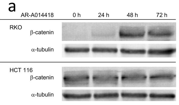
| Western blot | β-catenin β-catenin / GSK3α / GSK3β / Notch1 TAK1 / TAB1 / TAB2 / p-p65 / p65 |
|
26292722 |
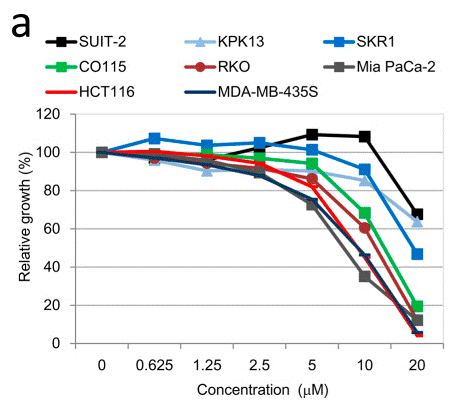
| Growth inhibition assay | Cell viability |
|
26292722 |
Tech Support
Tel: +1-832-582-8158 Ext:3
If you have any other enquiries, please leave a message.
Signaling Pathway Map
Products are for research use only. Not for human use. We do not sell to patients.
©Copyright 2013 Selleck Chemicals. All Rights Reserved.