-
 Australia
-
 Austria
-
 Belgium
-
 Brazil
-
 Canada
-
 China
China
-
 Czech Republic
-
 Denmark
-
 Finland
-
 France
-
 Germany
-
 Greece
-
 Hong Kong
-
 Hungary
-
 Iceland
-
 India
India
-
 Ireland
-
 Israel
-
 Italy
Italy
-
 Japan
Japan
-
 Korea
Korea
-
 Luxembourg
-
 Malaysia
-
 Netherlands
-
 New Zealand
-
 Norway
-
 Poland
-
 Qatar
Qatar
-
 Romania
-
 Saudi Arabia
-
 Singapore
-
 Spain
Spain
-
 Sweden
-
 Switzerland
-
 Taiwan
-
 Turkey
-
 United Kingdom
-
 United States
research use only
BIO-acetoxime GSK-3 inhibitor
Cat.No.S7915
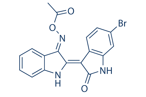
Chemical Structure
Molecular Weight: 398.21
Quality Control
| Related Targets | PI3K Akt mTOR ATM/ATR DNA-PK AMPK PDPK1 PTEN PP2A PDK |
|---|---|
| Other GSK-3 Inhibitors | CHIR-99021 (Laduviglusib) Laduviglusib (CHIR-99021) Hydrochloride SB216763 CHIR-98014 TWS119 GSK-3 Inhibitor IX (BIO) LY2090314 Tideglusib SB415286 AR-A014418 |
Solubility
|
In vitro |
DMSO
: 39 mg/mL
(97.93 mM)
Water : Insoluble Ethanol : Insoluble |
Molarity Calculator
|
In vivo |
|||||
In vivo Formulation Calculator (Clear solution)
Step 1: Enter information below (Recommended: An additional animal making an allowance for loss during the experiment)
Step 2: Enter the in vivo formulation (This is only the calculator, not formulation. Please contact us first if there is no in vivo formulation at the solubility Section.)
Calculation results:
Working concentration: mg/ml;
Method for preparing DMSO master liquid: mg drug pre-dissolved in μL DMSO ( Master liquid concentration mg/mL, Please contact us first if the concentration exceeds the DMSO solubility of the batch of drug. )
Method for preparing in vivo formulation: Take μL DMSO master liquid, next addμL PEG300, mix and clarify, next addμL Tween 80, mix and clarify, next add μL ddH2O, mix and clarify.
Method for preparing in vivo formulation: Take μL DMSO master liquid, next add μL Corn oil, mix and clarify.
Note: 1. Please make sure the liquid is clear before adding the next solvent.
2. Be sure to add the solvent(s) in order. You must ensure that the solution obtained, in the previous addition, is a clear solution before proceeding to add the next solvent. Physical methods such
as vortex, ultrasound or hot water bath can be used to aid dissolving.
Chemical Information, Storage & Stability
| Molecular Weight | 398.21 | Formula | C18H12BrN3O3 |
Storage (From the date of receipt) | |
|---|---|---|---|---|---|
| CAS No. | 667463-85-6 | Download SDF | Storage of Stock Solutions |
|
|
| Synonyms | GSK-3 Inhibitor X | Smiles | CC(=O)ON=C1C2=CC=CC=C2N=C1C3=C(NC4=C3C=CC(=C4)Br)O | ||
Mechanism of Action
| Targets/IC50/Ki |
GSK-3α
10 nM
GSK-3β
10 nM
|
|---|---|
| In vitro |
In human oral epithelial cells, BIO-acetoxime suppresses viral gene expression and protects oral epithelial cells from HSV-1 infection. In SY5Y-MYCN cells, this compound strongly reduces c-MYC expression and p-SMAD3 levels. It also decreases cell viability of KCN, KCNR, SY5Y, Kelly, and IMR32 cells by mediating apoptosis. In HEK 293T cells, this chemical is also found to reduce antiviral innate immunity downstream of IRF3 activation by inhibition of GSK3α/β activities.
|
| Kinase Assay |
Kinase Assays
|
|
Kinases activities are assayed in Buffer A or C, at 30 °C, at a final ATP concentration of 15 μM. Blank values are subtracted and activities calculated as pmoles of phosphate incorporated for a 10 min incubation. The activities are expressed in % of the maximal activity, i.e., in the absence of inhibitors. Controls are performed with appropriate dilutions of DMSO. GSK-3α/β is purified from porcine brain by affinity chromatography on immobilized axin. It is assayed, following a 1/100 dilution in 1 mg BSA/mL 10 mM DTT, with 5 μL 40 μM GS-1 peptide as a substrate, in buffer A, in the presence of 15 μM [γ-33P] ATP (3000 Ci/mmol; 1 mCi/mL) in a final volume of 30 μL. After 30 min incubation at 30 °C, 25 μL aliquots of supernatant are spotted onto 2.5 × 3 cm pieces of Whatman P81 phosphocellulose paper, and, 20 s later, the filters are washed five times (for at least 5 min each time) in a solution of 10 mL phosphoric acid/liter of water. The wet filters are counted in the presence of 1 mL of ACS scintillation fluid. CDK1/cyclin B is extracted in homogenization buffer from M phase starfish (Marthasterias glacialis) oocytes and purified by affinity chromatography on p9CKShs1-sepharose beads, from which it is eluted by free p9CKShs1. The kinase activity is assayed in buffer C, with 1 mg histone H1 /mL, in the presence of 15 μM [γ-32P] ATP (3000 Ci/mmol; 1 mCi/mL) in a final volume of 30 μL. After 10 min incubation at 30 °C, 25 μL aliquots of supernatant are spotted onto P81 phosphocellulose papers and treated as described above. CDK5/p25 is reconstituted by mixing equal amounts of recombinant mammalian CDK5 and p25 expressed in E. coli as GST (Glutathione-S-transferase) fusion proteins and purified by affinity chromatography on glutathione-agarose (p25 is a truncated version of p35, the 35 kDa CDK5 activator). Its activity is assayed in buffer C as described for CDK1/cyclin B.0
|
References |
|
Tech Support
Tel: +1-832-582-8158 Ext:3
If you have any other enquiries, please leave a message.
Signaling Pathway Map
Products are for research use only. Not for human use. We do not sell to patients.
©Copyright 2013 Selleck Chemicals. All Rights Reserved.