-
 Australia
-
 Austria
-
 Belgium
-
 Brazil
-
 Canada
-
 China
China
-
 Czech Republic
-
 Denmark
-
 Finland
-
 France
-
 Germany
-
 Greece
-
 Hong Kong
-
 Hungary
-
 Iceland
-
 India
India
-
 Ireland
-
 Israel
-
 Italy
Italy
-
 Japan
Japan
-
 Korea
Korea
-
 Luxembourg
-
 Malaysia
-
 Netherlands
-
 New Zealand
-
 Norway
-
 Poland
-
 Qatar
Qatar
-
 Romania
-
 Saudi Arabia
-
 Singapore
-
 Spain
Spain
-
 Sweden
-
 Switzerland
-
 Taiwan
-
 Turkey
-
 United Kingdom
-
 United States
research use only
SC79 Akt activator
Cat.No.S7863
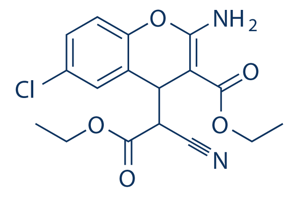
Chemical Structure
Molecular Weight: 364.78
Quality Control
Cell Culture, Treatment & Working Concentration
| Cell Lines | Assay Type | Concentration | Incubation Time | Formulation | Activity Description | PMID |
|---|---|---|---|---|---|---|
| A549 | Apoptosis assay | 5 μmol/L | SC80 blocks vitexin-induced apoptosis | 30797236 | ||
| HeLa | Function assay | decreased the number of autophagosomes induced by HVJ-E in cells | 30534001 | |||
| SH-SY5Y | Function assay | 10 μM | 30 min | pre-treatment with SC79 (10 μM) largely attenuated H2O2-induced survival reduction | 29560097 | |
| Sertoli | Function assay | 5.5 µM | 30 min | up-regulate the PFOS-induced down-regulation of p-Akt1 T308 and p-Akt1 S473 | 28439067 | |
| Click to View More Cell Line Experimental Data | ||||||
Solubility
|
In vitro |
DMSO
: 73 mg/mL
(200.12 mM)
Ethanol : 36 mg/mL Water : Insoluble |
Molarity Calculator
|
In vivo |
|||||
In vivo Formulation Calculator (Clear solution)
Step 1: Enter information below (Recommended: An additional animal making an allowance for loss during the experiment)
Step 2: Enter the in vivo formulation (This is only the calculator, not formulation. Please contact us first if there is no in vivo formulation at the solubility Section.)
Calculation results:
Working concentration: mg/ml;
Method for preparing DMSO master liquid: mg drug pre-dissolved in μL DMSO ( Master liquid concentration mg/mL, Please contact us first if the concentration exceeds the DMSO solubility of the batch of drug. )
Method for preparing in vivo formulation: Take μL DMSO master liquid, next addμL PEG300, mix and clarify, next addμL Tween 80, mix and clarify, next add μL ddH2O, mix and clarify.
Method for preparing in vivo formulation: Take μL DMSO master liquid, next add μL Corn oil, mix and clarify.
Note: 1. Please make sure the liquid is clear before adding the next solvent.
2. Be sure to add the solvent(s) in order. You must ensure that the solution obtained, in the previous addition, is a clear solution before proceeding to add the next solvent. Physical methods such
as vortex, ultrasound or hot water bath can be used to aid dissolving.
Chemical Information, Storage & Stability
| Molecular Weight | 364.78 | Formula | C17H17ClN2O5 |
Storage (From the date of receipt) | |
|---|---|---|---|---|---|
| CAS No. | 305834-79-1 | Download SDF | Storage of Stock Solutions |
|
|
| Synonyms | N/A | Smiles | CCOC(=O)C1=C(OC2=C(C1C(C#N)C(=O)OCC)C=C(C=C2)Cl)N | ||
Mechanism of Action
| Targets/IC50/Ki |
Akt
|
|---|---|
| In vitro |
SC79 suppresses PHAKTM-GFP plasma membrane translocation, and enhances phosphorylation of all three Akt isoforms in HEK293, HeLa, HL60, NB4, and HsSulton (B cells) cells. This compound reduces neuronal excitotoxicity and prevents stroke-induced neuronal death. It restores proliferation of BRAT1 knockdown cells, and reduces the production of superoxide in mitochondria of MitoSox positive cells. |
| Kinase Assay |
Cytosolic phosphorylation of Akt
|
|
Hela cells are serum starved for 1 hr and treated with IGF (100ng/mL) or SC79 (4 μg/mL) for 30 minutes. Cells are lysed in Lysis buffer containing 250 mM Sucrose, 20 mM HEPES, 10 mM KCl, 1.5 mM MgCl2, 1 mM EDTA, 1 mM EGTA supplemented with protease inhibitors. Cells are passed through 25G needle several times and kept on ice for 20 minutes. Total cell lysate is taken at this point. Cell lysates are centrifuged at 100,000g for 30 minutes. Supernatant is collected as the cytosolic fraction. Pellet is washed with lysis buffer and represents the membrane fraction. Total cell lysate, cytosolic and membrane fractions are resolved by SDS-PAGE and analyzed for phospho-Akt (S473), Total Akt, Tubulin (cytosolic marker) and Orai1 (membrane marker) by western blotting.
|
|
| In vivo |
In the permanent focal cerebral ischemia mouse model, SC79 (0.04 mg/g, i.p.) enables the cytosolic activation of Akt, and recapitulates the primary cellular function of Akt signaling, resulting in augmented neuronal survival. |
References |
|
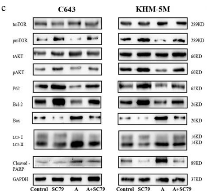
Applications
| Methods | Biomarkers | Images | PMID |
|---|---|---|---|
| Western blot | p-mTOR / mTOR / pAKT / AKT / P62 / Bcl-2 / Bax / LC3 / Cleaved PARP p-Akt / Akt / p-GSK3β / GSK3β / β-catenin / Cyclin D1 p4EBP1 / 4EBP1 / pS6 / RPS6 / ESRP1 / ESRP2 |
|
30301881 |
Tech Support
Tel: +1-832-582-8158 Ext:3
If you have any other enquiries, please leave a message.
Signaling Pathway Map
Products are for research use only. Not for human use. We do not sell to patients.
©Copyright 2013 Selleck Chemicals. All Rights Reserved.