-
 Australia
-
 Austria
-
 Belgium
-
 Brazil
-
 Canada
-
 China
China
-
 Czech Republic
-
 Denmark
-
 Finland
-
 France
-
 Germany
-
 Greece
-
 Hong Kong
-
 Hungary
-
 Iceland
-
 India
India
-
 Ireland
-
 Israel
-
 Italy
Italy
-
 Japan
Japan
-
 Korea
Korea
-
 Luxembourg
-
 Malaysia
-
 Netherlands
-
 New Zealand
-
 Norway
-
 Poland
-
 Qatar
Qatar
-
 Romania
-
 Saudi Arabia
-
 Singapore
-
 Spain
Spain
-
 Sweden
-
 Switzerland
-
 Taiwan
-
 Turkey
-
 United Kingdom
-
 United States
research use only
Go 6983 PKC inhibitor
Cat.No.S2911
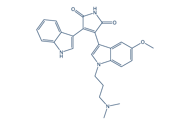
Chemical Structure
Molecular Weight: 442.51
Quality Control
Cell Culture, Treatment & Working Concentration
| Cell Lines | Assay Type | Concentration | Incubation Time | Formulation | Activity Description | PMID |
|---|---|---|---|---|---|---|
| PC12 | Function assay | 0.5 μM | GO6983 blocked the effect of PMA on the activation of Akt and MAPK induced by IGF-1 | 10788447 | ||
| PC-3 | Function assay | 1 μM | 2 h | Gö6983 abrogates the TPA-induced RGFR transactivation response | 15897236 | |
| HCT116 | Function assay | 2 μM | 8 h | attenuated PMA-induced FLIP mRNA expression | 16052516 | |
| HT29 | Function assay | 2 μM | 8 h | attenuated PMA-induced FLIP mRNA expression | 16052516 | |
| KM20 | Function assay | 2 μM | 8 h | attenuated PMA-induced FLIP mRNA expression | 16052516 | |
| KM12C | Function assay | 2 μM | 8 h | attenuated PMA-induced FLIP mRNA expression | 16052516 | |
| Caco-2 | Function assay | 2 μM | 8 h | completely attenuated PMA-induced FLIP mRNA expression | 16052516 | |
| A549 | Function assay | 10 μM | 1 h | markedly inhibited ATPγS-stimulated NADPH oxidase activity and H2O2 and/or ROS generation | 23326583 | |
| HeLa | Function assay | 2 μM | 48 h | suppressed the effect of PMA on apicularen A-induced cytotoxicity | 24447339 | |
| A673 | qHTS assay | qHTS of pediatric cancer cell lines to identify multiple opportunities for drug repurposing: Primary screen for A673 cells | 29435139 | |||
| NB-EBc1 | qHTS assay | qHTS of pediatric cancer cell lines to identify multiple opportunities for drug repurposing: Primary screen for NB-EBc1 cells | 29435139 | |||
| SK-N-SH | qHTS assay | qHTS of pediatric cancer cell lines to identify multiple opportunities for drug repurposing: Primary screen for SK-N-SH cells | 29435139 | |||
| LAN-5 | qHTS assay | qHTS of pediatric cancer cell lines to identify multiple opportunities for drug repurposing: Primary screen for LAN-5 cells | 29435139 | |||
| NB1643 | qHTS assay | qHTS of pediatric cancer cell lines to identify multiple opportunities for drug repurposing: Confirmatory screen for NB1643 cells | 29435139 | |||
| SK-N-MC | qHTS assay | qHTS of pediatric cancer cell lines to identify multiple opportunities for drug repurposing: Confirmatory screen for SK-N-MC cells | 29435139 | |||
| LAN-5 | qHTS assay | qHTS of pediatric cancer cell lines to identify multiple opportunities for drug repurposing: Confirmatory screen for LAN-5 cells | 29435139 | |||
| DAOY | qHTS assay | qHTS of pediatric cancer cell lines to identify multiple opportunities for drug repurposing: Confirmatory screen for DAOY cells | 29435139 | |||
| BT-37 | qHTS assay | qHTS of pediatric cancer cell lines to identify multiple opportunities for drug repurposing: Confirmatory screen for BT-37 cells | 29435139 | |||
| TC32 | qHTS assay | qHTS of pediatric cancer cell lines to identify multiple opportunities for drug repurposing: Confirmatory screen for TC32 cells | 29435139 | |||
| Rh41 | qHTS assay | qHTS of pediatric cancer cell lines to identify multiple opportunities for drug repurposing: Confirmatory screen for Rh41 cells | 29435139 | |||
| Rh30 | qHTS assay | qHTS of pediatric cancer cell lines to identify multiple opportunities for drug repurposing: Confirmatory screen for Rh30 cells | 29435139 | |||
| OHS-50 | qHTS assay | qHTS of pediatric cancer cell lines to identify multiple opportunities for drug repurposing: Confirmatory screen for OHS-50 cells | 29435139 | |||
| SK-N-SH | qHTS assay | qHTS of pediatric cancer cell lines to identify multiple opportunities for drug repurposing: Confirmatory screen for SK-N-SH cells | 29435139 | |||
| HEK293 | Function assay | Inhibition of Cav1.2 calcium current measured using whole cell patch clamp in human HEK293 cells transfected with rabbit L-type calcium channel subunits, IC50 = 20 μM. | ChEMBL | |||
| Click to View More Cell Line Experimental Data | ||||||
Solubility
|
In vitro |
DMSO
: 89 mg/mL
(201.12 mM)
Water : Insoluble Ethanol : Insoluble |
Molarity Calculator
|
In vivo |
|||||
In vivo Formulation Calculator (Clear solution)
Step 1: Enter information below (Recommended: An additional animal making an allowance for loss during the experiment)
Step 2: Enter the in vivo formulation (This is only the calculator, not formulation. Please contact us first if there is no in vivo formulation at the solubility Section.)
Calculation results:
Working concentration: mg/ml;
Method for preparing DMSO master liquid: mg drug pre-dissolved in μL DMSO ( Master liquid concentration mg/mL, Please contact us first if the concentration exceeds the DMSO solubility of the batch of drug. )
Method for preparing in vivo formulation: Take μL DMSO master liquid, next addμL PEG300, mix and clarify, next addμL Tween 80, mix and clarify, next add μL ddH2O, mix and clarify.
Method for preparing in vivo formulation: Take μL DMSO master liquid, next add μL Corn oil, mix and clarify.
Note: 1. Please make sure the liquid is clear before adding the next solvent.
2. Be sure to add the solvent(s) in order. You must ensure that the solution obtained, in the previous addition, is a clear solution before proceeding to add the next solvent. Physical methods such
as vortex, ultrasound or hot water bath can be used to aid dissolving.
Chemical Information, Storage & Stability
| Molecular Weight | 442.51 | Formula | C26H26N4O3 |
Storage (From the date of receipt) | |
|---|---|---|---|---|---|
| CAS No. | 133053-19-7 | Download SDF | Storage of Stock Solutions |
|
|
| Synonyms | GOE 6983, Gö 6983 | Smiles | CN(C)CCCN1C=C(C2=C1C=CC(=C2)OC)C3=C(C(=O)NC3=O)C4=CNC5=CC=CC=C54 | ||
Mechanism of Action
| Targets/IC50/Ki |
PKCγ
(Cell-free assay) 6 nM
PKCα
(Cell-free assay) 7 nM
PKCβ
(Cell-free assay) 7 nM
PKCδ
(Cell-free assay) 10 nM
PKCζ
(Cell-free assay) 60 nM
|
|---|---|
| In vitro |
Go 6983 (300 μM) suppresses PKCμ auto-phosphorylation by 20% reduction in NIH3T3 transfected with PKCμ. In hearts reperfused with PMNs and this compound (100 nM), left ventricular developed pressure (LVDP) and the rate of LVDP recoveres to 89% and 74% of baseline values, respectively, significantly higher than PMNs alone. This chemical (100 nM) significantly reduces PMNs adherence to the endothelium and infiltration into the myocardium compared with Ischemia followed by reperfusion (I/R)+ PMN hearts, and significantly inhibits superoxide release from PMNs by 90%. It attenuates post-I/R cardiac contractile dysfunction in the presence of PMNs, which may be related in part to decreased superoxide production. This inhibitor significantly inhibits antigen-induced superoxide release from leukocytes of patients previously sensitized to tree pollen. It inhibited intracellular Ca(2+) accumulation in human vascular tissue, suggesting a mechanism for its vasodilator properties. This compound (1 μM) combined with Ro-31-8425 (390 nM) slightly inhibits Angiotensin II–induced PLD2 activity in PGSMCs. It is isoform-specific PKC inhibitor that target the ATP binding site. It inhibits ΔPfPKB activity with an IC50 of 1 μM. In this chemical (5 μM)-treated cells, the number of rings in the following cycle is markedly less compared with the control cultures. This treatment (5 μM) results in an almost 60% decrease in formation of new rings in P. falciparum cultures. |
| Kinase Assay |
Binding assay
|
|
Phosphorylation reactions are carried out in a total volume of 100 μL, containing buffer C (50 mM Tris-HC1, pH 7.5, 10 mM β-mercaptoethanol), 4 mM MgCl2, 10 μg PS, 100 nM TPA, 5 μL of a Sf158 cell extract as a source of recombinant PKCμ or of Sf9 cell extracts as a source of other recombinant PKC isoenzymes, 10 μg of syntide 2 as substrate, and 35 μM ATP containing 1 μCi [γ-32P]ATP. In some experiments PS and TPA are omitted or various inhibitors at concentrations indicated in the text are added. After incubation for 10 min at 30℃, the reaction is terminated by transferring 50 μL of the assay mixture onto a 20 mm square piece of phosphocellulose paper, which is washed 3 times in deionized water and twice in acetone. The radioactivity on each paper is determined by liquid scintillation counting.
|
|
| In vivo |
Go6983 (22.0 μg/mouse, i.v.) strongly inhibits tumor metastasis by 51.2 % in a mouse pulmonary B16BL6 tumor model. |
References |
|
Applications
| Methods | Biomarkers | Images | PMID |
|---|---|---|---|
| Western blot | PKCη / PKCα / PKCδ / PKCε |
|
22892130 |
Tech Support
Tel: +1-832-582-8158 Ext:3
If you have any other enquiries, please leave a message.
Signaling Pathway Map
Products are for research use only. Not for human use. We do not sell to patients.
©Copyright 2013 Selleck Chemicals. All Rights Reserved.