-
 Australia
-
 Austria
-
 Belgium
-
 Brazil
-
 Canada
-
 China
China
-
 Czech Republic
-
 Denmark
-
 Finland
-
 France
-
 Germany
-
 Greece
-
 Hong Kong
-
 Hungary
-
 Iceland
-
 India
India
-
 Ireland
-
 Israel
-
 Italy
Italy
-
 Japan
Japan
-
 Korea
Korea
-
 Luxembourg
-
 Malaysia
-
 Netherlands
-
 New Zealand
-
 Norway
-
 Poland
-
 Qatar
Qatar
-
 Romania
-
 Saudi Arabia
-
 Singapore
-
 Spain
Spain
-
 Sweden
-
 Switzerland
-
 Taiwan
-
 Turkey
-
 United Kingdom
-
 United States
research use only
Sotrastaurin (AEB071) PKC inhibitor
Cat.No.S2791
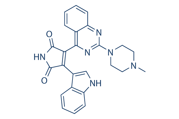
Chemical Structure
Molecular Weight: 438.48
Quality Control
Cell Culture, Treatment & Working Concentration
| Cell Lines | Assay Type | Concentration | Incubation Time | Formulation | Activity Description | PMID |
|---|---|---|---|---|---|---|
| T cell | Function Assay | 100 nM | 3 h | DMSO | inhibits rRNA synthesis | 25691158 |
| HUVECs | Function Assay | 500nM | 1 h | Reduces DTX-Triggered Endothelial Dysfunction | 25634538 | |
| A549 | Function Assay | 0.1 μM | 24 h | decreases the relative PKC-α level on cell membrane cotreated AS-IV | 25218161 | |
| A549 | Function Assay | 0.1 μM | 24 h | reduces the expression levels of MMP-2, MMP-9 and integrin β1 | 25218161 | |
| A549 | Growth Inhibition Assay | 0.1 μM | 24 h | enhances growth inhibition cotreated with AS-IV | 25218161 | |
| Mel202 | Growth Inhibition Assay | 0.5 μM | 3 h | DMSO | enhances IR-induced reduction in cell viability | 24595385 |
| 92.1 | Growth Inhibition Assay | 0.5 μM | 3 h | DMSO | enhances IR-induced reduction in cell viability | 24595385 |
| OCM3 | Growth Inhibition Assay | 0.5 μM | 3 h | DMSO | enhances IR-induced reduction in cell viability | 24595385 |
| Mel202 | Growth Inhibition Assay | 0.5 μM | 3 h | DMSO | increases IR-induced cell cycle arrest | 24595385 |
| 92.1 | Growth Inhibition Assay | 0.5 μM | 3 h | DMSO | increases IR-induced cell cycle arrest | 24595385 |
| OCM3 | Growth Inhibition Assay | 0.5 μM | 3 h | DMSO | increases IR-induced cell cycle arrest | 24595385 |
| Jeko-1 | Growth Inhibition Assay | 0-4 μM | DMSO | inhibits cell growth dose dependently | 24362935 | |
| Mino | Growth Inhibition Assay | 0-4 μM | DMSO | inhibits cell growth dose dependently | 24362935 | |
| Rec-1 | Growth Inhibition Assay | 0-4 μM | DMSO | inhibits cell growth dose dependently | 24362935 | |
| SP49 | Growth Inhibition Assay | 0-4 μM | DMSO | inhibits cell growth dose dependently | 24362935 | |
| Jeko-1 | Function Assay | 2.5 μM | 12 h | DMSO | downregulates NF-κB target genes | 24362935 |
| Mino | Function Assay | 2.5 μM | 12 h | DMSO | downregulates NF-κB target genes | 24362935 |
| Rec-1 | Function Assay | 2.5 μM | 12 h | DMSO | downregulates NF-κB target genes | 24362935 |
| SP49 | Function Assay | 2.5 μM | 12 h | DMSO | downregulates NF-κB target genes | 24362935 |
| CD3+ T | Function Assay | 0-500 nM | 1 h | inhibits NF-κB phosphorylation in a dose dependent manner | 23573283 | |
| Mel202 | Growth Inhibition Assay | 0-5 μM | 72 h | DMSO | inhibits cell growth dose dependently | 22653968 |
| Omm1.3 | Growth Inhibition Assay | 0-5 μM | 72 h | DMSO | inhibits cell growth dose dependently | 22653968 |
| 92.1 | Growth Inhibition Assay | 0-5 μM | 72 h | DMSO | inhibits cell growth dose dependently | 22653968 |
| Mel202 | Growth Inhibition Assay | 5 μM | 24 h | DMSO | induces G1 arrest | 22653968 |
| Omm1.3 | Growth Inhibition Assay | 5 μM | 24 h | DMSO | induces G1 arrest | 22653968 |
| 92.1 | Growth Inhibition Assay | 5 μM | 24 h | DMSO | induces G1 arrest | 22653968 |
| Mel202 | Apoptosis Assay | 5 μM | 72 h | DMSO | induces apoptosis slightly | 22653968 |
| Omm1.3 | Apoptosis Assay | 5 μM | 72 h | DMSO | induces apoptosis | 22653968 |
| 92.1 | Apoptosis Assay | 5 μM | 72 h | DMSO | induces apoptosis signifcantly | 22653968 |
| Mel202 | Function Assay | 5 μM | 24 h | inhibits expression and phosphorylation of PKC isoforms | 22653968 | |
| Omm1.3 | Function Assay | 5 μM | 24 h | inhibits expression and phosphorylation of PKC isoforms | 22653968 | |
| 92.1 | Function Assay | 5 μM | 24 h | inhibits expression and phosphorylation of PKC isoforms | 22653968 | |
| HBL1 | Growth Inhibition Assay | 0.16-20 μM | 5 d | IC50=0.5 μM | 21324920 | |
| TMD8 | Growth Inhibition Assay | 0.16-20 μM | 5 d | IC50=0.2 μM | 21324920 | |
| OCI-Ly10 | Growth Inhibition Assay | 0.16-20 μM | 5 d | IC50=1.3 μM | 21324920 | |
| U2932 | Growth Inhibition Assay | 0.16-20 μM | 5 d | IC50=10 μM | 21324920 | |
| OCI-Ly3 | Growth Inhibition Assay | 0.16-20 μM | 5 d | IC50>20 μM | 21324920 | |
| SuDHL2 | Growth Inhibition Assay | 0.16-20 μM | 5 d | IC50>20 μM | 21324920 | |
| SuDHL4 | Growth Inhibition Assay | 0.16-20 μM | 5 d | IC50>20 μM | 21324920 | |
| DB | Growth Inhibition Assay | 0.16-20 μM | 5 d | IC50>20 μM | 21324920 | |
| Jurkat IL-2 | Growth Inhibition Assay | IC50=6.71 ± 3.76 μM | 19940259 | |||
| PBMC IL-2 | Growth Inhibition Assay | IC50=4.84 ± 1.70 μM | 19940259 | |||
| Jurkat | Function assay | Inhibition of TCR/CD28-mediated human T cell activation in Jurkat cells expressing human IL2 promoter by luciferase reporter gene assay, IC50 = 0.054 μM. | 19827831 | |||
| Jurkat T | Function assay | 5 hrs | Inhibition of PKCtheta in human Jurkat T cells assessed as reduction in anti-CD3/CD28 antibody-induced T-cell activation by measuring decrease in IL-2 secretion after 5 hrs by luciferase reporter gene assay, IC50 = 0.081 μM. | 28131714 | ||
| B-cells | Function assay | Inhibition of PKCbeta in mouse B cells assessed as reduction in IgM-stimulated cell proliferation, IC50 = 0.234 μM. | 28131714 | |||
| bone marrow cells | Antiproliferative assay | 4 days | Antiproliferative activity against CBA mouse bone marrow cells assessed as inhibition of [3H]thymidine incorporation after 4 days, IC50 = 3.7 μM. | 19827831 | ||
| Click to View More Cell Line Experimental Data | ||||||
Solubility
|
In vitro |
DMSO
: 87 mg/mL
(198.41 mM)
Ethanol : 40 mg/mL Water : Insoluble |
Molarity Calculator
|
In vivo |
|||||
In vivo Formulation Calculator (Clear solution)
Step 1: Enter information below (Recommended: An additional animal making an allowance for loss during the experiment)
Step 2: Enter the in vivo formulation (This is only the calculator, not formulation. Please contact us first if there is no in vivo formulation at the solubility Section.)
Calculation results:
Working concentration: mg/ml;
Method for preparing DMSO master liquid: mg drug pre-dissolved in μL DMSO ( Master liquid concentration mg/mL, Please contact us first if the concentration exceeds the DMSO solubility of the batch of drug. )
Method for preparing in vivo formulation: Take μL DMSO master liquid, next addμL PEG300, mix and clarify, next addμL Tween 80, mix and clarify, next add μL ddH2O, mix and clarify.
Method for preparing in vivo formulation: Take μL DMSO master liquid, next add μL Corn oil, mix and clarify.
Note: 1. Please make sure the liquid is clear before adding the next solvent.
2. Be sure to add the solvent(s) in order. You must ensure that the solution obtained, in the previous addition, is a clear solution before proceeding to add the next solvent. Physical methods such
as vortex, ultrasound or hot water bath can be used to aid dissolving.
Chemical Information, Storage & Stability
| Molecular Weight | 438.48 | Formula | C25H22N6O2 |
Storage (From the date of receipt) | |
|---|---|---|---|---|---|
| CAS No. | 425637-18-9 | Download SDF | Storage of Stock Solutions |
|
|
| Synonyms | N/A | Smiles | CN1CCN(CC1)C2=NC3=CC=CC=C3C(=N2)C4=C(C(=O)NC4=O)C5=CNC6=CC=CC=C65 | ||
Mechanism of Action
| Features |
Unlike former PKC inhibitors, Sotrastaurin does not enhance apoptosis of murine T-cell blasts in a model of activation-induced cell death.
|
|---|---|
| Targets/IC50/Ki |
PKCθ
(Cell-free assay) 0.22 nM(Ki)
PKCβ1
(Cell-free assay) 0.64 nM(Ki)
PKCα
(Cell-free assay) 0.95 nM(Ki)
PKCη
(Cell-free assay) 1.8 nM(Ki)
PKCδ
(Cell-free assay) 2.1 nM(Ki)
PKCε
(Cell-free assay) 3.2 nM(Ki)
|
| In vitro |
Treatment with Sotrastaurin (AEB071) at concentrations below 10 μM effectively abrogated markers of early T-cell activation—such as interleukin-2 secretion and CD25 expression—in primary human and mouse T cells at low nanomolar levels. At 200 nM, it inhibits CD3/CD28 antibody- and alloantigen-induced T-cell proliferation without nonspecific antiproliferative effects. Furthermore, this compound (<3 μM) markedly impairs lymphocyte function-associated antigen-1-mediated T-cell adhesion. At concentrations under 20 μM, it selectively impairs the proliferation of CD79 mutant ABC DLBCL cell lines, correlating with reduced NF-κB signaling activity. A concentration of 5 μM induces G1 arrest and/or cell death in CD79 mutant cells. |
| Kinase Assay |
Protein Kinase Assays
|
|
Sotrastaurin (AEB071) was assayed for classical and novel PKC isotypes using scintillation proximity assay technology. In brief, the assay is performed in 20 mM Tris-HCl buffer, pH 7.4, and 0.1% bovine serum albumin by incubating 1.5 μM of the peptide substrate with 10 μM [33P]ATP, 10 mM Mg (NO3)2, 0.2 mM CaCl2, and PKC at a protein concentration varying from 25 to 400 ng/mL, and lipid vesicles containing 30 mol% phosphatidylserine, 5 mol% diacylglycerol (DAG), and 65 mol% phosphatidylcholine at a final lipid concentration of 0.5 μM. Incubation is performed for 60 min at room temperature. The reaction is stopped by adding 50 μl of a mixture containing 100 mM EDTA, 200 μM ATP, 0.1% Triton X-100, and 0.375 μg/well streptavidin-coated scintillation proximity assay beads in PBS without Ca2+ and Mg2+. Incorporated radioactivity is measured in a MicroBetaTrilux counter for 1 min.
|
|
| In vivo |
In a subcutaneous TMD8 xenograft model in SCID mice, Sotrastaurin (AEB071) (80 mg/kg) results in significant inhibition of in vivo tumor growth. When orally administered at 10 mg/kg and 30 mg/kg b.i.d., this compound shows a dose-dependent immunosuppressive effect leading to pronounced prolongation of heart allograft survival in rats. |
References |
|
Applications
| Methods | Biomarkers | Images | PMID |
|---|---|---|---|
| Western blot | pPKCδ/θ / phosphorylated MARCKS / p53 / MDM2 / PUMA / p21 Cyclin D1 / p27(Kip1) Bcl-xl / XIAP / Survivin PKCα / PKCδ / PKCβ / PKCε / PKCθ p-Marcks / p-ERK / p-AKT / p-S6 / Marcks / ERK / AKT / S6 |
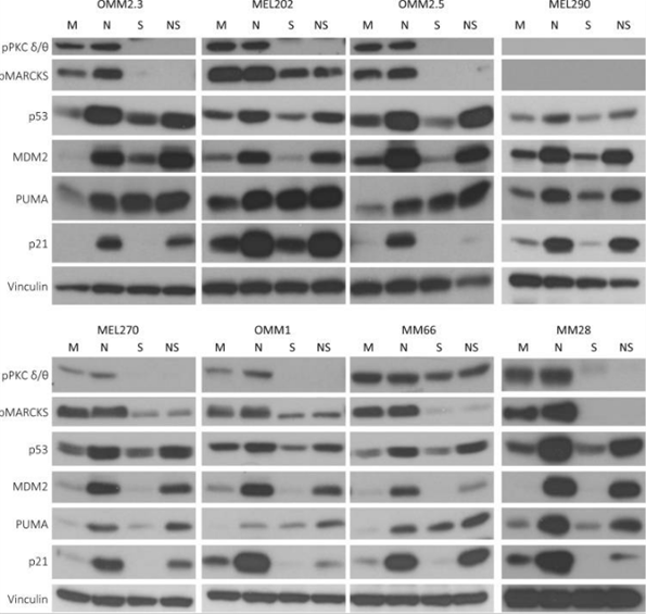
|
29593251 |
| Growth inhibition assay | Cell viability |
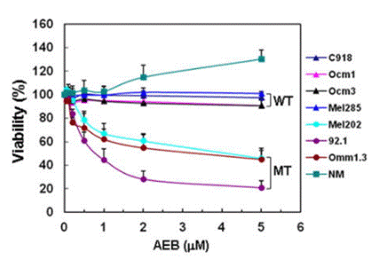
|
22653968 |
Clinical Trial Information
(data from https://clinicaltrials.gov, updated on 2024-05-22)
| NCT Number | Recruitment | Conditions | Sponsor/Collaborators | Start Date | Phases |
|---|---|---|---|---|---|
| NCT02273219 | Completed | Uveal Melanoma |
Columbia University |
November 19 2014 | Phase 1 |
| NCT01801358 | Terminated | Uveal Melanoma |
Array Biopharma now a wholly owned subsidiary of Pfizer|Array BioPharma |
August 2013 | Phase 1|Phase 2 |
| NCT01430416 | Completed | Uveal Melanoma |
Novartis Pharmaceuticals|Novartis |
December 20 2011 | Phase 1 |
| NCT01402440 | Terminated | Diffuse Large B-Cell Lymphoma |
Novartis Pharmaceuticals|Novartis |
November 2011 | Phase 1 |
Tech Support
Tel: +1-832-582-8158 Ext:3
If you have any other enquiries, please leave a message.
Frequently Asked Questions
Question 1:
Could you give me the information about how to prepare it for oral administration in mice?
Answer:
It can be dissolved in 2% DMSO/30% PEG 300/ddH2O at 10 mg/ml as a clear solution which can be used for injection, and in 2% DMSO/corn oil at 10 mg/ml as a suspension for oral administration.
Signaling Pathway Map
Products are for research use only. Not for human use. We do not sell to patients.
©Copyright 2013 Selleck Chemicals. All Rights Reserved.