-
 Australia
-
 Austria
-
 Belgium
-
 Brazil
-
 Canada
-
 China
China
-
 Czech Republic
-
 Denmark
-
 Finland
-
 France
-
 Germany
-
 Greece
-
 Hong Kong
-
 Hungary
-
 Iceland
-
 India
India
-
 Ireland
-
 Israel
-
 Italy
Italy
-
 Japan
Japan
-
 Korea
Korea
-
 Luxembourg
-
 Malaysia
-
 Netherlands
-
 New Zealand
-
 Norway
-
 Poland
-
 Qatar
Qatar
-
 Romania
-
 Saudi Arabia
-
 Singapore
-
 Spain
Spain
-
 Sweden
-
 Switzerland
-
 Taiwan
-
 Turkey
-
 United Kingdom
-
 United States
research use only
Capmatinib (INC280) c-Met inhibitor
Cat.No.S2788
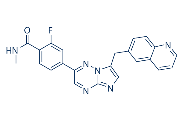
Chemical Structure
Molecular Weight: 412.42
Quality Control
| Related Targets | EGFR VEGFR PDGFR FGFR Src MEK CSF-1R FLT3 HER2 c-Kit |
|---|---|
| Other c-Met Inhibitors | Tepotinib (EMD-1214063) Dihexa SGX-523 PHA-665752 Foretinib (GSK1363089, XL880) SU11274 BMS-777607 JNJ-38877605 Tivantinib PF-04217903 |
Cell Culture, Treatment & Working Concentration
| Cell Lines | Assay Type | Concentration | Incubation Time | Formulation | Activity Description | PMID |
|---|---|---|---|---|---|---|
| Huh7-SR | Cell viability assay | 0, 2, 4, 8, 16 h | 48 h | significantly reduced the viability and strengthened the effects of sorafenib on sorafenib-resistant cells | 28164434 | |
| HepG2-SR | Cell viability assay | 0, 2, 4, 8, 16 h | 48 h | significantly reduced the viability and strengthened the effects of sorafenib on sorafenib-resistant cells | 28164434 | |
| NCI-H1993 | Growth inhibition assay | 72 hrs | Growth inhibition of human NCI-H1993 cells after 72 hrs by CCK-8 assay, IC50 = 0.0023 μM. | 28411455 | ||
| SNU5 | Growth inhibition assay | 72 hrs | Growth inhibition of human SNU5 cells after 72 hrs by CCK-8 assay, IC50 = 0.0027 μM. | 28411455 | ||
| Click to View More Cell Line Experimental Data | ||||||
Solubility
|
In vitro |
DMSO
: 6 mg/mL
(14.54 mM)
Water : Insoluble Ethanol : Insoluble |
Molarity Calculator
|
In vivo |
|||||
In vivo Formulation Calculator (Clear solution)
Step 1: Enter information below (Recommended: An additional animal making an allowance for loss during the experiment)
Step 2: Enter the in vivo formulation (This is only the calculator, not formulation. Please contact us first if there is no in vivo formulation at the solubility Section.)
Calculation results:
Working concentration: mg/ml;
Method for preparing DMSO master liquid: mg drug pre-dissolved in μL DMSO ( Master liquid concentration mg/mL, Please contact us first if the concentration exceeds the DMSO solubility of the batch of drug. )
Method for preparing in vivo formulation: Take μL DMSO master liquid, next addμL PEG300, mix and clarify, next addμL Tween 80, mix and clarify, next add μL ddH2O, mix and clarify.
Method for preparing in vivo formulation: Take μL DMSO master liquid, next add μL Corn oil, mix and clarify.
Note: 1. Please make sure the liquid is clear before adding the next solvent.
2. Be sure to add the solvent(s) in order. You must ensure that the solution obtained, in the previous addition, is a clear solution before proceeding to add the next solvent. Physical methods such
as vortex, ultrasound or hot water bath can be used to aid dissolving.
Chemical Information, Storage & Stability
| Molecular Weight | 412.42 | Formula | C23H17FN6O |
Storage (From the date of receipt) | |
|---|---|---|---|---|---|
| CAS No. | 1029712-80-8 | Download SDF | Storage of Stock Solutions |
|
|
| Synonyms | INC280, NVP-INC280, INCB28060 | Smiles | CNC(=O)C1=C(C=C(C=C1)C2=NN3C(=CN=C3N=C2)CC4=CC5=C(C=C4)N=CC=C5)F | ||
Mechanism of Action
| Features |
Inactive against RONβ, another member of the c-MET RTK family, as well as EGFR and HER-3 (members of the EGFR RTK family).
|
|---|---|
| Targets/IC50/Ki |
Wnt/β-catenin
c-Met
(Cell-free assay) 0.13 nM
|
| In vitro |
INCB28060 exhibits picomolar enzymatic potency and is highly specific for c-MET with more than 10,000-fold selectivity over a large panel of human kinases. This compound inhibits human c-MET phosphorylation and c-MET-mediated signaling in cancer cells. It inhibits c-MET-dependent cell proliferation and survival, and prevents anchorage-independent cancer cell growth and cell migration. |
| Kinase Assay |
c-Met Kinase Assay
|
|
The assay buffer contains 50 mM Tris-HCl, 10 mM MgCl2, 100 mM NaCl, 0.1 mg/ml BSA, 5mM DTT, pH 7.8. For HTS 0.8 μL of 5 mM of this compound dissolved in DMSO are dotted on 384-well plates. DMSO titration suggests that the maximum tolerated concentration of the solvent is 4%. To measure IC50s the INCB28060 plate is prepared by 3-fold and 11-point serial dilutions. 0.8 μL of this chemical in DMSO is transferred from INCB28060 plate to the assay plate. The final concentration of DMSO is 2%. Solutions of 8 nM unphosphorylated c-Met or 0.5 nM phosphorylated c-Met are prepared in assay buffer. A 1 mM stock solution of peptide substrate Biotin-EQEDEPEGDYFEWLE-amide dissolved in DMSO is diluted to 1 μM in assay buffer containing 400 μM ATP (unphosphorylated c-Met) or 160 uM ATP (phosphorylated c-Met). A 20 μL volume of enzyme solution (or assay buffer for the enzyme blank) is added to the appropriate wells in each plate and then 20 μL/well of substrate solution to initiate the reaction. The plate is protected from light and incubated at 25 °C for 90 minutes. The reaction is stopped by adding 20 μL of a solution containing 45 mM EDTA, 50 mM Tris-HCl, 50 mM NaCl, 0.4 mg/ml BSA, 200 nM SA-APC and 3 nM EUPy20. The plate is incubated for 15-30 minutes at room temperature and HTRF (homogenous time resolved fluorescence) is measured on a Perkin Elmer Fusion α-FP instrument. The HTRF program settings used are as follows: Primary excitation filter 330/30, Primary window: 200 uSec, Primary delay: 50 uSec, Number of flashes: 15, Well read time: 2000
|
|
| In vivo |
INCB28060 shows strong antitumor activity in c-MET-dependent mouse tumor models, even oral treatment of 0.03 mg/kg of this compound causes approximately 50% inhibition of c-MET-phosphorylation. Dose-dependent inhibition of tumor growth is observed in tumor-bearing mice. |
References |
|
Applications
| Methods | Biomarkers | Images | PMID |
|---|---|---|---|
| Western blot | p-Met / Met |
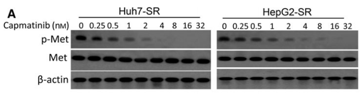
|
28164434 |
| Immunofluorescence | p-EGFR |
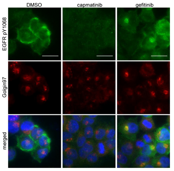
|
30390071 |
| Growth inhibition assay | Cell viability |
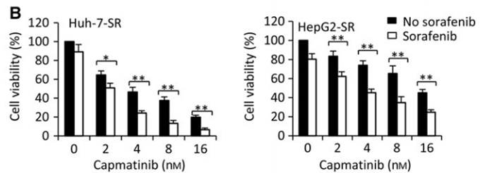
|
28164434 |
Tech Support
Tel: +1-832-582-8158 Ext:3
If you have any other enquiries, please leave a message.
Signaling Pathway Map
Products are for research use only. Not for human use. We do not sell to patients.
©Copyright 2013 Selleck Chemicals. All Rights Reserved.