-
 Australia
-
 Austria
-
 Belgium
-
 Brazil
-
 Canada
-
 China
China
-
 Czech Republic
-
 Denmark
-
 Finland
-
 France
-
 Germany
-
 Greece
-
 Hong Kong
-
 Hungary
-
 Iceland
-
 India
India
-
 Ireland
-
 Israel
-
 Italy
Italy
-
 Japan
Japan
-
 Korea
Korea
-
 Luxembourg
-
 Malaysia
-
 Netherlands
-
 New Zealand
-
 Norway
-
 Poland
-
 Qatar
Qatar
-
 Romania
-
 Saudi Arabia
-
 Singapore
-
 Spain
Spain
-
 Sweden
-
 Switzerland
-
 Taiwan
-
 Turkey
-
 United Kingdom
-
 United States
research use only
AS160 Antibody [L14F10]
Cat.No.: F3386
Application:
Reactivity:
-
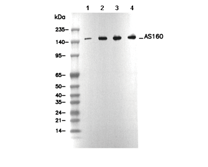 Lane 1: Hela, Lane 2: HEK-293, Lane 3: RAW 264.7, Lane 4: PC-12
Experiment Essentials
WB
Recommended SDS-PAGE separating gel concentration: 5%.
Recommended SDS-PAGE separating gel concentration: 5%.
Usage Information
| Dilution |
|---|
|
| Application |
|---|
| WB, IP |
| Reactivity |
|---|
| Human, Mouse, Rat |
| Source |
|---|
| Rabbit Monoclonal Antibody |
| Storage Buffer |
|---|
| PBS, pH 7.2+50% Glycerol+0.05% BSA+0.01% NaN3 |
| Storage (from the date of receipt) |
|---|
| -20°C (avoid freeze-thaw cycles), 2 years |
| Predicted MW |
|---|
| 146 kDa |
| Positive Control | Mouse brain; Rat brain; Human fetal heart; Human fetal kidney; A-673 cell; MCF7 cell; HeLa cell; HEK-293 cell; NIH/3T3 cell; RAW 264.7 cell; PC-12 cell |
|---|---|
| Negative Control |
Experimental Methods
| WB |
|---|
Experimental Protocol:
Sample preparation
1. Tissue: Lyse the tissue sample by adding an appropriate volume of ice-cold RIPA/Tris-Triton Lysis Buffer (containing Protease Inhibitor Cocktail),and homogenize the tissue at a low temperature. 2. Adherent cell: Aspirate the culture medium and wash the cells with ice-cold PBS twice. Lyse the cells by adding an appropriate volume of RIPA/Tris-Triton Lysis Buffer (containing Protease Inhibitor Cocktail) and put the sample on ice for 5 min. 3. Suspension cell: Transfer the culture medium to a pre-cooled centrifuge tube. Centrifuge and aspirate the supernatant. Wash the cells with ice-cold PBS twice. Lyse the cells by adding an appropriate volume of RIPA/Tris-Triton Lysis Buffer (containing Protease Inhibitor Cocktail) and put the sample on ice for 5 min. 4. Place the lysate into a pre-cooled microcentrifuge tube. Centrifuge at 4°C for 15 min. Collect the supernatant;
5. Remove a small volume of lysate to determine the protein concentration;
6. Combine the lysate with protein loading buffer. Boil 20 µL sample under 95-100°C for 5 min. Centrifuge for 5 min after cool down on ice.
Electrophoretic separation
1. According to the concentration of extracted protein, load appropriate amount of protein sample and marker onto SDS-PAGE gels for electrophoresis. Recommended separating gel (lower gel) concentration: 5%. Reference Table for Selecting SDS-PAGE Separation Gel Concentrations 2. Power up 80V for 30 minutes. Then the power supply is adjusted (110 V~150 V), the Marker is observed, and the electrophoresis can be stopped when the indicator band of the predyed protein Marker where the protein is located is properly separated. (Note that the current should not be too large when electrophoresis, too large current (more than 150 mA) will cause the temperature to rise, affecting the result of running glue. If high currents cannot be avoided, an ice bath can be used to cool the bath.)
Transfer membrane
1. Take out the converter, soak the clip and consumables in the pre-cooled converter;
2. Activate PVDF membrane with methanol for 1 min and rinse with transfer buffer;
3. Install it in the order of "black edge of clip - sponge - filter paper - filter paper - glue -PVDF membrane - filter paper - filter paper - sponge - white edge of clip"; 4. The protein was electrotransferred to PVDF membrane. ( 0.45 µm PVDF membrane is recommended ) Reference Table for Selecting PVDF Membrane Pore Size Specifications Recommended conditions for wet transfer: 200 mA, 120 min. ( Note that the transfer conditions can be adjusted according to the protein size. For high-molecular-weight proteins, a higher current and longer transfer time are recommended. However, ensure that the transfer tank remains at a low temperature to prevent gel melting.)
Block
1. After electrotransfer, wash the film with TBST at room temperature for 5 minutes;
2. Incubate the film in the blocking solution for 1 hour at room temperature;
3. Wash the film with TBST for 3 times, 5 minutes each time.
Antibody incubation
1. Use 5% skim milk powder to prepare the primary antibody working liquid (recommended dilution ratio for primary antibody 1:1000), gently shake and incubate with the film at 4°C overnight; 2. Wash the film with TBST 3 times, 5 minutes each time;
3. Add the secondary antibody to the blocking solution and incubate with the film gently at room temperature for 1 hour;
4. After incubation, wash the film with TBST 3 times for 5 minutes each time.
Antibody staining
1. Add the prepared ECL luminescent substrate (or select other color developing substrate according to the second antibody) and mix evenly;
2. Incubate with the film for 1 minute, remove excess substrate (keep the film moist), wrap with plastic film, and expose in the imaging system. |
Biological Description
| Specificity |
|---|
AS160 Antibody [L14F10] recognizes endogenous levels of total AS160 protein. |
| Subcellular Location |
|---|
| Cytoplasm |
| Uniprot ID |
|---|
| O60343 |
| Clone |
|---|
| L14F10 |
| Synonym(s) |
|---|
| AS160, KIAA0603, TBC1D4, TBC1 domain family member 4, Akt substrate of 160 kDa |
| Background |
|---|
| AS160 (also known as TBC1D4) is a 160 kDa Akt substrate and Rab GTPase-activating protein (Rab-GAP) that plays a crucial role in regulating insulin-stimulated glucose uptake by controlling the translocation of GLUT4 in adipocytes and muscle cells. AS160 contains a Rab-GAP domain, two phosphotyrosine-binding (PTB) domains, and a calmodulin-binding domain, with the latter mediating phosphorylation-independent glucose uptake in muscle. In the absence of insulin, non-phosphorylated AS160 binds to GLUT4 storage vesicles (GSVs) and inhibits their movement to the plasma membrane, limiting glucose entry. Upon insulin stimulation, Akt phosphorylates AS160 at key sites, inhibiting its GAP activity and allowing Rabs (such as RAB2A, RAB8A, RAB10, and RAB14) to remain in their active GTP-bound state. This promotes GLUT4 vesicle fusion with the plasma membrane, facilitating glucose uptake. Additionally, the second PTB domain contains a phospholipid-binding region, targeting AS160 to the plasma membrane, while a separate domain mediates binding to GSV cargo, underscoring its multifunctional role in glucose regulation. The gene encoding AS160 (TBC1D4) produces two isoforms: a long isoform found mainly in skeletal and cardiac muscle, and a short isoform with broader tissue distribution. AS160’s role as a regulatory switch is further emphasized by its involvement in the docking and fusion steps of GLUT4 vesicles, where phosphorylation converts it from an inhibitor to a facilitator of GLUT4 translocation. Mutations or dysregulation of AS160 can impair insulin-stimulated glucose uptake, contributing to insulin resistance and metabolic diseases. |
| References |
|---|
|
Tech Support
Tel: +1-832-582-8158 Ext:3
If you have any other enquiries, please leave a message.
Products are for research use only. Not for human use. We do not sell to patients.
©Copyright 2013 Selleck Chemicals. All Rights Reserved.