-
 Australia
-
 Austria
-
 Belgium
-
 Brazil
-
 Canada
-
 China
China
-
 Czech Republic
-
 Denmark
-
 Finland
-
 France
-
 Germany
-
 Greece
-
 Hong Kong
-
 Hungary
-
 Iceland
-
 India
India
-
 Ireland
-
 Israel
-
 Italy
Italy
-
 Japan
Japan
-
 Korea
Korea
-
 Luxembourg
-
 Malaysia
-
 Netherlands
-
 New Zealand
-
 Norway
-
 Poland
-
 Qatar
Qatar
-
 Romania
-
 Saudi Arabia
-
 Singapore
-
 Spain
Spain
-
 Sweden
-
 Switzerland
-
 Taiwan
-
 Turkey
-
 United Kingdom
-
 United States
research use only
IOX1 Histone Demethylase inhibitor
Cat.No.S7234
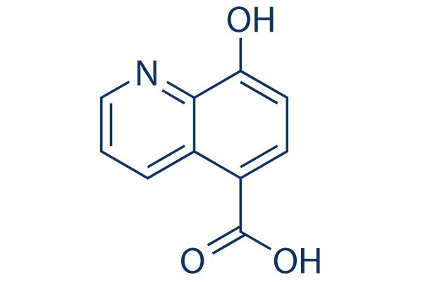
Chemical Structure
Molecular Weight: 189.17
Quality Control
| Related Targets | HDAC JAK BET Histone Methyltransferase PKC PARP HIF PRMT EZH2 AMPK |
|---|---|
| Other Histone Demethylase Inhibitors | GSK-J4 Hydrochloride SP2509 JIB-04 GSK-LSD1 2HCl Iadademstat (ORY-1001) Dihydrochloride CPI-455 HCl OG-L002 GSK J1 ML324 CP2 |
Cell Culture, Treatment & Working Concentration
| Cell Lines | Assay Type | Concentration | Incubation Time | Formulation | Activity Description | PMID |
|---|---|---|---|---|---|---|
| HCT116 cells | Cytotoxicity assay | 48 h | Cytotoxicity against human HCT116 cells assessed as cell viability after 48 hrs by MTT assay, IC50=28.1 μM | |||
| human A549 cells | Cytotoxicity assay | 48 h | Cytotoxicity against human A549 cells assessed as cell viability after 48 hrs by MTT assay, IC50=48.3 μM | |||
| human HeLa cells | Function assay | 72 h | Induction of histone methylation in human HeLa cells assessed as H3K9me2 level after 72 hrs by immunofluorescence assay | |||
| Click to View More Cell Line Experimental Data | ||||||
Solubility
|
In vitro |
DMSO
: 37 mg/mL
(195.59 mM)
Water : Insoluble Ethanol : Insoluble |
Molarity Calculator
|
In vivo |
|||||
In vivo Formulation Calculator (Clear solution)
Step 1: Enter information below (Recommended: An additional animal making an allowance for loss during the experiment)
Step 2: Enter the in vivo formulation (This is only the calculator, not formulation. Please contact us first if there is no in vivo formulation at the solubility Section.)
Calculation results:
Working concentration: mg/ml;
Method for preparing DMSO master liquid: mg drug pre-dissolved in μL DMSO ( Master liquid concentration mg/mL, Please contact us first if the concentration exceeds the DMSO solubility of the batch of drug. )
Method for preparing in vivo formulation: Take μL DMSO master liquid, next addμL PEG300, mix and clarify, next addμL Tween 80, mix and clarify, next add μL ddH2O, mix and clarify.
Method for preparing in vivo formulation: Take μL DMSO master liquid, next add μL Corn oil, mix and clarify.
Note: 1. Please make sure the liquid is clear before adding the next solvent.
2. Be sure to add the solvent(s) in order. You must ensure that the solution obtained, in the previous addition, is a clear solution before proceeding to add the next solvent. Physical methods such
as vortex, ultrasound or hot water bath can be used to aid dissolving.
Chemical Information, Storage & Stability
| Molecular Weight | 189.17 | Formula | C10H7NO3 |
Storage (From the date of receipt) | |
|---|---|---|---|---|---|
| CAS No. | 5852-78-8 | Download SDF | Storage of Stock Solutions |
|
|
| Synonyms | N/A | Smiles | C1=CC2=C(C=CC(=C2N=C1)O)C(=O)O | ||
Mechanism of Action
| Features |
Cell-permeant, broad-spectrum 2OG oxygenase inhibitor.
|
|---|---|
| Targets/IC50/Ki |
KDM3A
(Cell-free assay) 0.1 μM
KDM4C
(Cell-free assay) 0.6 μM
KDM6B
(Cell-free assay) 1.6 μM
KDM2A
(Cell-free assay) 1.8 μM
KDM4E
(Cell-free assay) 2.3 μM
KDM5C
(Cell-free assay) 19 μM
PHD2
(Cell-free assay) 33 μM
|
| In vitro |
IOX1 increases H3K9me3 levels in HeLa cells via KDM4A inhibition without significant effect on cell viability. This compound shows lower efficacy in HeLa cells due to low cell permeability, while the n-octyl ester derivative improves its cell permeability. |
| Kinase Assay |
AlphaScreen Assay
|
|
All reagents are diluted in 50 mM HEPES, 0.1% BSA, pH 7.5 supplemented with 0.01% Tween20 and allowed to equilibrate to room temperature prior to addition to plates. Catalytic turnover assays are run in 10 μL volumes in lowvolume 384-well plates at RT. The reaction consisted of enzyme (5 nM), biotinylated substrate peptide (30 nM), Fe(II) (1 μM), ascorbate (100 μM), 2OG (10 μM) and run at RT. For PHD2, the reaction consisted of enzyme (5 nM), biotinylated substrate peptide (60nM), Fe(II) (20 μM), ascorbate (200 μM), 2OG (2 μM) and run at RT. EDTA is used to quench the reaction (5 μL), AlphaScreen donor (Streptavidin-conjugated) and acceptor (Protein A-conjugated) beads preincubated with peptide product antibodies are added (5 μL). Plates are foil-sealed to protect from light, incubated at room temperature for 60 minutes and read on a PHERAstar FS plate reader using an AlphaScreen 680 excitation/570 emission filter set. The final bead concentration in 20 μL reaction is 20 μg/mL. IC50 values are calculated in Prism 6 after normalisation against corresponding DMSO controls.
|
References |
|
Tech Support
Tel: +1-832-582-8158 Ext:3
If you have any other enquiries, please leave a message.
Signaling Pathway Map
Products are for research use only. Not for human use. We do not sell to patients.
©Copyright 2013 Selleck Chemicals. All Rights Reserved.