-
 Australia
-
 Austria
-
 Belgium
-
 Brazil
-
 Canada
-
 China
China
-
 Czech Republic
-
 Denmark
-
 Finland
-
 France
-
 Germany
-
 Greece
-
 Hong Kong
-
 Hungary
-
 Iceland
-
 India
India
-
 Ireland
-
 Israel
-
 Italy
Italy
-
 Japan
Japan
-
 Korea
Korea
-
 Luxembourg
-
 Malaysia
-
 Netherlands
-
 New Zealand
-
 Norway
-
 Poland
-
 Qatar
Qatar
-
 Romania
-
 Saudi Arabia
-
 Singapore
-
 Spain
Spain
-
 Sweden
-
 Switzerland
-
 Taiwan
-
 Turkey
-
 United Kingdom
-
 United States
research use only
GSK-J4 Hydrochloride H3K27 Histone Demethylase Inhibitor
Cat.No.S7070
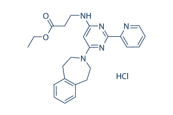
Chemical Structure
Molecular Weight: 453.96
Quality Control
| Related Targets | HDAC JAK BET Histone Methyltransferase PKC PARP HIF PRMT EZH2 AMPK |
|---|---|
| Other Histone Demethylase Inhibitors | SP2509 JIB-04 GSK-LSD1 2HCl Iadademstat (ORY-1001) Dihydrochloride CPI-455 HCl OG-L002 IOX1 GSK J1 ML324 CP2 |
Cell Culture, Treatment & Working Concentration
| Cell Lines | Assay Type | Concentration | Incubation Time | Formulation | Activity Description | PMID |
|---|---|---|---|---|---|---|
| CUTLL1 | Growth inhibitory assay | 2 μM | DMSO | affects cell growth | 25132549 | |
| CUTLL1 | Apoptosis assay | 2 μM | DMSO | induces apoptosis | 25132549 | |
| CUTLL1 | Function assay | 2 μM | DMSO | induces cell cycle arrest | 25132549 | |
| CUTLL1 | Kinase assay | 6 μM | DMSO | leads to increased H3K27me3 | 25132549 | |
| SF7761 | Kinase assay | 6 μM | DMSO | increases K27 methylation | 25401693 | |
| SF8628 | Kinase assay | 6 μM | DMSO | increases K28 methylation | 25401693 | |
| H3.3 | Kinase assay | 6 μM | DMSO | increases K29 methylation | 25401693 | |
| SF9012 | Kinase assay | 6 μM | DMSO | increases K30 methylation | 25401693 | |
| SF9402 | Kinase assay | 6 μM | DMSO | increases K31 methylation | 25401693 | |
| SF9427 | Kinase assay | 6 μM | DMSO | increases K32 methylation | 25401693 | |
| human astrocytes | Kinase assay | 6 μM | DMSO | increases K33 methylation | 25401693 | |
| SF7761 | Growth inhibitory assay | 6 μM | DMSO | inhibits K27M glioma cell growth | 25401693 | |
| SF8628 | Growth inhibitory assay | 6 μM | DMSO | inhibits K28M glioma cell growth | 25401693 | |
| H3.3 | Growth inhibitory assay | 6 μM | DMSO | inhibits K29M glioma cell growth | 25401693 | |
| SF9012 | Growth inhibitory assay | 6 μM | DMSO | inhibits K30M glioma cell growth | 25401693 | |
| SF9402 | Growth inhibitory assay | 6 μM | DMSO | inhibits K31M glioma cell growth | 25401693 | |
| SF9427 | Growth inhibitory assay | 6 μM | DMSO | inhibits K32M glioma cell growth | 25401693 | |
| human astrocytes | Growth inhibitory assay | 6 μM | DMSO | inhibits K33M glioma cell growth | 25401693 | |
| TG neurons | Function assay | 50 μM | DMSO | inhibits HSV-1 reactivation from sensory neurons | 25552720 | |
| Th17 | Function assay | 80 nM | DMSO | inhibits cell differentiation | 25840993 | |
| β-cells | Function assay | 20 μM | DMSO | blunts IFNγ, Il-1β, and TNFα-induced chemokine gene expression | 26505193 | |
| β-cells | Function assay | 20 μM | DMSO | induces β-cell dysfunction | 26505193 | |
| ESCs | Function assay | 1.8 µM | DMSO | induces DNA damage along with activation of the DNA damage response | 26759175 | |
| Raw 264.7 | Function assay | 0.8192 µM | DMSO | inhibits TNF-α production | 26776360 | |
| MCF7 | Function assay | 1 to 10 uM | 30 hrs | Inhibition of KDM5A in human MCF7 cells assessed as effect on H3K4me3 methylation levels at 1 to 10 uM after 30 hrs by Western blot analysis | 30392349 | |
| MCF7 | Function assay | 1 to 10 uM | 30 hrs | Inhibition of KDM5A in human MCF7 cells assessed as effect on H3K27me3 methylation levels at 1 to 10 uM after 30 hrs by Western blot analysis | 30392349 | |
| RAW264.7 | 0.82 uM | 24 hrs | Inhibition of LPS-induced TNFalpha production in mouse RAW264.7 cells 0.82 uM after 24 hrs by ELISA | 26776360 | ||
| RAW264.7 | 24 hrs | Inhibition of LPS-induced TNFalpha production in mouse RAW264.7 cells after 24 hrs by ELISA | 26776360 | |||
| Click to View More Cell Line Experimental Data | ||||||
Solubility
|
In vitro |
DMSO
: 91 mg/mL
(200.45 mM)
Ethanol : 91 mg/mL Water : 10 mg/mL |
Molarity Calculator
|
In vivo |
|||||
In vivo Formulation Calculator (Clear solution)
Step 1: Enter information below (Recommended: An additional animal making an allowance for loss during the experiment)
Step 2: Enter the in vivo formulation (This is only the calculator, not formulation. Please contact us first if there is no in vivo formulation at the solubility Section.)
Calculation results:
Working concentration: mg/ml;
Method for preparing DMSO master liquid: mg drug pre-dissolved in μL DMSO ( Master liquid concentration mg/mL, Please contact us first if the concentration exceeds the DMSO solubility of the batch of drug. )
Method for preparing in vivo formulation: Take μL DMSO master liquid, next addμL PEG300, mix and clarify, next addμL Tween 80, mix and clarify, next add μL ddH2O, mix and clarify.
Method for preparing in vivo formulation: Take μL DMSO master liquid, next add μL Corn oil, mix and clarify.
Note: 1. Please make sure the liquid is clear before adding the next solvent.
2. Be sure to add the solvent(s) in order. You must ensure that the solution obtained, in the previous addition, is a clear solution before proceeding to add the next solvent. Physical methods such
as vortex, ultrasound or hot water bath can be used to aid dissolving.
Chemical Information, Storage & Stability
| Molecular Weight | 453.96 | Formula | C24H27N5O2.HCl |
Storage (From the date of receipt) | |
|---|---|---|---|---|---|
| CAS No. | 1797983-09-5 | Download SDF | Storage of Stock Solutions |
|
|
Mechanism of Action
| Targets/IC50/Ki |
JMJD3
(Cell-free assay) 60 nM
|
|---|---|
| In vitro |
GSK J4 HCl is an ethyl ester derivative of the JMJD3 selective histone demethylase inhibitor GSK-J1 with an IC50 value greater than 50 μM in vitro. This compound is used to probe the consequences of demethylation of H3K27me3. In human primary macrophages, this chemical inhibits the lipopolysaccharide-induced production of cytokines, including pro-inflammatory tumour necrosis factor (TNF). In addition, it prevents the lipopolysaccharide-induced loss of H3K27me3 associated with the TNF transcription start sites and blocked the recruitment of RNA polymerase II. |
| Kinase Assay |
Histone Demethylase AlphaScreen
|
|
Inhibition of histone demethylases is assessed using the histone demethylase AlphaScreen assay (Amplified Luminescence Proximity Homogenous Assay). This assay uses a biotinylated peptide substrate and relies on detection of the product methyl mark using a specific antibody coupled to protein-A acceptor beads and a Steptavidin donor bead to capture the peptide. In brief, recombinant demethylase enzymes are incubated in the presence of Fe2+ in the form of Ferrous Ammonium Sulphate (FAS), -ketoglutarate (KG) and biotinylated peptide substrate. L-Ascorbic Acid is included to provide a reducing environment and prevent oxidation of Fe2+. After incubation with peptide substrate the presence of the product is detected using AlphaScreen technology. The demethylase AlphaScreen assays are performed in 384-well plate format using white proxiplates. All steps are carried out in assay buffer (50 mM HEPES pH 7.5, 0.1% (w/v) BSA and 0.01 % (v/v) Tween-20). FAS is dissolved fresh each day in 20 mM HCl to a concentration of 400 mM and diluted to 1.0 mM in deionized water. All other components are dissolved fresh each day in deionized water. For IC50 determinations 5 μL of assay buffer containing demethylase enzyme is transferred to wells of a 384-well proxiplate. Titrations of this compound (0.1 μL) are transferred to each well and the enzymes allowed to pre-incubate for 15 minutes with this chemical (final concentration of DMSO is 1%). The enzyme reaction is initiated by addition of 5 μL of a substrate mix consisting of α-KG, FAS, L-Ascorbic Acid and biotinylated peptide substrate and the reaction incubated for the indicated time at room temperature. The enzyme reaction is stopped after the indicated time by addinton of 5 μL of EDTA (7.5 mM final concentration in assay buffer). Streptavidin Donor beads (0.08 mg/ml) and Protein-A conjugated acceptor beads (0.08 mg/ml) are pre-incubated for 1 hour with an antibody to the product methyl mark and the presence of biotin-H3-product is detected by addition of 5 μL of the preincubated AlphaScreen beads (final concentrations of 0.02 mg/ml with respect to acceptor and donor beads). Detection is allowed to proceed for 1 hour at room temperature and the assay plates read in a BMG Labtech Pherastar FS plate reader. Data are normalized to the no enzyme control and the IC50 determined from the nonlinear regression curve fit using GraphPad Prism 5.
|
|
| In vivo |
GSK-J4 hydrochloride is a potent dual inhibitor of H3K27me3/me2-demethylases JMJD3/KDM6B and UTX/KDM6A. It inhibits LPS-induced TNF-α production in human primary macrophages. This compound is a cell permeable prodrug of GSK-J1. |
References |
|
Tech Support
Tel: +1-832-582-8158 Ext:3
If you have any other enquiries, please leave a message.
Signaling Pathway Map
Products are for research use only. Not for human use. We do not sell to patients.
©Copyright 2013 Selleck Chemicals. All Rights Reserved.