-
 Australia
-
 Austria
-
 Belgium
-
 Brazil
-
 Canada
-
 China
China
-
 Czech Republic
-
 Denmark
-
 Finland
-
 France
-
 Germany
-
 Greece
-
 Hong Kong
-
 Hungary
-
 Iceland
-
 India
India
-
 Ireland
-
 Israel
-
 Italy
Italy
-
 Japan
Japan
-
 Korea
Korea
-
 Luxembourg
-
 Malaysia
-
 Netherlands
-
 New Zealand
-
 Norway
-
 Poland
-
 Qatar
Qatar
-
 Romania
-
 Saudi Arabia
-
 Singapore
-
 Spain
Spain
-
 Sweden
-
 Switzerland
-
 Taiwan
-
 Turkey
-
 United Kingdom
-
 United States
research use only
NU7026 DNA-PK inhibitor
Cat.No.S2893
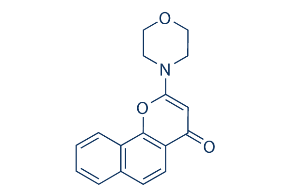
Chemical Structure
Molecular Weight: 281.31
Quality Control
| Related Targets | HDAC PARP ATM/ATR WRN DNA/RNA Synthesis Topoisomerase PPAR Sirtuin Casein Kinase eIF |
|---|---|
| Other DNA-PK Inhibitors | Nedisertib (M3814) AZD7648 NU7441 (KU-57788) KU-0060648 VX-984 CC-115 YU238259 LTURM34 Compound 401 |
Cell Culture, Treatment & Working Concentration
| Cell Lines | Assay Type | Concentration | Incubation Time | Formulation | Activity Description | PMID |
|---|---|---|---|---|---|---|
| HeLa cells | Function assay | Inhibition of DNA dependent protein kinase isolated from HeLa cells, IC50=0.23 μM | 15658870 | |||
| HeLa cells | Function assay | Inhibition of mTOR protein isolated from HeLa cells, IC50=6.4 μM | 15658870 | |||
| HeLa | Function assay | In vitro inhibition of DNA-dependent protein kinase(DNA-PK) from HeLa (human carcinoma) cells, IC50=0.23μM | 12941339 | |||
| Click to View More Cell Line Experimental Data | ||||||
Solubility
|
In vitro |
4-Methylpyridine : 5 mg/mL
DMSO
: 1 mg/mL
(3.55 mM)
Water : Insoluble |
Molarity Calculator
|
In vivo |
|||||
In vivo Formulation Calculator (Clear solution)
Step 1: Enter information below (Recommended: An additional animal making an allowance for loss during the experiment)
Step 2: Enter the in vivo formulation (This is only the calculator, not formulation. Please contact us first if there is no in vivo formulation at the solubility Section.)
Calculation results:
Working concentration: mg/ml;
Method for preparing DMSO master liquid: mg drug pre-dissolved in μL DMSO ( Master liquid concentration mg/mL, Please contact us first if the concentration exceeds the DMSO solubility of the batch of drug. )
Method for preparing in vivo formulation: Take μL DMSO master liquid, next addμL PEG300, mix and clarify, next addμL Tween 80, mix and clarify, next add μL ddH2O, mix and clarify.
Method for preparing in vivo formulation: Take μL DMSO master liquid, next add μL Corn oil, mix and clarify.
Note: 1. Please make sure the liquid is clear before adding the next solvent.
2. Be sure to add the solvent(s) in order. You must ensure that the solution obtained, in the previous addition, is a clear solution before proceeding to add the next solvent. Physical methods such
as vortex, ultrasound or hot water bath can be used to aid dissolving.
Chemical Information, Storage & Stability
| Molecular Weight | 281.31 | Formula | C17H15NO3 |
Storage (From the date of receipt) | |
|---|---|---|---|---|---|
| CAS No. | 154447-35-5 | Download SDF | Storage of Stock Solutions |
|
|
| Synonyms | LY293646 | Smiles | C1COCCN1C2=CC(=O)C3=C(O2)C4=CC=CC=C4C=C3 | ||
Mechanism of Action
| Targets/IC50/Ki |
DNA-PK
(Cell-free assay) 0.23 μM
PI3K
(Cell-free assay) 13 μM
|
|---|---|
| In vitro |
NU7026 potentiates ionizing radiation induced cytotoxicity in a concentration-dependent manner in V3YAC and PARP-1+/+ cells. This compound completely abolishes potentially lethal damage recovery in growth-arrested cells. It inhibits DNA DSB repair by 56% in the V3YAC cell line. This chemical (10 μM) potentiates the growth inhibitory effects of doxorubicin, mAMSA with PF50 values ranging from approximately 19 for mAMSA to approximately 2 in K562 cells. It (10 μM) also potentiates the growth inhibitory effect in this leukemia cell line with a PF50 value of 10.53. This compound (10 μM) enhances the -induced cell cycle G2 blockade in K562 cells. It potentiates topo II poisons involves inhibition of nonhomologous end joining and a G2/M checkpoint arrest. This compound (10 μM) exposure of 4 h in combination with 3 Gy radiation is required for a significant radiosensitisation effect in CH1 human ovarian cancer cells. It (< 10 μM) has synergistic cytotoxic activity at nontoxic doses of this chemical in a CLL cell line (I83) and in primary CLL-lymphocytes. This compound (10 μM) increases -induced G(2)/M arrest in I83 cells. It (10 μM) enhances -induced γH2AX throughout the cell cycle in the I83 cell line. This chemical (10 μM) Increases -Induced apoptosis in the I83 cell line. It (55 μM) results in a dramatic induction of telomere fusion in p53 null MEFs and significantly fewer telomere fusions in p53 and ligase IV double null MEFs. |
| Kinase Assay |
Recombinant kinase assay
|
|
Mammalian DNA-PK (500 ng/μL) is isolated from HeLa cell nuclear extract after chromatography using Q-Sepharose, S-Sepharose, and Heparin agarose. DNA-PK (250 ng) activity is measured at 30℃, in a final volume of 40 μL, in buffer containing 25 mM HEPES (pH 7.4), 12.5 mM MgCl2, 50 mM KCl, 1 mM DTT, 10% v/v Glycerol, 0.1% w/v NP-40, and 1 mg of the substrate GST-p53N66 in polypropylene 96-well plates. To the assay mix, varying concentrations of this compound (in DMSO at a final concentration of 1% v/v) are added. After 10 min of incubation, ATP is added to give a final concentration of 50 μM, along with a 30-mer double-stranded DNA oligonucleotide (final concentration of 0.5 ng/mL), to initiate the reaction. After 1 hour with shaking, 150 μL of PBS are added to the reaction, and 5 μL are then transferred to a 96-well opaque white plate containing 45 μl of PBS per well, where the GSTp53N66 substrate is allowed to bind to the wells for 1 hour. The IC50s for the compounds in all of the enzymes assays are derived from sigmoidal plots using the graphic package Prism, in which the enzyme activity in the varying concentration of compounds is plotted against the concentration of compound.
|
|
| In vivo |
NU7026 (20mg/kg, i.v.) undergoes rapid plasma clearance (0.108/hour) in mice and this is largely attributed to extensive metabolism. Bioavailability following interperitoneal (i.p.) and p.o. administration of this compound at dose of 20 mg/kg is 20 and 15%, respectively. |
References |
|
Applications
| Methods | Biomarkers | Images | PMID |
|---|---|---|---|
| Western blot | pDNA-PKcs S2056 / DNA-PKcs |
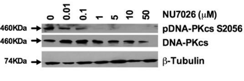
|
22131882 |
| Growth inhibition assay | Cell viability |
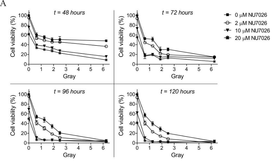
|
26716839 |
Tech Support
Tel: +1-832-582-8158 Ext:3
If you have any other enquiries, please leave a message.
Signaling Pathway Map
Products are for research use only. Not for human use. We do not sell to patients.
©Copyright 2013 Selleck Chemicals. All Rights Reserved.