-
 Australia
-
 Austria
-
 Belgium
-
 Brazil
-
 Canada
-
 China
China
-
 Czech Republic
-
 Denmark
-
 Finland
-
 France
-
 Germany
-
 Greece
-
 Hong Kong
-
 Hungary
-
 Iceland
-
 India
India
-
 Ireland
-
 Israel
-
 Italy
Italy
-
 Japan
Japan
-
 Korea
Korea
-
 Luxembourg
-
 Malaysia
-
 Netherlands
-
 New Zealand
-
 Norway
-
 Poland
-
 Qatar
Qatar
-
 Romania
-
 Saudi Arabia
-
 Singapore
-
 Spain
Spain
-
 Sweden
-
 Switzerland
-
 Taiwan
-
 Turkey
-
 United Kingdom
-
 United States
research use only
FCCP OXPHOS Uncoupler
Cat.No.S8276
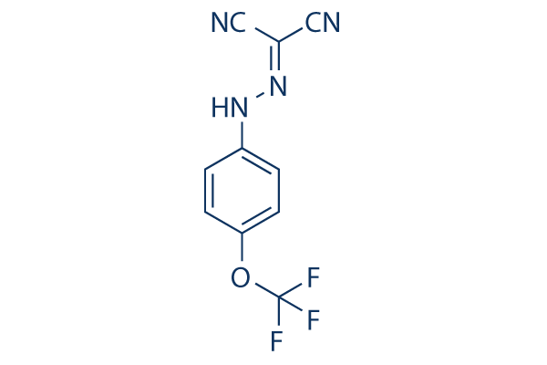
Chemical Structure
Molecular Weight: 254.17
Quality Control
Cell Culture, Treatment & Working Concentration
| Cell Lines | Assay Type | Concentration | Incubation Time | Formulation | Activity Description | PMID |
|---|---|---|---|---|---|---|
| T47D | Function assay | Inhibition of 1, 10-phenanthroline-induced HIF1 activation in human T47D cells by HRE3-TK-luciferase reporter gene assay, IC50=0.31μM | 20929261 | |||
| T47D | Function assay | Inhibition of hypoxia-induced HIF1 activation in human T47D cells by HRE3-TK-luciferase reporter gene assay, IC50=0.51μM | 20929261 | |||
| T47D | Function assay | 1 to 10 uM | Inhibition of HIF1-mediated induction of secreted VEGF level in 1, 10-phenanthroline-stimulated human T47D cells at 1 to 10 uM by ELISA | 20929261 | ||
| T47D | Function assay | 0.3 uM | 15 mins | Decrease in mitochondrial membrane potential in human T47D cells 0.3 uM after 15 mins by TMRM assay | 20929261 | |
| MDA-MB-231 | Cytotoxicity assay | 1 uM | Cytotoxicity against human MDA-MB-231 cells assessed as inhibition of cell proliferation/viability at 1 uM | 23245650 | ||
| MDA-MB-231 | Cytotoxicity assay | 0.1 to 3 uM | Cytotoxicity against human MDA-MB-231 cells assessed as inhibition of cell proliferation/viability at 0.1 to 3 uM in presence of 0.1 uM rotenone mitochondrial electron transport inhibitor | 23245650 | ||
| MDA-MB-231 | Function assay | 0.3 uM | Stimulation of oligomycin-induced state 4 respiration in human MDA-MB-231 cells at 0.3 uM | 23245650 | ||
| T47D | Function assay | 0.1 to 3 uM | Effect on cellular respiration in human T47D cells assessed as increase in oxygen consumption at 0.1 to 3 uM | 22938093 | ||
| T47D | Function assay | 10 to 30 uM | Stimulation of state 4 cellular respiration in human T47D cells at 10 to 30 uM in presence of oligomycin | 22938093 | ||
| T47D | Function assay | 0.3 to 1 uM | Stimulation of state 4 cellular respiration in human T47D cells at 0.3 to 1 uM in presence of oligomycin | 22938093 | ||
| Hep3B | Function assay | 10 uM | Stimulation of state 4 cellular respiration in human Hep3B cells at 10 uM in presence of oligomycin | 22938093 | ||
| Hep3B | Function assay | 1 uM | Stimulation of state 4 cellular respiration in human Hep3B cells at 1 uM in presence of oligomycin | 22938093 | ||
| T47D | Function assay | 3 uM | 15 to 20 mins | Decrease in mitochondrial membrane potential in human T47D cells at 3 uM after 15 to 20 mins by TMRM assay | 22938093 | |
| TA3/Ha | Function assay | 6 uM | Induction of NAD(P)H oxidation in mouse TA3/Ha cells assessed as reduction of NAD(P)H/NAD(P)+ ratio at 6 uM by spectrofluorometer analysis | 24568614 | ||
| SH-SY5Y | Function assay | 10 uM | 5 mins | Inhibition of SOC in human SH-SY5Y cells assessed as reduction in thapsigargin-induced Ca2+ influx at 10 uM pre-incubated for 5 mins with 0.2 uM CsA followed by compound addition by FURA-2AM dye based fluorescence assay | 25265024 | |
| SH-SY5Y | Function assay | 10 uM | 10 mins | Induction of mitochondrial membrane potential loss in human SH-SY5Y cells at 10 uM incubated for 10 mins in presence of 0.2 uM CsA by TMRE dye based assay | 25265024 | |
| T47D | Function assay | 0.3 uM | 3 to 12 mins | Increase in oxygen consumption rate of mitochondrial state 4 respiration in human T47D cells assessed as reinitiation of oligomycin-stalled cellular respiration at 0.3 uM incubated for 3 to 12 mins by Clark-type oxygen electrode assay | 26637046 | |
| T47D | Function assay | 0.3 uM | 30 mins | Effect on mitochondrial membrane potential in human T47D cells at 0.3 uM after 30 mins by TMRM dye based fluorescence microscopy | 26637046 | |
| T47D | Function assay | 0.3 uM | Increase in oxygen consumption rate in digitonin permeabilized human T47D cells assessed as reinitiation of sodium azide-stalled cellular respiration at 0.3 uM by oxytherm Clark-type electrode assay in presence of ascorbate | 26637046 | ||
| HCT116 | Function assay | 2 uM | 30 mins | Induction of AMPK phosphorylation at Thr-172 residue in human HCT116 cells at 2 uM after 30 mins in glucose supplemented media by immunoblot method | 28233680 | |
| HCT116 | Function assay | 2 uM | 30 mins | Induction of AMPK phosphorylation at Thr-172 residue in human HCT116 cells at 2 uM after 30 mins in absence of glucose by immunoblot method | 28233680 | |
| DLD1 | Function assay | 1 uM | Induction of mitochondrial dysfunction in human DLD1 cells assessed as reduction in mitochondrial ATP production at 1 uM by Seahorse XF real-time assay | 31774672 | ||
| DLD1 | Function assay | 1 uM | Induction of mitochondrial dysfunction in human DLD1 cells assessed as increase in glycolytic ATP production at 1 uM by Seahorse XF real-time assay | 31774672 | ||
| LS174T | Function assay | 1 uM | Induction of mitochondrial dysfunction in human LS174T cells assessed as reduction in mitochondrial ATP production at 1 uM by Seahorse XF real-time assay | 31774672 | ||
| LS174T | Function assay | 1 uM | Induction of mitochondrial dysfunction in human LS174T cells assessed as increase in glycolytic ATP production at 1 uM by Seahorse XF real-time assay | 31774672 | ||
| DLD1 | Function assay | 1 uM | Uncoupling of mitochondrial oxidative phosphorylation in human DLD1 cells at 1 uM in presence of oligomycin A by seahorse XFe96 analyser based assay | 31774672 | ||
| DLD1 | Function assay | 1 uM | Uncoupling of mitochondrial oxidative phosphorylation in human DLD1 cells assessed as increase in oxygen consumption rate at 1 uM in presence of oligomycin A by seahorse XFe96 analyser based assay | 31774672 | ||
| HEK293 | Function assay | 1 to 3 uM | Inhibition of LiCl-activated Wnt signaling in HEK293 cells at 1 to 3 uM by TOPFlash reporter gene assay | 31774672 | ||
| KOPN8 | Function assay | 10 uM | 0.3 hrs | Induction of mitochondrial membrane potential loss in human KOPN8 cells at 10 uM after 0.3 hrs by TMRM staining based flow cytometric analysis | 31084028 | |
| HepG2 | Function assay | Luciferase/luciferin-expressing antifolate-resistant parasites were used to infect a culture of HepG2 cells that were pre-incubated with compounds. Infected hepatocytes emit light due to the luciferase reaction. Assay results are presented as the percent , IC50=0.245μM | ChEMBL | |||
| Click to View More Cell Line Experimental Data | ||||||
Solubility
|
In vitro |
DMSO
: 6 mg/mL
(23.6 mM)
Water : Insoluble Ethanol : Insoluble |
Molarity Calculator
|
In vivo |
|||||
In vivo Formulation Calculator (Clear solution)
Step 1: Enter information below (Recommended: An additional animal making an allowance for loss during the experiment)
Step 2: Enter the in vivo formulation (This is only the calculator, not formulation. Please contact us first if there is no in vivo formulation at the solubility Section.)
Calculation results:
Working concentration: mg/ml;
Method for preparing DMSO master liquid: mg drug pre-dissolved in μL DMSO ( Master liquid concentration mg/mL, Please contact us first if the concentration exceeds the DMSO solubility of the batch of drug. )
Method for preparing in vivo formulation: Take μL DMSO master liquid, next addμL PEG300, mix and clarify, next addμL Tween 80, mix and clarify, next add μL ddH2O, mix and clarify.
Method for preparing in vivo formulation: Take μL DMSO master liquid, next add μL Corn oil, mix and clarify.
Note: 1. Please make sure the liquid is clear before adding the next solvent.
2. Be sure to add the solvent(s) in order. You must ensure that the solution obtained, in the previous addition, is a clear solution before proceeding to add the next solvent. Physical methods such
as vortex, ultrasound or hot water bath can be used to aid dissolving.
Chemical Information, Storage & Stability
| Molecular Weight | 254.17 | Formula | C10H5F3N4O |
Storage (From the date of receipt) | |
|---|---|---|---|---|---|
| CAS No. | 370-86-5 | Download SDF | Storage of Stock Solutions |
|
|
| Synonyms | Trifluoromethoxy carbonylcyanide phenylhydrazone, Carbonyl cyanide 4-(trifluoromethoxy)phenylhydrazone | Smiles | C1=CC(=CC=C1NN=C(C#N)C#N)OC(F)(F)F | ||
Mechanism of Action
| Targets/IC50/Ki |
OXPHOS
ATP synthase
|
|---|---|
| In vitro |
FCCP treatment induces a very rapid 2-fold increase in intracellular Ca2+ concentration that is accompanied by a strong protein synthesis rate inhibition. The translation inhibition correlates with an increased phosphorylation of the α subunit of eIF2 (eIF2α) and a 1.7-fold increase in the double-stranded RNA-dependent protein kinase activity. This compound also mildly decreases ATP and reactive oxygen species levels. It increases the expression of mitochondrial genes such as Tfam and COXIV while inducing morphological features of quiescent mouse HSCs and abrogating TGF-β signal transduction. |
| In vivo |
FCCP significantly reduces mitochondrial membrane potential and ATP production in 8-cell mouse embryos and the number of inner cell mass cells within blastocysts with unchanged blastocyst development. This perturbed embryonic mitochondrial function is concomitant with reduced birth weight in female offspring following embryo transfer, which persists until weaning. Although this compound-treated males also exhibits reduced glucose tolerance as female, but their insulin sensitivity and adiposity gain between 4 and 14 weeks is unchanged. Reducing mitochondrial function and, thus, decreasing ATP output in the precompacting embryo can influence offspring phenotype. |
References |
|
Tech Support
Tel: +1-832-582-8158 Ext:3
If you have any other enquiries, please leave a message.
Products are for research use only. Not for human use. We do not sell to patients.
©Copyright 2013 Selleck Chemicals. All Rights Reserved.