-
 Australia
-
 Austria
-
 Belgium
-
 Brazil
-
 Canada
-
 China
China
-
 Czech Republic
-
 Denmark
-
 Finland
-
 France
-
 Germany
-
 Greece
-
 Hong Kong
-
 Hungary
-
 Iceland
-
 India
India
-
 Ireland
-
 Israel
-
 Italy
Italy
-
 Japan
Japan
-
 Korea
Korea
-
 Luxembourg
-
 Malaysia
-
 Netherlands
-
 New Zealand
-
 Norway
-
 Poland
-
 Qatar
Qatar
-
 Romania
-
 Saudi Arabia
-
 Singapore
-
 Spain
Spain
-
 Sweden
-
 Switzerland
-
 Taiwan
-
 Turkey
-
 United Kingdom
-
 United States
research use only
AG-1024 IGF-1R inhibitor
Cat.No.S1234
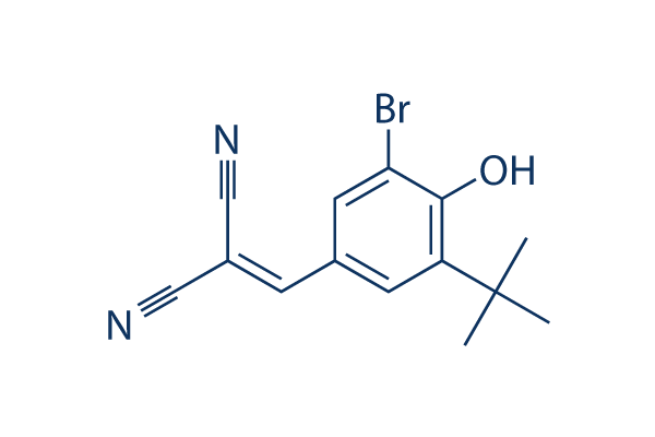
Chemical Structure
Molecular Weight: 305.17
Quality Control
| Related Targets | EGFR VEGFR FGFR PDGFR c-Met Src MEK CSF-1R FLT3 HER2 |
|---|---|
| Other IGF-1R Inhibitors | Linsitinib (OSI-906) BMS-536924 NVP-AEW541 BMS-754807 Picropodophyllin (AXL1717) GSK1904529A NVP-ADW742 NT157 PQ 401 Ginsenoside Rg5 |
Solubility
|
In vitro |
DMSO
: 61 mg/mL
(199.88 mM)
Water : Insoluble Ethanol : Insoluble |
Molarity Calculator
|
In vivo |
|||||
In vivo Formulation Calculator (Clear solution)
Step 1: Enter information below (Recommended: An additional animal making an allowance for loss during the experiment)
Step 2: Enter the in vivo formulation (This is only the calculator, not formulation. Please contact us first if there is no in vivo formulation at the solubility Section.)
Calculation results:
Working concentration: mg/ml;
Method for preparing DMSO master liquid: mg drug pre-dissolved in μL DMSO ( Master liquid concentration mg/mL, Please contact us first if the concentration exceeds the DMSO solubility of the batch of drug. )
Method for preparing in vivo formulation: Take μL DMSO master liquid, next addμL PEG300, mix and clarify, next addμL Tween 80, mix and clarify, next add μL ddH2O, mix and clarify.
Method for preparing in vivo formulation: Take μL DMSO master liquid, next add μL Corn oil, mix and clarify.
Note: 1. Please make sure the liquid is clear before adding the next solvent.
2. Be sure to add the solvent(s) in order. You must ensure that the solution obtained, in the previous addition, is a clear solution before proceeding to add the next solvent. Physical methods such
as vortex, ultrasound or hot water bath can be used to aid dissolving.
Chemical Information, Storage & Stability
| Molecular Weight | 305.17 | Formula | C14H13BrN2O |
Storage (From the date of receipt) | |
|---|---|---|---|---|---|
| CAS No. | 65678-07-1 | Download SDF | Storage of Stock Solutions |
|
|
| Synonyms | Tyrphostin, AGS 200 | Smiles | CC(C)(C)C1=C(C(=CC(=C1)C=C(C#N)C#N)Br)O | ||
Mechanism of Action
| Targets/IC50/Ki |
IGF-1R
(NIH-3T3 fibroblasts ) 7 μM
Insulin Receptor
(NIH-3T3 fibroblasts ) 57 μM
|
|---|---|
| In vitro |
AG-1024 blocks the IGF-1 receptor and IR autophosphorylation with IC50 of 7 μM and 57 μM, respectively. This compound also inhibits the receptor tyrosine kinase activity towards exogenous substrates (TKA) with IC50 values of 18 μM and 80 μM, respectively. This chemical (10 μM) inhibits cell proliferation in a time-dependent manner, and induces apoptosis in MCF-7 cells at 48 hours by 20.1% and >40% when combined with irradiation (10 Gy), more potently than that of irradiation (10 Gy) alone by 11.8%, which is associated with a down-regulation of phospho-Akt1 and bcl-2, and up-regulation of Bax, p53 and p21. It significantly inhibits melanoma cell proliferation with an IC50 of <50 nM in the absence of serum, by blocking MAPK/ERK2 signaling, subsequently rapidly inducing pRb dephosphorylation and activation, and eventually the formation of growth suppressive pRb-E2F complexes. Treatment with this compound down-regulates the expression of Bcr-Abl and P-Akt, and up-regulates DNA-PKcs expression in UT7-9 and Ba/F3-p210 cells, leading to decreased clonogenic survival and proliferation. It also significantly inhibits the proliferation of cells resistant to the BCR-ABL inhibitor STI571, which correlates with a dose-dependent decrease in Bcr-Abl protein expression.
|
| Kinase Assay |
Inhibition of IGF-1- and insulin-stimulated cellular proliferation
|
|
NIH-3T3 cells overexpressing IGF-1 or insulin receptors are plated on 96-well plates (2,000-5,000 cells/well) and maintained overnight in complete medium. Cells are then changed to DMEM containing 1% FBS in the presence of 10 nM IGF-1 or insulin and various concentrations of AG-1024 for 120 hours. Medium is replaced every 48 hours. At the indicated periods of time, the medium is aspirated from the wells and 100 μL MTT is added to each well. The cells are then incubated for 4 hours at 37 °C and lysed by addition of 100 μL isoamylic alcohol and shaking for 20 minutes. The plate is then read in the ELISA reader at 570 and 690 nm. The IC50 value is calculated at the 120-hour time point.
|
|
| In vivo |
Administration of AG-1024 at a dose of 30 μg for 10 days significantly inhibits the tumor growth of Ba/F3-p210 xenograft in mice.
|
References |
|
Tech Support
Tel: +1-832-582-8158 Ext:3
If you have any other enquiries, please leave a message.
Signaling Pathway Map
Products are for research use only. Not for human use. We do not sell to patients.
©Copyright 2013 Selleck Chemicals. All Rights Reserved.