-
 Australia
-
 Austria
-
 Belgium
-
 Brazil
-
 Canada
-
 China
China
-
 Czech Republic
-
 Denmark
-
 Finland
-
 France
-
 Germany
-
 Greece
-
 Hong Kong
-
 Hungary
-
 Iceland
-
 India
India
-
 Ireland
-
 Israel
-
 Italy
Italy
-
 Japan
Japan
-
 Korea
Korea
-
 Luxembourg
-
 Malaysia
-
 Netherlands
-
 New Zealand
-
 Norway
-
 Poland
-
 Qatar
Qatar
-
 Romania
-
 Saudi Arabia
-
 Singapore
-
 Spain
Spain
-
 Sweden
-
 Switzerland
-
 Taiwan
-
 Turkey
-
 United Kingdom
-
 United States
research use only
PP121 PDGFR inhibitor
Cat.No.S2622
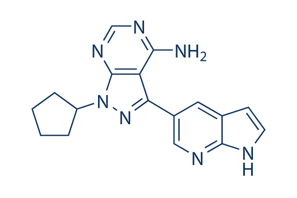
Chemical Structure
Molecular Weight: 319.36
Quality Control
| Related Targets | EGFR VEGFR FGFR c-Met Src MEK CSF-1R HER2 FLT3 c-Kit |
|---|---|
| Other PDGFR Inhibitors | CP-673451 Orantinib (SU6668) Tyrphostin AG 1296 Trapidil AZD2932 Sennoside B Tyrphostin AG1433 AG 1295 N-(p-Coumaroyl) Serotonin Seralutinib (GB002) |
Solubility
|
In vitro |
DMSO
: 32 mg/mL
(100.2 mM)
Ultrasonicated;
Water : Insoluble Ethanol : Insoluble |
Molarity Calculator
|
In vivo |
|||||
In vivo Formulation Calculator (Clear solution)
Step 1: Enter information below (Recommended: An additional animal making an allowance for loss during the experiment)
Step 2: Enter the in vivo formulation (This is only the calculator, not formulation. Please contact us first if there is no in vivo formulation at the solubility Section.)
Calculation results:
Working concentration: mg/ml;
Method for preparing DMSO master liquid: mg drug pre-dissolved in μL DMSO ( Master liquid concentration mg/mL, Please contact us first if the concentration exceeds the DMSO solubility of the batch of drug. )
Method for preparing in vivo formulation: Take μL DMSO master liquid, next addμL PEG300, mix and clarify, next addμL Tween 80, mix and clarify, next add μL ddH2O, mix and clarify.
Method for preparing in vivo formulation: Take μL DMSO master liquid, next add μL Corn oil, mix and clarify.
Note: 1. Please make sure the liquid is clear before adding the next solvent.
2. Be sure to add the solvent(s) in order. You must ensure that the solution obtained, in the previous addition, is a clear solution before proceeding to add the next solvent. Physical methods such
as vortex, ultrasound or hot water bath can be used to aid dissolving.
Chemical Information, Storage & Stability
| Molecular Weight | 319.36 | Formula | C17H17N7 |
Storage (From the date of receipt) | |
|---|---|---|---|---|---|
| CAS No. | 1092788-83-4 | Download SDF | Storage of Stock Solutions |
|
|
| Synonyms | N/A | Smiles | C1CCC(C1)N2C3=NC=NC(=C3C(=N2)C4=CN=C5C(=C4)C=CN5)N | ||
Mechanism of Action
| Targets/IC50/Ki |
PDGFR
2 nM
Hck
8 nM
VEGFR2
12 nM
mTOR
13 nM
Src
14 nM
Abl
18 nM
p110α
52 nM
DNA-PK
60 nM
p110δ
150 nM
EphB4
190 nM
|
|---|---|
| In vitro |
PP-121 selectivity interacts within a hydrophobic pocket that is conserved between both tyrosine kinases and PI3Ks, not serine-threonine kinases. PP-121 makes a hydrogen bond to Glu310 in Src, effectively substituting for the structural role of the catalytic lysine and resulting in the ordering of helix C and stabilization of an active conformation. PP-121 also inhibits other PI3Ks including p110α and DNA-PK with IC50 of 52 nM and 60 nM, respectively. PP-121 potently and dose-dependently blocks the phosphorylation of Akt, p70S6K and S6 in two glioblastoma cell lines, U87 and LN229. PP-121 potently inhibits the proliferation of a subset of the tumor cell lines by direct inhibition of PI3Ks and mTOR. PP-121 induces a G0/G1 arrest in LN220, U87 and Seg1 cells. PP-121 also blocks tyrosine phosphorylation induced by v-Src in NIH3T3 cells transformed with v-Src(Thr338). PP-121 could restore actin stress fiber staining in NIH3T3 cells transformed with v-Src(Thr338). PP-121 at a low concentration of 40 nM inhibits Ret autophosphorylation in TT thyroid carcinoma cells that express the C634W oncogenic Ret mutant35. PP-121 inhibits cell proliferation with IC50 of 50 nM in TT thyroid carcinoma cells. PP-121 inhibits cell proliferating stimulated only with VEGF with IC50 of 41 nM in human umbilical vein endothelial cells (HUVECs). PP-121 directly inhibits Bcr-Abl induced tyrosine phosphorylation, resulting in drug-induced apoptosis in K562 cells and a combination of apoptosis and cell cycle arrest in Bcr-Abl expressing BaF3 cells. |
| Kinase Assay |
Kinase assays
|
|
Purified kinase domains are incubated with PP-121 at 2- or 4-fold dilutions over a concentration range of 1nM-50 µM or with vehicle (0.1% DMSO) in the presence of 10 µM ATP, 2.5 µCi of γ-32P-ATP and substrate. Reactions are terminated by spotting onto nitrocellulose or phosphocellulose membranes, depending on the substrate; this membrane is then washed 5–6 times to remove unbound radioactivity and dried. Transferred radioactivity is quantitated by phosphorimaging and IC50 values are calculated by fitting the data to a sigmoidal doseresponse using Prism software.
|
References |
Tech Support
Tel: +1-832-582-8158 Ext:3
If you have any other enquiries, please leave a message.
Signaling Pathway Map
Products are for research use only. Not for human use. We do not sell to patients.
©Copyright 2013 Selleck Chemicals. All Rights Reserved.