-
 Australia
-
 Austria
-
 Belgium
-
 Brazil
-
 Canada
-
 China
China
-
 Czech Republic
-
 Denmark
-
 Finland
-
 France
-
 Germany
-
 Greece
-
 Hong Kong
-
 Hungary
-
 Iceland
-
 India
India
-
 Ireland
-
 Israel
-
 Italy
Italy
-
 Japan
Japan
-
 Korea
Korea
-
 Luxembourg
-
 Malaysia
-
 Netherlands
-
 New Zealand
-
 Norway
-
 Poland
-
 Qatar
Qatar
-
 Romania
-
 Saudi Arabia
-
 Singapore
-
 Spain
Spain
-
 Sweden
-
 Switzerland
-
 Taiwan
-
 Turkey
-
 United Kingdom
-
 United States
research use only
GNF-2 Bcr-Abl inhibitor
Cat.No.S2899
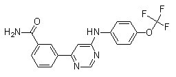
Chemical Structure
Molecular Weight: 374.32
Quality Control
| Related Targets | EGFR VEGFR JAK FGFR PDGFR Src HIF FLT3 FLT HER2 |
|---|---|
| Other Bcr-Abl Inhibitors | Degrasyn (WP1130) Bafetinib (INNO-406) Rebastinib (DCC-2036) GNF-5 Berbamine Olverembatinib dimesylate (GZD824) PD173955 GNF-7 Berbamine dihydrochloride Radotinib |
Solubility
|
In vitro |
DMSO
: 74 mg/mL
(197.69 mM)
Water : Insoluble Ethanol : Insoluble |
Molarity Calculator
|
In vivo |
|||||
In vivo Formulation Calculator (Clear solution)
Step 1: Enter information below (Recommended: An additional animal making an allowance for loss during the experiment)
Step 2: Enter the in vivo formulation (This is only the calculator, not formulation. Please contact us first if there is no in vivo formulation at the solubility Section.)
Calculation results:
Working concentration: mg/ml;
Method for preparing DMSO master liquid: mg drug pre-dissolved in μL DMSO ( Master liquid concentration mg/mL, Please contact us first if the concentration exceeds the DMSO solubility of the batch of drug. )
Method for preparing in vivo formulation: Take μL DMSO master liquid, next addμL PEG300, mix and clarify, next addμL Tween 80, mix and clarify, next add μL ddH2O, mix and clarify.
Method for preparing in vivo formulation: Take μL DMSO master liquid, next add μL Corn oil, mix and clarify.
Note: 1. Please make sure the liquid is clear before adding the next solvent.
2. Be sure to add the solvent(s) in order. You must ensure that the solution obtained, in the previous addition, is a clear solution before proceeding to add the next solvent. Physical methods such
as vortex, ultrasound or hot water bath can be used to aid dissolving.
Chemical Information, Storage & Stability
| Molecular Weight | 374.32 | Formula | C18H13F3N4O2 |
Storage (From the date of receipt) | |
|---|---|---|---|---|---|
| CAS No. | 778270-11-4 | Download SDF | Storage of Stock Solutions |
|
|
| Synonyms | N/A | Smiles | C1=CC(=CC(=C1)C(=O)N)C2=CC(=NC=N2)NC3=CC=C(C=C3)OC(F)(F)F | ||
Mechanism of Action
| Targets/IC50/Ki |
Bcr-Abl (SUP-B15 cell line)
268 nM
Bcr-Abl (K562 cell line)
273 nM
|
|---|---|
| In vitro |
GNF-2 causes a dose-dependent growth inhibition of the Bcr-abl–positive cell lines with IC50 values of 273 nM (K562) and 268 nM (SUP-B15). This compound inhibits the growth of Ba/F3.p210E255V and Ba/F3.p185Y253H cells with IC50 values of 268 nM and 194 nM respectively. This chemical (1 μM) induces apoptosis of Ba/F3.p210 cells as well as Ba/F3.p210E255V cells. It inhibits the cellular tyrosine phosphorylation of Bcr-abl in a dose-dependent manner with IC50 of 267 nM. This compound (1 μM) induces a significant decrease in the levels of phospho-Stat5 in Ba/F3.p210 cells. It binds to the myristic binding pocket of Bcr-abl. This inhibitor inhibits the kinase activity of non-myristoylated c-Abl more potently than that of myristoylated c-Abl by binding to the myristate-binding pocket in the C-lobe of the kinase domain. This agent (10 μM) requires BCR and/or the c-Abl SH3 and/or SH2 domains to inhibit BCR-Abl-dependent cell proliferation. It, but not a methylated GNF-2 analog, binds c-Abl in cellular extracts derived from 3T3 fibroblasts. This compound (10 μM), in a dose-dependent manner, clearly inhibits tyrosine phosphorylation of CrkII. It inhibits the phosphorylation of CrkII in c-AblG2A-expressing cells with IC50 of 0.051 μM. This chemical binds in an extended conformation in the myristate pocket with the CF3-group buried at the same depth as the final two carbons of the myristate ligand. This compound (10 µM) combined with imatinib (1 µM) reduces the number of resistant clones to 1 µM imatinib by at least 90%. It inhibits the auto-phosphorylation and proliferation of BafF3 cells expressing p210Bcr–Abl and p210Bcr–Abl mutants. This agent (8 nM) in combination with GNF-5 (20 nM) results in additive effects with respect to inhibition of the Abl64–515 kinase activity.
|
| Kinase Assay |
Binding assay
|
|
Recombinant proteins (100 nM for each construct) or immunoprecipitated proteins are diluted in kinase buffer (20 mM HEPES (pH 7.4), 50 mM KCl, 0.1% CHAPS, 30 mM MgCl2, 2 mM MnCl2, 1 mM DTT, and 1% glycerol). Aliquots of the diluted proteins are preincubated with either DMSO or this compound for 30 min at room temperature and then added to K-LISA PTK EAY reaction plates. The kinase reaction is initiated by adding 0.1 mM ATP and is allowed to proceed for 30 min at room temperature. The phosphorylation of GST-Abltide is monitored by SDS-PAGE and phosphorimaging analysis or autoradiography.
|
References |
|
Tech Support
Tel: +1-832-582-8158 Ext:3
If you have any other enquiries, please leave a message.
Signaling Pathway Map
Products are for research use only. Not for human use. We do not sell to patients.
©Copyright 2013 Selleck Chemicals. All Rights Reserved.