
- Bioactive Compounds
- By Signaling Pathways
- PI3K/Akt/mTOR
- Epigenetics
- Methylation
- Immunology & Inflammation
- Protein Tyrosine Kinase
- Angiogenesis
- Apoptosis
- Autophagy
- ER stress & UPR
- JAK/STAT
- MAPK
- Cytoskeletal Signaling
- Cell Cycle
- TGF-beta/Smad
- DNA Damage/DNA Repair
- Compound Libraries
- Antibodies
- Bioreagents
- qPCR
- 2x SYBR Green qPCR Master Mix
- 2x SYBR Green qPCR Master Mix(Low ROX)
- 2x SYBR Green qPCR Master Mix(High ROX)
- Protein Assay
- Protein A/G Magnetic Beads for IP
- Anti-Flag magnetic beads
- Anti-Flag Affinity Gel
- Anti-Myc magnetic beads
- Anti-HA magnetic beads
- Magnetic Separator
- Poly DYKDDDDK Tag Peptide lyophilized powder
- Protease Inhibitor Cocktail
- Protease Inhibitor Cocktail (EDTA-Free, 100X in DMSO)
- Phosphatase Inhibitor Cocktail (2 Tubes, 100X)
- Cell Biology
- Cell Counting Kit-8 (CCK-8)
- Animal Experiment
- Mouse Direct PCR Kit (For Genotyping)
- New Products
- Contact Us
-
 Australia
-
 Austria
-
 Belgium
-
 Canada
-
 China
China
-
 Czech Republic
-
 Denmark
-
 Finland
-
 France
-
 Germany
-
 Greece
-
 Hong Kong
-
 Hungary
-
 Iceland
-
 India
India
-
 Ireland
-
 Israel
-
 Italy
Italy
-
 Japan
Japan
-
 Korea
Korea
-
 Luxembourg
-
 Malaysia
-
 Netherlands
-
 New Zealand
-
 Norway
-
 Poland
-
 Qatar
Qatar
-
 Romania
-
 Saudi Arabia
-
 Singapore
-
 Spain
Spain
-
 Sweden
-
 Switzerland
-
 Taiwan
-
 Turkey
-
 United Kingdom
-
 United States
-
 Other Countries
Anti-TSH Receptor/TSH-R Rabbit Antibody [L10M22]
Catalog No.: F3696
Application:
Reactivity:
-
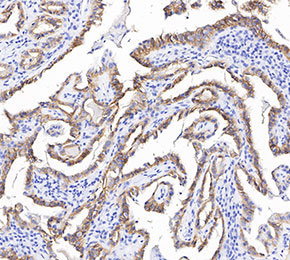 Immunohistochemical analysis of formalin fixed paraffin embedded human thyroid gland tissue with F3696 at 1:1000 dilution.
Usage Information
| Dilution |
|---|
|
| Application |
|---|
| IHC |
| Reactivity |
|---|
| Human |
| Source |
|---|
| Rabbit |
| Storage Buffer |
|---|
| PBS, pH 7.2+50% Glycerol+0.05% BSA+0.01% NaN3 |
| Storage (from the date of receipt) |
|---|
| -20°C (avoid freeze-thaw cycles), 2 years |
| Positive Control | Human thyroid; Human follicular thyroid adenocarcinoma; Human papillary thyroid adenocarcinoma |
|---|---|
| Negative Control | Human colon; Human gastric cancer |
Exprimental Methods
| IHC |
|---|
Experimental Protocol:
Deparaffinization/Rehydration
1. Deparaffinize/hydrate sections:
2. Incubate sections in three washes of xylene for 5 min each.
3. Incubate sections in two washes of 100% ethanol for 10 min each.
4. Incubate sections in two washes of 95% ethanol for 10 min each.
5. Wash sections two times in dH2O for 5 min each.
6.Antigen retrieval: For Citrate: Heat slides in a microwave submersed in 1X citrate unmasking solution until boiling is initiated; continue with 10 min at a sub-boiling temperature (95°-98°C). Cool slides on bench top for 30 min.
Staining
1. Wash sections in dH2O three times for 5 min each.
2. Incubate sections in 3% hydrogen peroxide for 10 min.
3. Wash sections in dH2O two times for 5 min each.
4. Wash sections in wash buffer for 5 min.
5. Block each section with 100–400 µl of blocking solution for 1 hr at room temperature.
6. Remove blocking solution and add 100–400 µl primary antibody diluent in to each section. Incubate overnight at 4°C.
7. Remove antibody solution and wash sections with wash buffer three times for 5 min each.
8. Cover section with 1–3 drops HRPas needed. Incubate in a humidified chamber for 30 min at room temperature.
9. Wash sections three times with wash buffer for 5 min each.
10. Add DAB Chromogen Concentrate to DAB Diluent and mix well before use.
11. Apply 100–400 µl DAB to each section and monitor closely. 1–10 min generally provides an acceptable staining intensity.
12. Immerse slides in dH2O.
13. If desired, counterstain sections with hematoxylin.
14. Wash sections in dH2O two times for 5 min each.
15. Dehydrate sections: Incubate sections in 95% ethanol two times for 10 sec each; Repeat in 100% ethanol, incubating sections two times for 10 sec each; Repeat in xylene, incubating sections two times for 10 sec each.
16. Mount sections with coverslips and mounting medium.
|
Biological Description
| Specificity |
|---|
| TSH Receptor/TSH-R Rabbit mAb detects endogenous levels of total TSH Receptor/TSH-R protein. |
| Subcellular Location |
|---|
| Cell membrane, Membrane |
| Uniprot ID |
|---|
| P16473 |
| Clone |
|---|
| L10M22 |
| Synonym(s) |
|---|
| LGR3, TSHR, Thyrotropin receptor, Thyroid-stimulating hormone receptor, TSH-R |
| Background |
|---|
| The thyroid-stimulating hormone receptor (TSHR) is a key regulator of thyroid hormone metabolism and serves as a primary controller of thyroid cell function and growth. It belongs to the family of G-protein-coupled receptors with seven transmembrane domains and is positioned on the basolateral membrane of thyroid follicular cells. The TSHR protein is composed of 764 amino acids, has a molecular weight of approximately 87 kDa, and mediates its effects through interaction with multiple G protein subtypes—most notably Gαs and Gαq. Upon activation by TSH, the receptor initiates intracellular signaling through these G proteins, thereby modulating the activity of downstream effector molecules. The Gαs pathway stimulates the cyclic adenosine monophosphate (cAMP) cascade, while the Gαq pathway activates the phospholipase C (PLC) cascade. At elevated TSH concentrations, cAMP binds to protein kinase A (PKA), which phosphorylates various target effectors, enhancing their catalytic activity. In parallel, PLC activation generates inositol 1,4,5-triphosphate (IP₃) and diacylglycerol (DAG), further amplifying cellular responses. TSHR expression is positively regulated by physiological TSH levels but downregulated at persistently high TSH concentrations. Chronic overstimulation of TSHR, particularly through the cAMP pathway, can lead to excessive thyroid hormone secretion, thyroid follicular hyperplasia, and clinical hyperthyroidism. Mutations in the TSHR gene may affect the receptor’s protein structure or its post-translational modifications, thereby altering receptor function. Although TSHR does not directly initiate carcinogenesis, it can significantly contribute to tumor growth when oncogenes are already activated. |
| References |
|---|
|
Tech Support
Answers to questions you may have can be found in the inhibitor handling instructions. Topics include how to prepare stock solutions, how to store inhibitors, and issues that need special attention for cell-based assays and animal experiments.
Tel: +1-832-582-8158 Ext:3
If you have any other enquiries, please leave a message.
* Indicates a Required Field
Products are for research use only. Not for human use. We do not sell to patients.
©Copyright 2013 Selleck Chemicals. All Rights Reserved.