|
Toll Free: (877) 796-6397 -- USA and Canada only -- |
Fax: +1-832-582-8590 Orders: +1-832-582-8158 |
Tech Support: +1-832-582-8158 Ext:3 Please provide your Order Number in the email. |
Biological Description
| Specificity | Anti-TSH Receptor/TSH-R Rabbit Antibody [L10M22] detects endogenous levels of total TSH Receptor/TSH-R protein. |
|---|---|
| Background | The thyroid-stimulating hormone receptor (TSHR) is a key regulator of thyroid hormone metabolism and serves as a primary controller of thyroid cell function and growth. It belongs to the family of G-protein-coupled receptors with seven transmembrane domains and is positioned on the basolateral membrane of thyroid follicular cells. The TSHR protein is composed of 764 amino acids, has a molecular weight of approximately 87 kDa, and mediates its effects through interaction with multiple G protein subtypes—most notably Gαs and Gαq. Upon activation by TSH, the receptor initiates intracellular signaling through these G proteins, thereby modulating the activity of downstream effector molecules. The Gαs pathway stimulates the cyclic adenosine monophosphate (cAMP) cascade, while the Gαq pathway activates the phospholipase C (PLC) cascade. At elevated TSH concentrations, cAMP binds to protein kinase A (PKA), which phosphorylates various target effectors, enhancing their catalytic activity. In parallel, PLC activation generates inositol 1,4,5-triphosphate (IP₃) and diacylglycerol (DAG), further amplifying cellular responses. TSHR expression is positively regulated by physiological TSH levels but downregulated at persistently high TSH concentrations. Chronic overstimulation of TSHR, particularly through the cAMP pathway, can lead to excessive thyroid hormone secretion, thyroid follicular hyperplasia, and clinical hyperthyroidism. Mutations in the TSHR gene may affect the receptor’s protein structure or its post-translational modifications, thereby altering receptor function. Although TSHR does not directly initiate carcinogenesis, it can significantly contribute to tumor growth when oncogenes are already activated. |
Usage Information
| Application | IHC | Dilution |
|
||
|---|---|---|---|---|---|
| Reactivity | Human | ||||
| Source | Rabbit | MW | |||
| Storage Buffer | PBS, pH 7.2+50% Glycerol+0.05% BSA+0.01% NaN3 | Storage (from the date of receipt) |
-20°C (avoid freeze-thaw cycles), 2 years | ||
| IHC |
Experimental Protocol:
Deparaffinization/Rehydration
1. Deparaffinize/hydrate sections:
2. Incubate sections in three washes of xylene for 5 min each.
3. Incubate sections in two washes of 100% ethanol for 10 min each.
4. Incubate sections in two washes of 95% ethanol for 10 min each.
5. Wash sections two times in dH2O for 5 min each.
6.Antigen retrieval: For Citrate: Heat slides in a microwave submersed in 1X citrate unmasking solution until boiling is initiated; continue with 10 min at a sub-boiling temperature (95°-98°C). Cool slides on bench top for 30 min.
Staining
1. Wash sections in dH2O three times for 5 min each.
2. Incubate sections in 3% hydrogen peroxide for 10 min.
3. Wash sections in dH2O two times for 5 min each.
4. Wash sections in wash buffer for 5 min.
5. Block each section with 100–400 µl of blocking solution for 1 hr at room temperature.
6. Remove blocking solution and add 100–400 µl primary antibody diluent in to each section. Incubate overnight at 4°C.
7. Remove antibody solution and wash sections with wash buffer three times for 5 min each.
8. Cover section with 1–3 drops HRPas needed. Incubate in a humidified chamber for 30 min at room temperature.
9. Wash sections three times with wash buffer for 5 min each.
10. Add DAB Chromogen Concentrate to DAB Diluent and mix well before use.
11. Apply 100–400 µl DAB to each section and monitor closely. 1–10 min generally provides an acceptable staining intensity.
12. Immerse slides in dH2O.
13. If desired, counterstain sections with hematoxylin.
14. Wash sections in dH2O two times for 5 min each.
15. Dehydrate sections: Incubate sections in 95% ethanol two times for 10 sec each; Repeat in 100% ethanol, incubating sections two times for 10 sec each; Repeat in xylene, incubating sections two times for 10 sec each.
16. Mount sections with coverslips and mounting medium.
|
References
|
Application Data
IHC
Validated by Selleck
-
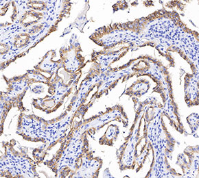 Immunohistochemical analysis of formalin fixed paraffin embedded human thyroid gland tissue with F3696 at 1:1000 dilution.
IHC
Validated by Selleck
-
Immunohistochemical analysis of formalin fixed paraffin embedded human thyroid gland tissue with F3696 at 1:1000 dilution.