
- Bioactive Compounds
- By Signaling Pathways
- PI3K/Akt/mTOR
- Epigenetics
- Methylation
- Immunology & Inflammation
- Protein Tyrosine Kinase
- Angiogenesis
- Apoptosis
- Autophagy
- ER stress & UPR
- JAK/STAT
- MAPK
- Cytoskeletal Signaling
- Cell Cycle
- TGF-beta/Smad
- DNA Damage/DNA Repair
- Compound Libraries
- Antibodies
- Bioreagents
- qPCR
- 2x SYBR Green qPCR Master Mix
- 2x SYBR Green qPCR Master Mix(Low ROX)
- 2x SYBR Green qPCR Master Mix(High ROX)
- Protein Assay
- Protein A/G Magnetic Beads for IP
- Anti-Flag magnetic beads
- Anti-Flag Affinity Gel
- Anti-Myc magnetic beads
- Anti-HA magnetic beads
- Magnetic Separator
- Poly DYKDDDDK Tag Peptide lyophilized powder
- Protease Inhibitor Cocktail
- Protease Inhibitor Cocktail (EDTA-Free, 100X in DMSO)
- Phosphatase Inhibitor Cocktail (2 Tubes, 100X)
- Cell Biology
- Cell Counting Kit-8 (CCK-8)
- Animal Experiment
- Mouse Direct PCR Kit (For Genotyping)
- New Products
- Contact Us
-
 Australia
-
 Austria
-
 Belgium
-
 Canada
-
 China
China
-
 Czech Republic
-
 Denmark
-
 Finland
-
 France
-
 Germany
-
 Greece
-
 Hong Kong
-
 Hungary
-
 Iceland
-
 India
India
-
 Ireland
-
 Israel
-
 Italy
Italy
-
 Japan
Japan
-
 Korea
Korea
-
 Luxembourg
-
 Malaysia
-
 Netherlands
-
 New Zealand
-
 Norway
-
 Poland
-
 Qatar
Qatar
-
 Romania
-
 Saudi Arabia
-
 Singapore
-
 Spain
Spain
-
 Sweden
-
 Switzerland
-
 Taiwan
-
 Turkey
-
 United Kingdom
-
 United States
-
 Other Countries
Anti-RANK Mouse Antibody [E4N12]
Catalog No.: F2425
Application:
Reactivity:
-
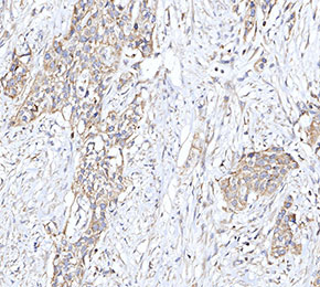 Immunohistochemical analysis of formalin fixed paraffin embedded human gastric cancer tissue with F2425 at 1:50 dilution.,
 -
Immunohistochemical analysis of formalin fixed paraffin embedded human gastric cancer tissue with F2425 at 1:50 dilution.,
Usage Information
| Dilution |
|---|
|
| Application |
|---|
| IP, IHC |
| Reactivity |
|---|
| Human, Mouse |
| Source |
|---|
| Mouse |
| Storage Buffer |
|---|
| PBS, pH 7.2+50% Glycerol+0.05% BSA+0.01% NaN3 |
| Storage (from the date of receipt) |
|---|
| -20°C (avoid freeze-thaw cycles), 2 years |
| Positive Control | Human skin; Human t cell lymphoma; RAW cell |
|---|---|
| Negative Control |
Exprimental Methods
| IHC |
|---|
Experimental Protocol:
Deparaffinization/Rehydration
1. Deparaffinize/hydrate sections:
2. Incubate sections in three washes of xylene for 5 min each.
3. Incubate sections in two washes of 100% ethanol for 10 min each.
4. Incubate sections in two washes of 95% ethanol for 10 min each.
5. Wash sections two times in dH2O for 5 min each.
6.Antigen retrieval: For Citrate: Heat slides in a microwave submersed in 1X citrate unmasking solution until boiling is initiated; continue with 10 min at a sub-boiling temperature (95°-98°C). Cool slides on bench top for 30 min.
Staining
1. Wash sections in dH2O three times for 5 min each.
2. Incubate sections in 3% hydrogen peroxide for 10 min.
3. Wash sections in dH2O two times for 5 min each.
4. Wash sections in wash buffer for 5 min.
5. Block each section with 100–400 µl of blocking solution for 1 hr at room temperature.
6. Remove blocking solution and add 100–400 µl primary antibody diluent in to each section. Incubate overnight at 4°C.
7. Remove antibody solution and wash sections with wash buffer three times for 5 min each.
8. Cover section with 1–3 drops HRPas needed. Incubate in a humidified chamber for 30 min at room temperature.
9. Wash sections three times with wash buffer for 5 min each.
10. Add DAB Chromogen Concentrate to DAB Diluent and mix well before use.
11. Apply 100–400 µl DAB to each section and monitor closely. 1–10 min generally provides an acceptable staining intensity.
12. Immerse slides in dH2O.
13. If desired, counterstain sections with hematoxylin.
14. Wash sections in dH2O two times for 5 min each.
15. Dehydrate sections: Incubate sections in 95% ethanol two times for 10 sec each; Repeat in 100% ethanol, incubating sections two times for 10 sec each; Repeat in xylene, incubating sections two times for 10 sec each.
16. Mount sections with coverslips and mounting medium.
|
Biological Description
| Specificity |
|---|
Anti-RANK Mouse Antibody [E4N12] detects endogenous levels of total RANK protein. |
| Subcellular Location |
|---|
| Cell membrane, Membrane |
| Uniprot ID |
|---|
| Q9Y6Q6 |
| Clone |
|---|
| E4N12 |
| Synonym(s) |
|---|
| CD265, RANK, TNFRSF11A, Tumor necrosis factor receptor superfamily member 11A, Osteoclast differentiation factor receptor, Receptor activator of NF-KB, ODFR |
| Background |
|---|
| RANK (Receptor Activator of Nuclear Factor κB) is a type I transmembrane glycoprotein of 616 amino acids, comprising a 28-amino-acid signal peptide, a 184-amino-acid extracellular domain, a 21-amino-acid transmembrane region, and a 383-amino-acid intracellular domain containing three functional cytoplasmic motifs—PFQEP(369–373), PVQEET(559–564), and PVQEQG(604–609)—that mediate interactions with adaptor proteins such as TRAF6 to activate NF-κB, MAPK (JNK, ERK, p38), and Akt/PKB pathways. Encoded by the TNFRSF11A gene, RANK is expressed in osteoclast precursors, dendritic cells, monocytes, macrophages, activated T cells, natural killer cells, thymic medullary epithelial cells, mammary epithelial cells, and certain epithelial stem cell populations. Functionally, RANK regulates osteoclast differentiation, activation, and survival in bone remodeling; supports lymph node organogenesis, thymic epithelial development, and immune tolerance; maintains epithelial stem cell homeostasis; mediates progesterone-driven mammary epithelial proliferation; and, when dysregulated, contributes to tumorigenesis, metastasis, and cancer stem cell expansion. |
| References |
|---|
|
Tech Support
Answers to questions you may have can be found in the inhibitor handling instructions. Topics include how to prepare stock solutions, how to store inhibitors, and issues that need special attention for cell-based assays and animal experiments.
Tel: +1-832-582-8158 Ext:3
If you have any other enquiries, please leave a message.
* Indicates a Required Field
Products are for research use only. Not for human use. We do not sell to patients.
©Copyright 2013 Selleck Chemicals. All Rights Reserved.