- Bioactive Compounds
- By Signaling Pathways
- PI3K/Akt/mTOR
- Epigenetics
- Methylation
- Immunology & Inflammation
- Protein Tyrosine Kinase
- Angiogenesis
- Apoptosis
- Autophagy
- ER stress & UPR
- JAK/STAT
- MAPK
- Cytoskeletal Signaling
- Cell Cycle
- TGF-beta/Smad
- DNA Damage/DNA Repair
- Compound Libraries
- Popular Compound Libraries
- Customize Library
- Clinical and FDA-approved Related
- Bioactive Compound Libraries
- Inhibitor Related
- Natural Product Related
- Metabolism Related
- Cell Death Related
- By Signaling Pathway
- By Disease
- Anti-infection and Antiviral Related
- Neuronal and Immunology Related
- Fragment and Covalent Related
- FDA-approved Drug Library
- FDA-approved & Passed Phase I Drug Library
- Preclinical/Clinical Compound Library
- Bioactive Compound Library-I
- Bioactive Compound Library-Ⅱ
- Kinase Inhibitor Library
- Express-Pick Library
- Natural Product Library
- Human Endogenous Metabolite Compound Library
- Alkaloid Compound LibraryNew
- Angiogenesis Related compound Library
- Anti-Aging Compound Library
- Anti-alzheimer Disease Compound Library
- Antibiotics compound Library
- Anti-cancer Compound Library
- Anti-cancer Compound Library-Ⅱ
- Anti-cancer Metabolism Compound Library
- Anti-Cardiovascular Disease Compound Library
- Anti-diabetic Compound Library
- Anti-infection Compound Library
- Antioxidant Compound Library
- Anti-parasitic Compound Library
- Antiviral Compound Library
- Apoptosis Compound Library
- Autophagy Compound Library
- Calcium Channel Blocker LibraryNew
- Cambridge Cancer Compound Library
- Carbohydrate Metabolism Compound LibraryNew
- Cell Cycle compound library
- CNS-Penetrant Compound Library
- Covalent Inhibitor Library
- Cytokine Inhibitor LibraryNew
- Cytoskeletal Signaling Pathway Compound Library
- DNA Damage/DNA Repair compound Library
- Drug-like Compound Library
- Endoplasmic Reticulum Stress Compound Library
- Epigenetics Compound Library
- Exosome Secretion Related Compound LibraryNew
- FDA-approved Anticancer Drug LibraryNew
- Ferroptosis Compound Library
- Flavonoid Compound Library
- Fragment Library
- Glutamine Metabolism Compound Library
- Glycolysis Compound Library
- GPCR Compound Library
- Gut Microbial Metabolite Library
- HIF-1 Signaling Pathway Compound Library
- Highly Selective Inhibitor Library
- Histone modification compound library
- HTS Library for Drug Discovery
- Human Hormone Related Compound LibraryNew
- Human Transcription Factor Compound LibraryNew
- Immunology/Inflammation Compound Library
- Inhibitor Library
- Ion Channel Ligand Library
- JAK/STAT compound library
- Lipid Metabolism Compound LibraryNew
- Macrocyclic Compound Library
- MAPK Inhibitor Library
- Medicine Food Homology Compound Library
- Metabolism Compound Library
- Methylation Compound Library
- Mouse Metabolite Compound LibraryNew
- Natural Organic Compound Library
- Neuronal Signaling Compound Library
- NF-κB Signaling Compound Library
- Nucleoside Analogue Library
- Obesity Compound Library
- Oxidative Stress Compound LibraryNew
- Plant Extract Library
- Phenotypic Screening Library
- PI3K/Akt Inhibitor Library
- Protease Inhibitor Library
- Protein-protein Interaction Inhibitor Library
- Pyroptosis Compound Library
- Small Molecule Immuno-Oncology Compound Library
- Mitochondria-Targeted Compound LibraryNew
- Stem Cell Differentiation Compound LibraryNew
- Stem Cell Signaling Compound Library
- Natural Phenol Compound LibraryNew
- Natural Terpenoid Compound LibraryNew
- TGF-beta/Smad compound library
- Traditional Chinese Medicine Library
- Tyrosine Kinase Inhibitor Library
- Ubiquitination Compound Library
-
Cherry Picking
You can personalize your library with chemicals from within Selleck's inventory. Build the right library for your research endeavors by choosing from compounds in all of our available libraries.
Please contact us at info@selleckchem.com to customize your library.
You could select:
- Antibodies
- Bioreagents
- qPCR
- 2x SYBR Green qPCR Master Mix
- 2x SYBR Green qPCR Master Mix(Low ROX)
- 2x SYBR Green qPCR Master Mix(High ROX)
- Protein Assay
- Protein A/G Magnetic Beads for IP
- Anti-Flag magnetic beads
- Anti-Flag Affinity Gel
- Anti-Myc magnetic beads
- Anti-HA magnetic beads
- Poly DYKDDDDK Tag Peptide lyophilized powder
- Protease Inhibitor Cocktail
- Protease Inhibitor Cocktail (EDTA-Free, 100X in DMSO)
- Phosphatase Inhibitor Cocktail (2 Tubes, 100X)
- Cell Biology
- Cell Counting Kit-8 (CCK-8)
- Animal Experiment
- Mouse Direct PCR Kit (For Genotyping)
- New Products
- Contact Us
research use only
BIO GSK-3 inhibitor
BIO (GSK-3 Inhibitor IX, 6-bromoindirubin-3-oxime, 6-Bromoindirubin-3'-oxime, MLS 2052) is a specific inhibitor of GSK-3 with IC50 of 5 nM for GSK-3α/β in a cell-free assay, shows >16-fold selectivity over CDK5, also a pan-JAK inhibitor with IC50 of 30 nM for Tyk2. This compound induces apoptosis in human melanoma cells.
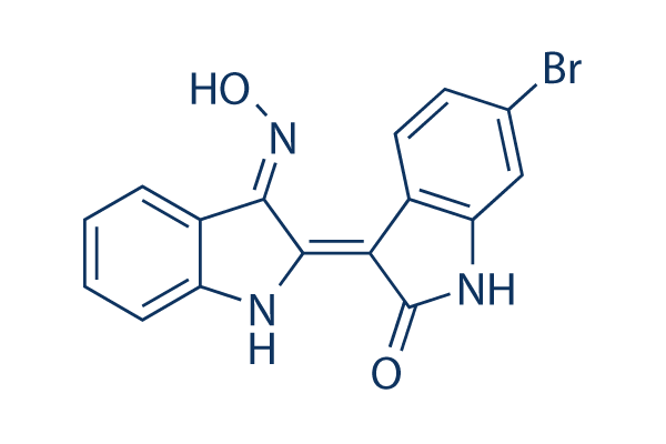
Chemical Structure
Molecular Weight: 356.17
Purity & Quality Control
Batch:
Purity:
99.93%
99.93
Related Products
| Related Targets | GSK-3α GSK-3β | Click to Expand |
|---|---|---|
| Related Products | CHIR-99021 (Laduviglusib) Laduviglusib (CHIR-99021) Hydrochloride SB216763 TWS119 CHIR-98014 LY2090314 Tideglusib SB415286 AR-A014418 1-Azakenpaullone IM-12 CP21R7 TDZD-8 Indirubin (NSC 105327) AZD2858 AZD1080 BIO-acetoxime Bikinin | Click to Expand |
| Related Compound Libraries | Kinase Inhibitor Library PI3K/Akt Inhibitor Library Apoptosis Compound Library Cell Cycle compound library NF-κB Signaling Compound Library | Click to Expand |
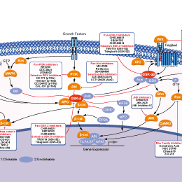
Signaling Pathway
Cell Culture and Working Concentration
| Cell Lines | Assay Type | Concentration | Incubation Time | Formulation | Activity Description | PMID |
|---|---|---|---|---|---|---|
| HEI-OC1 | Function assay | Protection against cisplatin-induced cell death in neonatal mouse HEI-OC1 cells assessed as reduction in caspase-3/7 activity, EC50 = 0.192 μM. | 30091915 | |||
| SH-SY5Y | Function assay | Inhibition of GSK3-mediated beta-casein phosphorylation in human SH-SY5Y cells in presence of MG132 by Western blot analysis, IC50 = 0.29 μM. | 18816110 | |||
| K562 | Antiproliferative assay | 72 hrs | Antiproliferative activity against human K562 cells after 72 hrs by MTT assay, IC50 = 1.3 μM. | 19783149 | ||
| IMR90 | Antiproliferative assay | 72 hrs | Antiproliferative activity against human IMR90 cells after 72 hrs by MTT assay, IC50 = 1.9 μM. | 19783149 | ||
| HCT116 | Antiproliferative assay | 72 hrs | Antiproliferative activity against human HCT116 cells after 72 hrs by MTT assay, IC50 = 5.2 μM. | 19783149 | ||
| HepG2 | Cytotoxicity assay | 24 hrs | Cytotoxicity against human HepG2 cells assessed as cell growth inhibition after 24 hrs by alamar blue assay, IC50 = 5.3 μM. | 28743492 | ||
| HL60 | Antiproliferative assay | 5 days | Antiproliferative activity against human HL60 cells after 5 days by MTT assay, IC50 = 5.4 μM. | 19783149 | ||
| HuH7 | Antiproliferative assay | 72 hrs | Antiproliferative activity against human HuH7 cells after 72 hrs by MTT assay, IC50 = 6.2 μM. | 19783149 | ||
| SH-SY5Y | Cytotoxicity assay | 48 hrs | Cytotoxicity against human SH-SY5Y cells after 48 hrs by MTS reduction assay, IC50 = 9 μM. | 18816110 | ||
| SH-SY5Y | Function assay | 48 hrs | Survival of human SH-SY5Y cells after 48 hrs by MTS reduction assay, IC50 = 9.5 μM. | 16854069 | ||
| SH-SY5Y | Function assay | Death of human SH-SY5Y cells in absence of 20 uM Q-VD-OPh by MTS reduction assay, IC50 = 10 μM. | 16854069 | |||
| SH-SY5Y | Function assay | Death of human SH-SY5Y cells in presence of 20 uM Q-VD-OPh by MTS reduction assay, IC50 = 13 μM. | 16854069 | |||
| SH-SY5Y | Function assay | 24 hrs | Survival of human SH-SY5Y cells after 24 hrs by MTS reduction assay, IC50 = 18 μM. | 16854069 | ||
| IMR32 | Cytotoxicity assay | 48 hrs | Cytotoxicity against human IMR32 cells assessed as cell viability after 48 hrs by MTT assay | 21802947 | ||
| SK-N-SH | Cytotoxicity assay | 48 hrs | Cytotoxicity against human SK-N-SH cells assessed as cell viability after 48 hrs by MTT assay | 21802947 | ||
| NB39 | Cytotoxicity assay | 48 hrs | Cytotoxicity against human NB39 cells assessed as cell viability after 48 hrs by MTT assay | 21802947 | ||
| A673 | qHTS assay | qHTS of pediatric cancer cell lines to identify multiple opportunities for drug repurposing: Primary screen for A673 cells | 29435139 | |||
| DAOY | qHTS assay | qHTS of pediatric cancer cell lines to identify multiple opportunities for drug repurposing: Primary screen for DAOY cells | 29435139 | |||
| Saos-2 | qHTS assay | qHTS of pediatric cancer cell lines to identify multiple opportunities for drug repurposing: Primary screen for Saos-2 cells | 29435139 | |||
| BT-37 | qHTS assay | qHTS of pediatric cancer cell lines to identify multiple opportunities for drug repurposing: Primary screen for BT-37 cells | 29435139 | |||
| RD | qHTS assay | qHTS of pediatric cancer cell lines to identify multiple opportunities for drug repurposing: Primary screen for RD cells | 29435139 | |||
| SK-N-SH | qHTS assay | qHTS of pediatric cancer cell lines to identify multiple opportunities for drug repurposing: Primary screen for SK-N-SH cells | 29435139 | |||
| BT-12 | qHTS assay | qHTS of pediatric cancer cell lines to identify multiple opportunities for drug repurposing: Primary screen for BT-12 cells | 29435139 | |||
| MG 63 (6-TG R) | qHTS assay | qHTS of pediatric cancer cell lines to identify multiple opportunities for drug repurposing: Primary screen for MG 63 (6-TG R) cells | 29435139 | |||
| NB1643 | qHTS assay | qHTS of pediatric cancer cell lines to identify multiple opportunities for drug repurposing: Primary screen for NB1643 cells | 29435139 | |||
| OHS-50 | qHTS assay | qHTS of pediatric cancer cell lines to identify multiple opportunities for drug repurposing: Primary screen for OHS-50 cells | 29435139 | |||
| Rh41 | qHTS assay | qHTS of pediatric cancer cell lines to identify multiple opportunities for drug repurposing: Primary screen for Rh41 cells | 29435139 | |||
| Rh30 | qHTS assay | qHTS of pediatric cancer cell lines to identify multiple opportunities for drug repurposing: Primary screen for Rh30 cells | 29435139 | |||
| SJ-GBM2 | qHTS assay | qHTS of pediatric cancer cell lines to identify multiple opportunities for drug repurposing: Primary screen for SJ-GBM2 cells | 29435139 | |||
| SK-N-MC | qHTS assay | qHTS of pediatric cancer cell lines to identify multiple opportunities for drug repurposing: Primary screen for SK-N-MC cells | 29435139 | |||
| NB-EBc1 | qHTS assay | qHTS of pediatric cancer cell lines to identify multiple opportunities for drug repurposing: Primary screen for NB-EBc1 cells | 29435139 | |||
| LAN-5 | qHTS assay | qHTS of pediatric cancer cell lines to identify multiple opportunities for drug repurposing: Primary screen for LAN-5 cells | 29435139 | |||
| Rh18 | qHTS assay | qHTS of pediatric cancer cell lines to identify multiple opportunities for drug repurposing: Primary screen for Rh18 cells | 29435139 | |||
| A549 | Function assay | 10 uM | 15 mins | Inhibition of human mPGES1 from human A549 cells assessed as PGE2 at 10 uM after 15 mins by HPLC | 24697244 | |
| HEK293 | Function assay | 0.5 to 1 uM | Inhibition of human GSK3 activity in HEK293 cells containing the Wnt/beta-catenin activated reporter pSuperTOPFLASH (STF293 cells) at 0.5 to 1 uM by Wnt reporter gene assay | 24697244 | ||
| sf9 | Function assay | 1 uM | Inhibition of recombinant human N-terminal GST-tagged CDK4 (S4 to E303 residues)/Cyclin D1 (Q4 to I295 residues) expressed in sf9 cells at 1 uM using RB-CTF as substrate by filter binding assay | 28557430 | ||
| sf9 | Function assay | 1 uM | Inhibition of human N-terminal GST/His6-tagged GSK3beta (M1 to T420 residues) expressed in baculovirus infected sf9 cells at 1 uM using RBER-IRStide as substrate by filter binding assay | 28557430 | ||
| sf9 | Function assay | 1 uM | Inhibition of recombinant human N-terminal GST-tagged CDK2 (M1 to L298 residues)/Cyclin A2 (M1 to L432 residues) expressed in baculovirus infected sf9 cells at 1 uM using Histone H1 as substrate by filter binding assay | 28557430 | ||
| sf9 | Function assay | 1 uM | Inhibition of recombinant human N-terminal GST-tagged CDK5 (M1 to P292 residues)/p35NCK (M1 to R307 residues) expressed in baculovirus infected sf9 cells at 1 uM using RB-CTF as substrate by filter binding assay | 28557430 | ||
| sf9 | Function assay | 1 uM | Inhibition of human N-terminal GST/His6-tagged Aurora B (A2 to A344 residues) expressed in sf9 cells at 1 uM using tetra(LRRLSLG) as substrate by filter binding assay | 28557430 | ||
| sf9 | Function assay | 1 uM | Inhibition of human N-terminal GST/His6-tagged FGFR1 (G400 to R820 residues) expressed in baculovirus infected sf9 cells at 1 uM using Poly(Glu,Tyr)4:1 as substrate by filter binding assay | 28557430 | ||
| sf9 | Function assay | 1 uM | Inhibition of recombinant human N-terminal GST/His6-tagged CDK1 (M1 to M297 residues)/Cyclin B1 (M1 to V433 residues) expressed in sf9 cells at 1 uM using RB-CTF as substrate by filter binding assay | 28557430 | ||
| sf9 | Function assay | 1 uM | Inhibition of recombinant human N-terminal GST-tagged CDK2 (M1 to L298 residues)/Cyclin E1 (M1 to A395 residues) expressed in sf9 cells at 1 uM using RB-CTF as substrate by filter binding assay | 28557430 | ||
| sf9 | Function assay | 1 uM | Inhibition of human N-terminal GST/His6-tagged Aurora A (M1 to S403 residues) expressed in baculovirus infected sf9 cells at 1 uM using tetra(LRRLSLG) as substrate by filter binding assay | 28557430 | ||
| sf9 | Function assay | 1 uM | Inhibition of human N-terminal GST-tagged Aurora B (M1 to S27 residues) expressed in sf9 cells at 1 uM using CDC25C-derived peptide as substrate by filter binding assay | 28557430 | ||
| HT22 | Function assay | 10 uM | 24 hrs | Inhibition of GSK3-mediated beta casein phosphorylation in mouse HT22 cells at 10 uM after 24 hrs by Western blot analysis in presence of MG132 | 22998443 | |
| Click to View More Cell Line Experimental Data | ||||||
Mechanism of Action
| Features | The first pharmacological agent shown to maintain self-renewal in human and mouse embryonic stem cells. | |||||||||||
|---|---|---|---|---|---|---|---|---|---|---|---|---|
| Targets |
|
In vitro |
||||
| In vitro | BIO (6-bromoindirubin-3'-oxime) is a specific inhibitor of glycogen synthase kinase-3 (GSK-3), with IC50 of 5 nM for GSK-3α/β, shows >16-fold selectivity over CDK5. This compound interacts within the ATP binding pocket of these kinases, reduces β-catenin phosphorylation on a GSK-3-specific site in cellular models, closely mimicks Wnt signaling in Xenopus embryos. [1] In human and mouse embryonic stem cells, this chemical maintains the undifferentiated phenotype and sustains expression of the pluripotent state-specific transcription factors Oct-3/4, Rex-1 and Nanog. This compound-mediated Wnt activation is functionally reversible, as withdrawal of the compound leads to normal multidifferentiation programs in both human and mouse embryonic stem cells. [2] It promotes proliferation in mammalian cardiomyocytes. [3]6BIO is also a pan-JAK inhibitor, with IC50 values of 0.03, 1.5, 8.0, 0.5 μM for TYK2, JAK1, JAK2 and JAK3. This compound selectively inhibits phosphorylation of STAT3 and induces apoptosis of human melanoma cells. [4] | |||
|---|---|---|---|---|
| Kinase Assay | Kinase assay | |||
| Kinase activities are assayed in Buffer A or C at 30°C, at a final ATP concentration of 15 μM. Blank values are subtracted and activities calculated as pmoles of phosphate incorporated during a 10 min incubation. Controls are performed with appropriate dilutions of dimethylsulfoxide. In a few cases phosphorylation of the substrate is assessed by autoradiography after SDS-PAGE. GSK-3α/β is purified from porcine brain by affinity chromatography on immobilized axin. It is assayed, following a 1/100 dilution in 1 mg BSA/ml 10 mM DTT, with 5 μl 40 μM GS-1 peptide, a specific GSK-3 substrate, (YRRAAVPPSPSLSRHSSPHQSpEDEEE), in buffer A, in the presence of 15 μM [γ-32P] ATP (3,000 Ci/mmol; 1 mCi/ml) in a final volume of 30 μl. After 30 min incubation at 30°C, 25 μl aliquots of supernatant are spotted onto 2.5 × 3 cm pieces of Whatman P81 phosphocellulose paper, and 20 seconds later, the filters are washed five times (for at least 5 min each time) in a solution of 10 ml phosphoric acid/liter of water. The wet filters are counted in the presence of 1 ml ACS scintillation fluid. | ||||
| Cell Research | Cell lines | COS1, Hepa or SH-SY5Y cells | ||
| Concentrations | ~10 μM | |||
| Incubation Time | 12 or 24 h | |||
| Method | COS1, Hepa (wild-type, CEM/LM AhR deficient and ELB1 ARNT deficient), or SH-SY5Y cells are grown in 6 cm culture dishes in Dulbecco's Modified Medium (DMEM) containing 10% fetal bovine serum. For treatment, IO (5 μM), BIO (5 or 10 μM), MeBIO (5 or 50 μM), LiCl (20 or 40 mM), or mock solution (DMSO, 0.5% final concentration) is added to medium when cell density reaches ∼70% confluence. After 12 (SH-SY5Y) or 24 hours, the cells, while still in plate, are lysed with lysis buffer (1% SDS, 1 mM sodium orthovanadate, 10 mM Tris [pH 7.4]). The lysate is passed several times through a 26G needle, centrifuged at 10,000 × g for 5 min, and adjusted to equal protein concentration. About 8 μg of each sample is loaded for immunoblotting. Enhanced chemiluminescence is used for detection. The following primary antibodies are used: mouse anti-β-catenin CT (Upstate Biotechnolgies, Clone 7D8, recognizes total β-catenin), mouse anti-phospho-β-catenin (Upstate Biotechnologies, Clone 8E7, recognizes dephosphorylated β-catenin), mouse anti-GSK-3 β, mouse anti-GSK-3 phosphoTyr216, rabbit anti-AhR (Aryl hydrocarbon receptor), and rabbit anti-actin. | |||
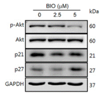
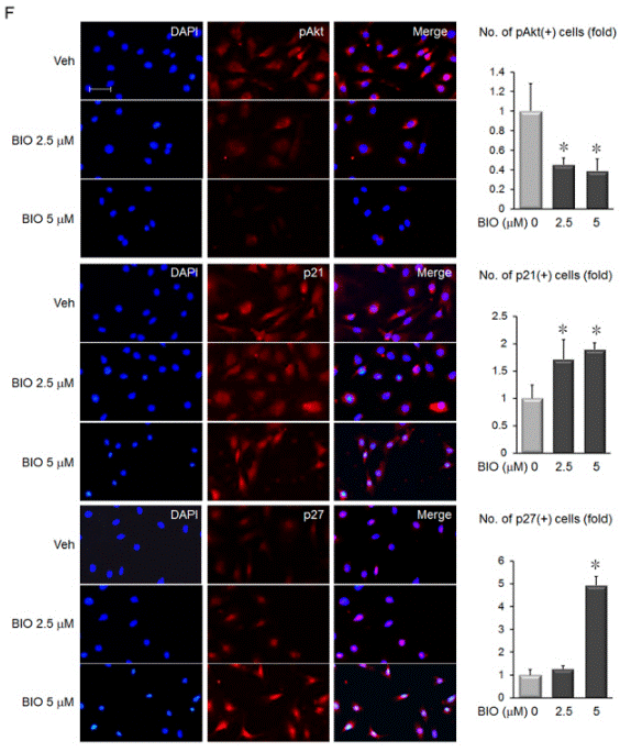
| Experimental Result Images | Methods | Biomarkers | Images | PMID |
| Western blot | p-AKT / AKT / p21 / p27 p-β-catenin / β-catenin FoxO3a / FoxO1 / p-FoxO3a / p-FoxO1 |
|
27510556 | |
| Immunofluorescence | pAKT / p21 / p27 TNF-α E-cadherin / Nanog Oct3/4 |
|
27510556 | |
| Growth inhibition assay | Cell proliferation |
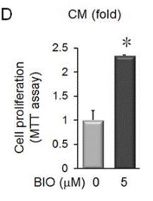
|
27510556 | |
In Vivo |
||
| In vivo | BIO suppresses melanoma tumor growth in a mouse xenograft model. [4] | |
|---|---|---|
| Animal Research | Animal Models | mouse |
| Dosages | 50 mg/kg | |
| Administration | Oral gavage | |
| NCT Number | Recruitment | Conditions | Sponsor/Collaborators | Start Date | Phases |
|---|---|---|---|---|---|
| NCT06337422 | Not yet recruiting | Healthy Volunteer |
International Bio service |
September 23 2024 | Phase 1 |
| NCT04276857 | Not yet recruiting | Locally Advanced Pancreatic Cancer|Irreversible Electroporation |
University of Saskatchewan |
September 1 2024 | Not Applicable |
| NCT06359041 | Not yet recruiting | Generalized Myasthenia Gravis (gMG) |
Cabaletta Bio |
August 2024 | Phase 1|Phase 2 |
References |
|
Chemical Information
| Molecular Weight | 356.17 | Formula | C16H10BrN3O2 |
| CAS No. | 667463-62-9 | SDF | Download SDF |
| Synonyms | GSK-3 Inhibitor IX, 6-bromoindirubin-3-oxime, 6-Bromoindirubin-3'-oxime, MLS 2052 | ||
| Smiles | C1=CC=C2C(=C1)C(=C(N2)C3=C(NC4=C3C=CC(=C4)Br)O)N=O | ||
Storage and Stability
| Storage (From the date of receipt) | |||
|
In vitro |
DMSO : 71 mg/mL ( (199.34 mM) Moisture-absorbing DMSO reduces solubility. Please use fresh DMSO.) Ethanol : 7 mg/mL Water : Insoluble |
Molecular Weight Calculator |
|
In vivo Add solvents to the product individually and in order. |
In vivo Formulation Calculator |
|||||
Preparing Stock Solutions
Molarity Calculator
In vivo Formulation Calculator (Clear solution)
Step 1: Enter information below (Recommended: An additional animal making an allowance for loss during the experiment)
mg/kg
g
μL
Step 2: Enter the in vivo formulation (This is only the calculator, not formulation. Please contact us first if there is no in vivo formulation at the solubility Section.)
% DMSO
%
% Tween 80
% ddH2O
%DMSO
%
Calculation results:
Working concentration: mg/ml;
Method for preparing DMSO master liquid: mg drug pre-dissolved in μL DMSO ( Master liquid concentration mg/mL, Please contact us first if the concentration exceeds the DMSO solubility of the batch of drug. )
Method for preparing in vivo formulation: Take μL DMSO master liquid, next addμL PEG300, mix and clarify, next addμL Tween 80, mix and clarify, next add μL ddH2O, mix and clarify.
Method for preparing in vivo formulation: Take μL DMSO master liquid, next add μL Corn oil, mix and clarify.
Note: 1. Please make sure the liquid is clear before adding the next solvent.
2. Be sure to add the solvent(s) in order. You must ensure that the solution obtained, in the previous addition, is a clear solution before proceeding to add the next solvent. Physical methods such
as vortex, ultrasound or hot water bath can be used to aid dissolving.
Tech Support
Answers to questions you may have can be found in the inhibitor handling instructions. Topics include how to prepare stock solutions, how to store inhibitors, and issues that need special attention for cell-based assays and animal experiments.
Tel: +1-832-582-8158 Ext:3
If you have any other enquiries, please leave a message.
* Indicates a Required Field


