
- Bioactive Compounds
- By Signaling Pathways
- PI3K/Akt/mTOR
- Epigenetics
- Methylation
- Immunology & Inflammation
- Protein Tyrosine Kinase
- Angiogenesis
- Apoptosis
- Autophagy
- ER stress & UPR
- JAK/STAT
- MAPK
- Cytoskeletal Signaling
- Cell Cycle
- TGF-beta/Smad
- DNA Damage/DNA Repair
- Compound Libraries
- Antibodies
- Bioreagents
- qPCR
- 2x SYBR Green qPCR Master Mix
- 2x SYBR Green qPCR Master Mix(Low ROX)
- 2x SYBR Green qPCR Master Mix(High ROX)
- Protein Assay
- Protein A/G Magnetic Beads for IP
- Anti-Flag magnetic beads
- Anti-Flag Affinity Gel
- Anti-Myc magnetic beads
- Anti-HA magnetic beads
- Magnetic Separator
- Poly FLAG Peptide lyophilized powder
- Protease Inhibitor Cocktail
- Protease Inhibitor Cocktail (EDTA-Free, 100X in DMSO)
- Phosphatase Inhibitor Cocktail (2 Tubes, 100X)
- Cell Biology
- Cell Counting Kit-8 (CCK-8)
- Animal Experiment
- Mouse Direct PCR Kit (For Genotyping)
- New Products
- Contact Us
-
 Australia
-
 Austria
-
 Belgium
-
 Canada
-
 China
China
-
 Czech Republic
-
 Denmark
-
 Finland
-
 France
-
 Germany
-
 Greece
-
 Hong Kong
-
 Hungary
-
 Iceland
-
 India
India
-
 Ireland
-
 Israel
-
 Italy
Italy
-
 Japan
Japan
-
 Korea
Korea
-
 Luxembourg
-
 Malaysia
-
 Netherlands
-
 New Zealand
-
 Norway
-
 Poland
-
 Qatar
Qatar
-
 Romania
-
 Saudi Arabia
-
 Singapore
-
 Spain
Spain
-
 Sweden
-
 Switzerland
-
 Taiwan
-
 Turkey
-
 United Kingdom
-
 United States
-
 Other Countries
Tozasertib (VX-680)
Synonyms: MK-0457
Tozasertib (VX-680) is a pan-Aurora inhibitor, mostly against Aurora A with Kiapp of 0.6 nM in a cell-free assay, less potent towards Aurora B/Aurora C and 100-fold more selective for Aurora A than 55 other kinases. The only exceptions are Fms-related tyrosine kinase-3 (FLT-3) and BCR-ABL tyrosine kinase, which are inhibited by the Tozasertib with both Ki of 30 nM. Tozasertib induces apoptosis and autophagy. Phase 2.
Tozasertib (VX-680) Chemical Structure
CAS No. 639089-54-6
Purity & Quality Control
Batch:
Purity:
99.99%
99.99
Tozasertib (VX-680) Related Products
| Related Targets | Aurora A Aurora B Aurora C Aurora B | Click to Expand |
|---|---|---|
| Related Products | Alisertib (MLN8237) Barasertib (AZD1152-HQPA) ZM 447439 MLN8054 Hesperadin Danusertib (PHA-739358) MK-5108 TCS7010 (Aurora A Inhibitor I) AMG-900 PHA-680632 SNS-314 CCT137690 GSK1070916 CYC116 TAK-901 CCT129202 SNS-314 Mesylate LY3295668 SP-96 | Click to Expand |
| Related Compound Libraries | Kinase Inhibitor Library PI3K/Akt Inhibitor Library MAPK Inhibitor Library DNA Damage/DNA Repair compound Library Cell Cycle compound library | Click to Expand |
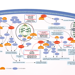
Signaling Pathway
Cell Data
| Cell Lines | Assay Type | Concentration | Incubation Time | Formulation | Activity Description | PMID |
|---|---|---|---|---|---|---|
| BE-13 | Growth Inhibition Assay | IC50=0.00338 μM | SANGRER | |||
| RS4-11 | Growth Inhibition Assay | IC50=0.00404 μM | SANGRER | |||
| MFH-ino | Growth Inhibition Assay | IC50=0.0099 μM | SANGRER | |||
| NTERA-S-cl-D1 | Growth Inhibition Assay | IC50=0.01434 μM | SANGRER | |||
| 697 | Growth Inhibition Assay | IC50=0.02471 μM | SANGRER | |||
| NALM-6 | Growth Inhibition Assay | IC50=0.02552 μM | SANGRER | |||
| ES8 | Growth Inhibition Assay | IC50=0.04613 μM | SANGRER | |||
| HUTU-80 | Growth Inhibition Assay | IC50=0.05299 μM | SANGRER | |||
| MV-4-11 | Growth Inhibition Assay | IC50=0.07782 μM | SANGRER | |||
| MONO-MAC-6 | Growth Inhibition Assay | IC50=0.07879 μM | SANGRER | |||
| LC-2-ad | Growth Inhibition Assay | IC50=0.08789 μM | SANGRER | |||
| BL-41 | Growth Inhibition Assay | IC50=0.10445 μM | SANGRER | |||
| A4-Fuk | Growth Inhibition Assay | IC50=0.11563 μM | SANGRER | |||
| SW954 | Growth Inhibition Assay | IC50=0.12229 μM | SANGRER | |||
| BV-173 | Growth Inhibition Assay | IC50=0.12641 μM | SANGRER | |||
| TE-11 | Growth Inhibition Assay | IC50=0.14982 μM | SANGRER | |||
| SK-UT-1 | Growth Inhibition Assay | IC50=0.15965 μM | SANGRER | |||
| SIG-M5 | Growth Inhibition Assay | IC50=0.16707 μM | SANGRER | |||
| OCUB-M | Growth Inhibition Assay | IC50=0.16983 μM | SANGRER | |||
| K052 | Growth Inhibition Assay | IC50=0.1948 μM | SANGRER | |||
| VA-ES-BJ | Growth Inhibition Assay | IC50=0.20086 μM | SANGRER | |||
| SW982 | Growth Inhibition Assay | IC50=0.2138 μM | SANGRER | |||
| LB647-SCLC | Growth Inhibition Assay | IC50=0.21523 μM | SANGRER | |||
| PSN1 | Growth Inhibition Assay | IC50=0.22026 μM | SANGRER | |||
| BB30-HNC | Growth Inhibition Assay | IC50=0.22591 μM | SANGRER | |||
| ST486 | Growth Inhibition Assay | IC50=0.23087 μM | SANGRER | |||
| MOLT-4 | Growth Inhibition Assay | IC50=0.23337 μM | SANGRER | |||
| EW-16 | Growth Inhibition Assay | IC50=0.23768 μM | SANGRER | |||
| KS-1 | Growth Inhibition Assay | IC50=0.23785 μM | SANGRER | |||
| SR | Growth Inhibition Assay | IC50=0.24564 μM | SANGRER | |||
| KM12 | Growth Inhibition Assay | IC50=0.2636 μM | SANGRER | |||
| EM-2 | Growth Inhibition Assay | IC50=0.26641 μM | SANGRER | |||
| MEG-01 | Growth Inhibition Assay | IC50=0.27849 μM | SANGRER | |||
| NB13 | Growth Inhibition Assay | IC50=0.27984 μM | SANGRER | |||
| RKO | Growth Inhibition Assay | IC50=0.30813 μM | SANGRER | |||
| CESS | Growth Inhibition Assay | IC50=0.31328 μM | SANGRER | |||
| EoL-1-cell | Growth Inhibition Assay | IC50=0.33459 μM | SANGRER | |||
| DOHH-2 | Growth Inhibition Assay | IC50=0.33781 μM | SANGRER | |||
| A388 | Growth Inhibition Assay | IC50=0.34086 μM | SANGRER | |||
| LAMA-84 | Growth Inhibition Assay | IC50=0.35178 μM | SANGRER | |||
| IMR-5 | Growth Inhibition Assay | IC50=0.3554 μM | SANGRER | |||
| KARPAS-422 | Growth Inhibition Assay | IC50=0.37272 μM | SANGRER | |||
| MRK-nu-1 | Growth Inhibition Assay | IC50=0.3813 μM | SANGRER | |||
| BL-70 | Growth Inhibition Assay | IC50=0.38974 μM | SANGRER | |||
| LXF-289 | Growth Inhibition Assay | IC50=0.40406 μM | SANGRER | |||
| RL95-2 | Growth Inhibition Assay | IC50=0.40567 μM | SANGRER | |||
| QIMR-WIL | Growth Inhibition Assay | IC50=0.42676 μM | SANGRER | |||
| K-562 | Growth Inhibition Assay | IC50=0.43472 μM | SANGRER | |||
| NCI-H510A | Growth Inhibition Assay | IC50=0.43823 μM | SANGRER | |||
| NCI-H524 | Growth Inhibition Assay | IC50=0.51147 μM | SANGRER | |||
| KE-37 | Growth Inhibition Assay | IC50=0.52102 μM | SANGRER | |||
| KP-N-YS | Growth Inhibition Assay | IC50=0.54392 μM | SANGRER | |||
| LS-411N | Growth Inhibition Assay | IC50=0.57752 μM | SANGRER | |||
| CTV-1 | Growth Inhibition Assay | IC50=0.58773 μM | SANGRER | |||
| NCI-SNU-16 | Growth Inhibition Assay | IC50=0.63571 μM | SANGRER | |||
| HT-144 | Growth Inhibition Assay | IC50=0.63798 μM | SANGRER | |||
| NCI-H187 | Growth Inhibition Assay | IC50=0.6413 μM | SANGRER | |||
| OCI-AML2 | Growth Inhibition Assay | IC50=0.64403 μM | SANGRER | |||
| CCRF-CEM | Growth Inhibition Assay | IC50=0.65346 μM | SANGRER | |||
| ONS-76 | Growth Inhibition Assay | IC50=0.66458 μM | SANGRER | |||
| IST-SL2 | Growth Inhibition Assay | IC50=0.71982 μM | SANGRER | |||
| NB6 | Growth Inhibition Assay | IC50=0.77254 μM | SANGRER | |||
| SK-PN-DW | Growth Inhibition Assay | IC50=0.7914 μM | SANGRER | |||
| HCC1599 | Growth Inhibition Assay | IC50=0.80874 μM | SANGRER | |||
| MC116 | Growth Inhibition Assay | IC50=0.85011 μM | SANGRER | |||
| TE-15 | Growth Inhibition Assay | IC50=0.85098 μM | SANGRER | |||
| HOP-62 | Growth Inhibition Assay | IC50=0.86329 μM | SANGRER | |||
| TGBC24TKB | Growth Inhibition Assay | IC50=0.86385 μM | SANGRER | |||
| HCE-4 | Growth Inhibition Assay | IC50=0.88063 μM | SANGRER | |||
| ALL-PO | Growth Inhibition Assay | IC50=0.88175 μM | SANGRER | |||
| KGN | Growth Inhibition Assay | IC50=0.89995 μM | SANGRER | |||
| ML-2 | Growth Inhibition Assay | IC50=0.90259 μM | SANGRER | |||
| ES4 | Growth Inhibition Assay | IC50=0.91128 μM | SANGRER | |||
| SF126 | Growth Inhibition Assay | IC50=0.94819 μM | SANGRER | |||
| SK-N-DZ | Growth Inhibition Assay | IC50=0.96189 μM | SANGRER | |||
| HCC1187 | Growth Inhibition Assay | IC50=1.00505 μM | SANGRER | |||
| DU-4475 | Growth Inhibition Assay | IC50=1.01756 μM | SANGRER | |||
| NKM-1 | Growth Inhibition Assay | IC50=1.02775 μM | SANGRER | |||
| HL-60 | Growth Inhibition Assay | IC50=1.06574 μM | SANGRER | |||
| SBC-1 | Growth Inhibition Assay | IC50=1.12542 μM | SANGRER | |||
| TE-10 | Growth Inhibition Assay | IC50=1.12946 μM | SANGRER | |||
| ETK-1 | Growth Inhibition Assay | IC50=1.13613 μM | SANGRER | |||
| HAL-01 | Growth Inhibition Assay | IC50=1.16709 μM | SANGRER | |||
| BB65-RCC | Growth Inhibition Assay | IC50=1.18005 μM | SANGRER | |||
| EW-1 | Growth Inhibition Assay | IC50=1.18562 μM | SANGRER | |||
| SK-NEP-1 | Growth Inhibition Assay | IC50=1.21111 μM | SANGRER | |||
| SK-LMS-1 | Growth Inhibition Assay | IC50=1.22212 μM | SANGRER | |||
| DEL | Growth Inhibition Assay | IC50=1.25643 μM | SANGRER | |||
| GT3TKB | Growth Inhibition Assay | IC50=1.28057 μM | SANGRER | |||
| MOLT-16 | Growth Inhibition Assay | IC50=1.35405 μM | SANGRER | |||
| CMK | Growth Inhibition Assay | IC50=1.42117 μM | SANGRER | |||
| NB5 | Growth Inhibition Assay | IC50=1.64229 μM | SANGRER | |||
| NCI-H1963 | Growth Inhibition Assay | IC50=1.70583 μM | SANGRER | |||
| KURAMOCHI | Growth Inhibition Assay | IC50=1.78911 μM | SANGRER | |||
| TE-8 | Growth Inhibition Assay | IC50=1.80368 μM | SANGRER | |||
| NCI-H1304 | Growth Inhibition Assay | IC50=1.83073 μM | SANGRER | |||
| A101D | Growth Inhibition Assay | IC50=1.87395 μM | SANGRER | |||
| SCLC-21H | Growth Inhibition Assay | IC50=1.97057 μM | SANGRER | |||
| GB-1 | Growth Inhibition Assay | IC50=2.01647 μM | SANGRER | |||
| KARPAS-45 | Growth Inhibition Assay | IC50=2.02654 μM | SANGRER | |||
| ATN-1 | Growth Inhibition Assay | IC50=2.02858 μM | SANGRER | |||
| NCI-H720 | Growth Inhibition Assay | IC50=2.06244 μM | SANGRER | |||
| RPMI-6666 | Growth Inhibition Assay | IC50=2.16207 μM | SANGRER | |||
| NB17 | Growth Inhibition Assay | IC50=2.2927 μM | SANGRER | |||
| IST-SL1 | Growth Inhibition Assay | IC50=2.29765 μM | SANGRER | |||
| SH-4 | Growth Inhibition Assay | IC50=2.32469 μM | SANGRER | |||
| K5 | Growth Inhibition Assay | IC50=2.40319 μM | SANGRER | |||
| OVCAR-4 | Growth Inhibition Assay | IC50=2.4613 μM | SANGRER | |||
| ACN | Growth Inhibition Assay | IC50=2.50213 μM | SANGRER | |||
| TGW | Growth Inhibition Assay | IC50=2.65832 μM | SANGRER | |||
| NCI-H2107 | Growth Inhibition Assay | IC50=2.83711 μM | SANGRER | |||
| NCI-H82 | Growth Inhibition Assay | IC50=2.83838 μM | SANGRER | |||
| SK-N-FI | Growth Inhibition Assay | IC50=2.86868 μM | SANGRER | |||
| LB1047-RCC | Growth Inhibition Assay | IC50=2.88126 μM | SANGRER | |||
| LU-134-A | Growth Inhibition Assay | IC50=2.8926 μM | SANGRER | |||
| NCI-H209 | Growth Inhibition Assay | IC50=2.91253 μM | SANGRER | |||
| NOMO-1 | Growth Inhibition Assay | IC50=3.02274 μM | SANGRER | |||
| RH-1 | Growth Inhibition Assay | IC50=3.17291 μM | SANGRER | |||
| LOUCY | Growth Inhibition Assay | IC50=3.18693 μM | SANGRER | |||
| TE-9 | Growth Inhibition Assay | IC50=3.26736 μM | SANGRER | |||
| PF-382 | Growth Inhibition Assay | IC50=3.35778 μM | SANGRER | |||
| RPMI-8402 | Growth Inhibition Assay | IC50=3.58603 μM | SANGRER | |||
| HEL | Growth Inhibition Assay | IC50=3.632 μM | SANGRER | |||
| NOS-1 | Growth Inhibition Assay | IC50=3.84754 μM | SANGRER | |||
| ES1 | Growth Inhibition Assay | IC50=3.92293 μM | SANGRER | |||
| NCI-H2171 | Growth Inhibition Assay | IC50=3.92423 μM | SANGRER | |||
| NCI-H747 | Growth Inhibition Assay | IC50=3.94221 μM | SANGRER | |||
| MHH-NB-11 | Growth Inhibition Assay | IC50=3.95312 μM | SANGRER | |||
| MZ1-PC | Growth Inhibition Assay | IC50=3.9924 μM | SANGRER | |||
| MMAC-SF | Growth Inhibition Assay | IC50=4.02467 μM | SANGRER | |||
| NMC-G1 | Growth Inhibition Assay | IC50=4.22723 μM | SANGRER | |||
| SW872 | Growth Inhibition Assay | IC50=4.3434 μM | SANGRER | |||
| TE-12 | Growth Inhibition Assay | IC50=4.56394 μM | SANGRER | |||
| LU-139 | Growth Inhibition Assay | IC50=4.61835 μM | SANGRER | |||
| HC-1 | Growth Inhibition Assay | IC50=4.69494 μM | SANGRER | |||
| COR-L279 | Growth Inhibition Assay | IC50=4.75891 μM | SANGRER | |||
| SF268 | Growth Inhibition Assay | IC50=4.79916 μM | SANGRER | |||
| MC-CAR | Growth Inhibition Assay | IC50=5.06757 μM | SANGRER | |||
| TK10 | Growth Inhibition Assay | IC50=5.35469 μM | SANGRER | |||
| TE-1 | Growth Inhibition Assay | IC50=5.49004 μM | SANGRER | |||
| NCI-H2126 | Growth Inhibition Assay | IC50=5.64574 μM | SANGRER | |||
| Daudi | Growth Inhibition Assay | IC50=5.6912 μM | SANGRER | |||
| NCI-H1648 | Growth Inhibition Assay | IC50=5.81454 μM | SANGRER | |||
| OS-RC-2 | Growth Inhibition Assay | IC50=5.98597 μM | SANGRER | |||
| DJM-1 | Growth Inhibition Assay | IC50=6.34666 μM | SANGRER | |||
| LS-1034 | Growth Inhibition Assay | IC50=6.7566 μM | SANGRER | |||
| NCI-H1581 | Growth Inhibition Assay | IC50=6.78405 μM | SANGRER | |||
| UACC-257 | Growth Inhibition Assay | IC50=7.04512 μM | SANGRER | |||
| KM-H2 | Growth Inhibition Assay | IC50=7.18457 μM | SANGRER | |||
| NCI-H1436 | Growth Inhibition Assay | IC50=7.69932 μM | SANGRER | |||
| IA-LM | Growth Inhibition Assay | IC50=7.859 μM | SANGRER | |||
| NCI-H526 | Growth Inhibition Assay | IC50=8.25637 μM | SANGRER | |||
| GCIY | Growth Inhibition Assay | IC50=8.36965 μM | SANGRER | |||
| CP67-MEL | Growth Inhibition Assay | IC50=8.5326 μM | SANGRER | |||
| KALS-1 | Growth Inhibition Assay | IC50=8.83851 μM | SANGRER | |||
| NCI-H1770 | Growth Inhibition Assay | IC50=8.90265 μM | SANGRER | |||
| 8-MG-BA | Growth Inhibition Assay | IC50=9.32844 μM | SANGRER | |||
| KY821 | Growth Inhibition Assay | IC50=9.77484 μM | SANGRER | |||
| SNB75 | Growth Inhibition Assay | IC50=10.076 μM | SANGRER | |||
| NCCIT | Growth Inhibition Assay | IC50=11.0582 μM | SANGRER | |||
| SJSA-1 | Growth Inhibition Assay | IC50=11.2891 μM | SANGRER | |||
| LB373-MEL-D | Growth Inhibition Assay | IC50=11.3827 μM | SANGRER | |||
| TALL-1 | Growth Inhibition Assay | IC50=11.4058 μM | SANGRER | |||
| NB69 | Growth Inhibition Assay | IC50=11.7705 μM | SANGRER | |||
| NCI-H1355 | Growth Inhibition Assay | IC50=11.9426 μM | SANGRER | |||
| DMS-153 | Growth Inhibition Assay | IC50=12.0426 μM | SANGRER | |||
| OPM-2 | Growth Inhibition Assay | IC50=12.1596 μM | SANGRER | |||
| NB1 | Growth Inhibition Assay | IC50=12.29 μM | SANGRER | |||
| A3-KAW | Growth Inhibition Assay | IC50=12.3236 μM | SANGRER | |||
| NCI-H1882 | Growth Inhibition Assay | IC50=12.4066 μM | SANGRER | |||
| KG-1 | Growth Inhibition Assay | IC50=12.6545 μM | SANGRER | |||
| LC4-1 | Growth Inhibition Assay | IC50=12.7706 μM | SANGRER | |||
| HCE-T | Growth Inhibition Assay | IC50=13.0049 μM | SANGRER | |||
| NEC8 | Growth Inhibition Assay | IC50=13.1038 μM | SANGRER | |||
| IST-MEL1 | Growth Inhibition Assay | IC50=13.5788 μM | SANGRER | |||
| EW-3 | Growth Inhibition Assay | IC50=13.7402 μM | SANGRER | |||
| CTB-1 | Growth Inhibition Assay | IC50=14.0329 μM | SANGRER | |||
| LS-123 | Growth Inhibition Assay | IC50=14.1588 μM | SANGRER | |||
| NCI-H1417 | Growth Inhibition Assay | IC50=14.3052 μM | SANGRER | |||
| MZ7-mel | Growth Inhibition Assay | IC50=14.4433 μM | SANGRER | |||
| JiyoyeP-2003 | Growth Inhibition Assay | IC50=15.6326 μM | SANGRER | |||
| ES6 | Growth Inhibition Assay | IC50=16.2361 μM | SANGRER | |||
| HH | Growth Inhibition Assay | IC50=17.1963 μM | SANGRER | |||
| SF539 | Growth Inhibition Assay | IC50=17.9922 μM | SANGRER | |||
| Calu-6 | Growth Inhibition Assay | IC50=19.239 μM | SANGRER | |||
| SK-MM-2 | Growth Inhibition Assay | IC50=19.555 μM | SANGRER | |||
| IST-MES1 | Growth Inhibition Assay | IC50=19.6663 μM | SANGRER | |||
| GI-ME-N | Growth Inhibition Assay | IC50=19.8227 μM | SANGRER | |||
| CAL-148 | Growth Inhibition Assay | IC50=20.9934 μM | SANGRER | |||
| EVSA-T | Growth Inhibition Assay | IC50=21.1499 μM | SANGRER | |||
| LP-1 | Growth Inhibition Assay | IC50=21.3432 μM | SANGRER | |||
| BOKU | Growth Inhibition Assay | IC50=21.4533 μM | SANGRER | |||
| KLE | Growth Inhibition Assay | IC50=22.1903 μM | SANGRER | |||
| LB831-BLC | Growth Inhibition Assay | IC50=25.1526 μM | SANGRER | |||
| NCI-H889 | Growth Inhibition Assay | IC50=25.1931 μM | SANGRER | |||
| REH | Growth Inhibition Assay | IC50=25.4671 μM | SANGRER | |||
| KP-N-RT-BM-1 | Growth Inhibition Assay | IC50=25.4752 μM | SANGRER | |||
| MPP-89 | Growth Inhibition Assay | IC50=25.5314 μM | SANGRER | |||
| no-11 | Growth Inhibition Assay | IC50=25.747 μM | SANGRER | |||
| NCI-H748 | Growth Inhibition Assay | IC50=25.7627 μM | SANGRER | |||
| LB2518-MEL | Growth Inhibition Assay | IC50=27.1773 μM | SANGRER | |||
| TGBC1TKB | Growth Inhibition Assay | IC50=27.5585 μM | SANGRER | |||
| MHH-PREB-1 | Growth Inhibition Assay | IC50=28.0734 μM | SANGRER | |||
| MZ2-MEL | Growth Inhibition Assay | IC50=28.6143 μM | SANGRER | |||
| U-266 | Growth Inhibition Assay | IC50=28.6366 μM | SANGRER | |||
| SNU-C1 | Growth Inhibition Assay | IC50=28.943 μM | SANGRER | |||
| SW962 | Growth Inhibition Assay | IC50=30.2747 μM | SANGRER | |||
| Raji | Growth Inhibition Assay | IC50=30.5592 μM | SANGRER | |||
| KNS-42 | Growth Inhibition Assay | IC50=30.8956 μM | SANGRER | |||
| LB996-RCC | Growth Inhibition Assay | IC50=31.1702 μM | SANGRER | |||
| CHP-126 | Growth Inhibition Assay | IC50=31.1984 μM | SANGRER | |||
| RXF393 | Growth Inhibition Assay | IC50=32.497 μM | SANGRER | |||
| COLO-684 | Growth Inhibition Assay | IC50=32.6438 μM | SANGRER | |||
| A704 | Growth Inhibition Assay | IC50=33.5538 μM | SANGRER | |||
| A253 | Growth Inhibition Assay | IC50=33.5852 μM | SANGRER | |||
| KNS-81-FD | Growth Inhibition Assay | IC50=34.5456 μM | SANGRER | |||
| TE-441-T | Growth Inhibition Assay | IC50=34.6371 μM | SANGRER | |||
| HCC2157 | Growth Inhibition Assay | IC50=35.4619 μM | SANGRER | |||
| ES3 | Growth Inhibition Assay | IC50=36.675 μM | SANGRER | |||
| NCI-H1155 | Growth Inhibition Assay | IC50=37.815 μM | SANGRER | |||
| SNU-C2B | Growth Inhibition Assay | IC50=38.1654 μM | SANGRER | |||
| JAR | Growth Inhibition Assay | IC50=38.2449 μM | SANGRER | |||
| GDM-1 | Growth Inhibition Assay | IC50=38.9116 μM | SANGRER | |||
| KU812 | Growth Inhibition Assay | IC50=41.507 μM | SANGRER | |||
| BC-1 | Growth Inhibition Assay | IC50=42.6731 μM | SANGRER | |||
| GI-1 | Growth Inhibition Assay | IC50=42.9192 μM | SANGRER | |||
| NCI-H1694 | Growth Inhibition Assay | IC50=44.9472 μM | SANGRER | |||
| DG-75 | Growth Inhibition Assay | IC50=45.1577 μM | SANGRER | |||
| COR-L88 | Growth Inhibition Assay | IC50=45.2778 μM | SANGRER | |||
| LS-513 | Growth Inhibition Assay | IC50=45.9156 μM | SANGRER | |||
| HD-MY-Z | Growth Inhibition Assay | IC50=46.4612 μM | SANGRER | |||
| L-363 | Growth Inhibition Assay | IC50=46.881 μM | SANGRER | |||
| TE-6 | Growth Inhibition Assay | IC50=48.446 μM | SANGRER | |||
| NCI-H345 | Growth Inhibition Assay | IC50=48.468 μM | SANGRER | |||
| TE-5 | Growth Inhibition Assay | IC50=49.7118 μM | SANGRER | |||
| MCF7 | Antiproliferative assay | Antiproliferative activity against human MCF7 cells, IC50=0.048μM | 18630890 | |||
| HCT116 | Antiproliferative assay | Antiproliferative activity against human HCT116 cells, IC50=0.053μM | 18630890 | |||
| HT29 | Antiproliferative assay | Antiproliferative activity against human HT29 cells, IC50=0.15μM | 18630890 | |||
| HCT | Function assay | Inhibition of Aurora A in human HCT cells assessed as loss of autophosphorylation of Aurora A, IC50=0.31μM | 18630890 | |||
| HCT | Function assay | Inhibition of Aurora A assessed as separation of centrosomes in human HCT cells, IC50=0.31μM | 18630890 | |||
| HCT | Function assay | Inhibition of Aurora B assessed as loss of phospho histone H3 in human HCT cells, IC50=0.31μM | 18630890 | |||
| H29 | Function assay | Inhibition of Aurora B assessed as loss of phospho histone H3 in human H29 cells, IC50=0.74μM | 18630890 | |||
| HCT116 | Cytotoxicity assay | 10 to 14 days | Cytotoxicity against human HCT116 cells assessed as number of colonies after 10 to 14 days by colony forming assay, IC50=0.024μM | 19143567 | ||
| HCT116 | Cytotoxicity assay | 72 hrs | Cytotoxicity against human HCT116 cells assessed as distinct polyploidy phenotype after 72 hrs, Activity=0.03μM | 19143567 | ||
| COLO205 | Antiproliferative assay | Antiproliferative activity against human COLO205 cells by [3H]thymidine uptake assay, IC50=0.019μM | 19447622 | |||
| HCT116 | Cytotoxicity assay | Cytotoxicity against human HCT116 cells by MTS assay, IC50=0.12μM | 20550212 | |||
| HeLa | Function assay | Inhibition of phosphotransferase activity of recombinant aurora A expressed in HeLa cells, Ki=0.002μM | 20573509 | |||
| BA/F3 | Antiproliferative assay | Antiproliferative activity against mouse BA/F3 cells expressing Bcr-Abl T315I mutant, IC50=0.03μM | 20604564 | |||
| BA/F3 | Function assay | Inhibition of Bcr-Abl T315I mutant autophosphorylation in mouse BA/F3 cells, IC50=5μM | 20604564 | |||
| insect | Function assay | Inhibition of human recombinant His-tagged Aurora A kinase expressed in insect cells, IC50=0.0007μM | 21194953 | |||
| HCT116 | Antiproliferative assay | Antiproliferative activity against human HCT116 cells, IC50=0.028μM | 21802948 | |||
| HCT116 | Function assay | Inhibition of Aurora B kinase assessed as reduction in histone H3 phosphorylation in human HCT116 cells, IC50=0.055μM | 21802948 | |||
| HCT116 | Cytotoxicity assay | 48 hrs | Cytotoxicity against human HCT116 cells after 48 hrs by MTT assay, IC50=0.3μM | 22572580 | ||
| MCF7 | Cytotoxicity assay | 48 hrs | Cytotoxicity against human MCF7 cells after 48 hrs by MTT assay, IC50=0.38μM | 22572580 | ||
| sf9 | Function assay | 15 mins | Inhibition of GST-tagged Aurora kinase A catalytic domain (123 to 401 amino acids) (unknown origin) expressed in sf9 cells using tetra(LRRWSLG) as substrate preincubated for 15 mins prior to substrate addition measured after 90 mins by luminescence assay, IC50=0.02μM | 23808327 | ||
| HCT116 | Antiproliferative assay | 96 hrs | Antiproliferative activity against human HCT116 cells after 96 hrs by MTS assay, EC50=0.12μM | 23808327 | ||
| HCT116 | Antiproliferative assay | Antiproliferative activity against human HCT116 cells, IC50=0.12μM | 23808327 | |||
| HCT116 | Antitumor assay | 50 mg/kg | 5 days | Antitumor activity against human HCT116 cells xenografted in athymic nu/nu mouse assessed as reduction in tumor size at 50 mg/kg, iv qd for 5 days per week for 2 weeks measured twice a week | 23808327 | |
| HeLa | Function assay | 10 to 1000 nM | 48 hrs | Inhibition of Aurora kinase B in human HeLa cells assessed as delay in mitotic arrest by accumulation of multinucleated cells with 4N/8N DNA content at 10 to 1000 nM after 48 hrs by propidium iodide staining-based FACS flow cytometric analysis | 23808327 | |
| MOLT4 | Antiproliferative assay | 72 hrs | Antiproliferative activity against human MOLT4 cells assessed as cell viability after 72 hrs by CCK8 assay, IC50=0.0212μM | 24681066 | ||
| U937 | Antiproliferative assay | 72 hrs | Antiproliferative activity against human U937 cells assessed as cell viability after 72 hrs by CCK8 assay, IC50=0.036μM | 24681066 | ||
| HL60 | Antiproliferative assay | 72 hrs | Antiproliferative activity against human HL60 cells assessed as cell viability after 72 hrs by CCK8 assay, IC50=0.0382μM | 24681066 | ||
| K562 | Antiproliferative assay | 72 hrs | Antiproliferative activity against human K562 cells assessed as cell viability after 72 hrs by CCK8 assay, IC50=0.0791μM | 24681066 | ||
| MDA-MB-231 | Antiproliferative assay | 72 hrs | Antiproliferative activity against human MDA-MB-231 cells assessed as cell viability after 72 hrs by CCK8 assay, IC50=0.127μM | 24681066 | ||
| A431 | Antiproliferative assay | 72 hrs | Antiproliferative activity against human A431 cells assessed as cell viability after 72 hrs by CCK8 assay, IC50=0.24μM | 24681066 | ||
| DU145 | Antiproliferative assay | 72 hrs | Antiproliferative activity against human DU145 cells assessed as cell viability after 72 hrs by CCK8 assay, IC50=0.4μM | 24681066 | ||
| MCF7 | Antiproliferative assay | 72 hrs | Antiproliferative activity against human MCF7 cells assessed as cell viability after 72 hrs by CCK8 assay, IC50=1.3μM | 24681066 | ||
| HeLa | Antiproliferative assay | 72 hrs | Antiproliferative activity against human HeLa cells assessed as cell viability after 72 hrs by CCK8 assay, IC50=2.93μM | 24681066 | ||
| A549 | Antiproliferative assay | 72 hrs | Antiproliferative activity against human A549 cells assessed as cell viability after 72 hrs by CCK8 assay, IC50=3.05μM | 24681066 | ||
| PANC1 | Antiproliferative assay | 72 hrs | Antiproliferative activity against human PANC1 cells assessed as cell viability after 72 hrs by CCK8 assay, IC50=4.13μM | 24681066 | ||
| HCT116 | Antiproliferative assay | 72 hrs | Antiproliferative activity against human HCT116 cells assessed as cell viability after 72 hrs by CCK8 assay, IC50=4.32μM | 24681066 | ||
| PC3 | Antiproliferative assay | 72 hrs | Antiproliferative activity against human PC3 cells assessed as cell viability after 72 hrs by CCK8 assay, IC50=5.81μM | 24681066 | ||
| SKBR3 | Antiproliferative assay | 72 hrs | Antiproliferative activity against human SKBR3 cells assessed as cell viability after 72 hrs by CCK8 assay, IC50=9.99μM | 24681066 | ||
| NCI-N87 | Antiproliferative assay | 72 hrs | Antiproliferative activity against human NCI-N87 cells assessed as cell viability after 72 hrs by CCK8 assay, IC50=11.6μM | 24681066 | ||
| HepG2 | Cytotoxicity assay | 48 hrs | Cytotoxicity against human HepG2 cells after 48 hrs by MTT assay, TC50=3.3μM | 24910766 | ||
| HeLa | Function assay | 12 hrs | Inhibition of Aurora A in human HeLa cells after 12 hrs by ELISA method, IC50=0.261μM | 25812967 | ||
| HeLa | Function assay | 12 hrs | Inhibition of Aurora B in human HeLa cells after 12 hrs by ELISA method, IC50=0.453μM | 25812967 | ||
| A549 | Cytotoxicity assay | 48 hrs | Cytotoxicity against human A549 cells after 48 hrs by MTT assay, IC50=19.4μM | 25812967 | ||
| HeLa | Cytotoxicity assay | 48 hrs | Cytotoxicity against human HeLa cells after 48 hrs by MTT assay, IC50=27.3μM | 25812967 | ||
| HCT8 | Cytotoxicity assay | 48 hrs | Cytotoxicity against human HCT8 cells after 48 hrs by MTT assay, IC50=44.6μM | 25812967 | ||
| Hela | Function assay | Inhibition of recombinant Aurora A kinase derived from human Hela cells using kemptide as substrate in presence of [c-32P]ATP, Ki=0.002μM | 27884697 | |||
| NCI-H23 | Antiproliferative assay | Antiproliferative activity against human NCI-H23 cells harboring KRAS G12C mutant at | 28038940 | |||
| NCI-H358 | Antiproliferative assay | Antiproliferative activity against human NCI-H358 cells harboring KRAS G12C mutant at | 28038940 | |||
| U937 | Antiproliferative assay | 48 hrs | Antiproliferative activity against human U937 cells after 48 hrs by CCK8 assay, IC50=0.036μM | 29358147 | ||
| K562 | Antiproliferative assay | 48 hrs | Antiproliferative activity against human K562 cells after 48 hrs by CCK8 assay, IC50=0.079μM | 29358147 | ||
| MDA-MB-231 | Antiproliferative assay | 48 hrs | Antiproliferative activity against human MDA-MB-231 cells after 48 hrs by CCK8 assay, IC50=0.127μM | 29358147 | ||
| A431 | Antiproliferative assay | 48 hrs | Antiproliferative activity against human A431 cells after 48 hrs by CCK8 assay, IC50=0.24μM | 29358147 | ||
| MCF7 | Antiproliferative assay | 48 hrs | Antiproliferative activity against human MCF7 cells after 48 hrs by CCK8 assay, IC50=1.3μM | 29358147 | ||
| HeLa | Antiproliferative assay | 48 hrs | Antiproliferative activity against human HeLa cells after 48 hrs by CCK8 assay, IC50=2.93μM | 29358147 | ||
| HCT116 | Antiproliferative assay | 48 hrs | Antiproliferative activity against human HCT116 cells after 48 hrs by CCK8 assay, IC50=4.32μM | 29358147 | ||
| TC32 | qHTS assay | qHTS of pediatric cancer cell lines to identify multiple opportunities for drug repurposing: Primary screen for TC32 cells | 29435139 | |||
| A673 | qHTS assay | qHTS of pediatric cancer cell lines to identify multiple opportunities for drug repurposing: Primary screen for A673 cells | 29435139 | |||
| SK-N-MC | qHTS assay | qHTS of pediatric cancer cell lines to identify multiple opportunities for drug repurposing: Primary screen for SK-N-MC cells | 29435139 | |||
| NB-EBc1 | qHTS assay | qHTS of pediatric cancer cell lines to identify multiple opportunities for drug repurposing: Primary screen for NB-EBc1 cells | 29435139 | |||
| SK-N-SH | qHTS assay | qHTS of pediatric cancer cell lines to identify multiple opportunities for drug repurposing: Primary screen for SK-N-SH cells | 29435139 | |||
| NB1643 | qHTS assay | qHTS of pediatric cancer cell lines to identify multiple opportunities for drug repurposing: Primary screen for NB1643 cells | 29435139 | |||
| LAN-5 | qHTS assay | qHTS of pediatric cancer cell lines to identify multiple opportunities for drug repurposing: Primary screen for LAN-5 cells | 29435139 | |||
| BT-12 | qHTS assay | qHTS of pediatric cancer cell lines to identify multiple opportunities for drug repurposing: Primary screen for BT-12 cells | 29435139 | |||
| Rh18 | qHTS assay | qHTS of pediatric cancer cell lines to identify multiple opportunities for drug repurposing: Primary screen for Rh18 cells | 29435139 | |||
| OHS-50 | qHTS assay | qHTS of pediatric cancer cell lines to identify multiple opportunities for drug repurposing: Primary screen for OHS-50 cells | 29435139 | |||
| RD | qHTS assay | qHTS of pediatric cancer cell lines to identify multiple opportunities for drug repurposing: Primary screen for RD cells | 29435139 | |||
| MG 63 (6-TG R) | qHTS assay | qHTS of pediatric cancer cell lines to identify multiple opportunities for drug repurposing: Primary screen for MG 63 (6-TG R) cells | 29435139 | |||
| Rh30 | qHTS assay | qHTS of pediatric cancer cell lines to identify multiple opportunities for drug repurposing: Primary screen for Rh30 cells | 29435139 | |||
| Rh41 | qHTS assay | qHTS of pediatric cancer cell lines to identify multiple opportunities for drug repurposing: Primary screen for Rh41 cells | 29435139 | |||
| NB1643 | qHTS assay | qHTS of pediatric cancer cell lines to identify multiple opportunities for drug repurposing: Confirmatory screen for NB1643 cells | 29435139 | |||
| A673 | qHTS assay | qHTS of pediatric cancer cell lines to identify multiple opportunities for drug repurposing: Confirmatory screen for A673 cells) | 29435139 | |||
| SK-N-MC | qHTS assay | qHTS of pediatric cancer cell lines to identify multiple opportunities for drug repurposing: Confirmatory screen for SK-N-MC cells | 29435139 | |||
| DAOY | qHTS assay | qHTS of pediatric cancer cell lines to identify multiple opportunities for drug repurposing: Confirmatory screen for DAOY cells | 29435139 | |||
| TC32 | qHTS assay | qHTS of pediatric cancer cell lines to identify multiple opportunities for drug repurposing: Confirmatory screen for TC32 cells | 29435139 | |||
| MG 63 (6-TG R) | qHTS assay | qHTS of pediatric cancer cell lines to identify multiple opportunities for drug repurposing: Confirmatory screen for MG 63 (6-TG R) cells | 29435139 | |||
| U-2 OS | qHTS assay | qHTS of pediatric cancer cell lines to identify multiple opportunities for drug repurposing: Confirmatory screen for U-2 OS cells | 29435139 | |||
| Rh41 | qHTS assay | qHTS of pediatric cancer cell lines to identify multiple opportunities for drug repurposing: Confirmatory screen for Rh41 cells | 29435139 | |||
| Rh30 | qHTS assay | qHTS of pediatric cancer cell lines to identify multiple opportunities for drug repurposing: Confirmatory screen for Rh30 cells | 29435139 | |||
| Saos-2 | qHTS assay | qHTS of pediatric cancer cell lines to identify multiple opportunities for drug repurposing: Confirmatory screen for Saos-2 cells | 29435139 | |||
| OHS-50 | qHTS assay | qHTS of pediatric cancer cell lines to identify multiple opportunities for drug repurposing: Confirmatory screen for OHS-50 cells | 29435139 | |||
| SK-N-SH | qHTS assay | qHTS of pediatric cancer cell lines to identify multiple opportunities for drug repurposing: Confirmatory screen for SK-N-SH cells | 29435139 | |||
| L929sA | Function assay | 24 hrs | Inhibition of Aurk mouse L929sA cells transfected with human Fas assessed as reduction in cell growth after 24 hrs in presence of TNF by Hoechst 33342 staining-based microscopic assay, IC50=0.97μM | 29437386 | ||
| L929sA | Function assay | 0.5 hrs | Inhibition of RIPK1 in mouse L929sA cells transfected with human Fas assessed as inhibition in TNF-induced necroptosis pretreated for 0.5 hrs followed by TNF and zVAD.fmk addition measured after 3 hrs by Hoechst 33342/propidium iodide staining-based assay, IC50=0.98μM | 29437386 | ||
| L929sA | Function assay | 24 hrs | Inhibition of RIPK1 in mouse L929sA cells transfected with human Fas assessed as inhibition in TNF-induced necroptosis pretreated for 24 hrs followed by TNF and zVAD.fmk addition measured after 3 hrs by Hoechst 33342/propidium iodide staining-based assay, IC50=1.02μM | 29437386 | ||
| L929sA | Function assay | 24 hrs | Inhibition of Aurk mouse L929sA cells transfected with human Fas assessed as increase in nuclear area after 24 hrs in presence of TNF by Hoechst 33342 staining-based microscopic assay, IC50=1.06μM | 29437386 | ||
| HCT116 | Cytotoxicity assay | 48 hrs | Cytotoxicity against human HCT116 cells assessed as reduction in cell viability after 48 hrs by MTT assay, IC50=0.45μM | 30143423 | ||
| HCT15 | Cytotoxicity assay | 48 hrs | Cytotoxicity against human HCT15 cells assessed as reduction in cell viability after 48 hrs by MTT assay, IC50=1.23μM | 30143423 | ||
| HCT116 | Function assay | 100 nM | 24 hrs | Inhibition of cytokinesis in human HCT116 cells at 100 nM after 24 hrs by propidium iodide/RNase staining-based flow cytometric analysis | 30143423 | |
| HCT116 | Function assay | 1 uM | 24 hrs | Inhibition of cytokinesis in human HCT116 cells at 1 uM after 24 hrs by propidium iodide/RNase staining-based flow cytometric analysis | 30143423 | |
| HCT116 | Function assay | 1 uM | 3 hrs | Inhibition of Aurora B in human HCT116 cells assessed as reduction in histone H3 phosphorylation at Ser10 residue up to 1 uM after 3 hrs by Western blot analysis | 30143423 | |
| HCT116 | Function assay | 3 hrs | Inhibition of Aurora A phosphorylation at Thr288 residue in human HCT116 cells up to 1 uM after 3 hrs by Western blot analysis | 30143423 | ||
| HCT116 | Function assay | 3 hrs | Inhibition of Aurora B phosphorylation at Thr232 residue in human HCT116 cells up to 1 uM after 3 hrs by Western blot method | 30143423 | ||
| Sf9 | Function assay | Inhibition of GST-tagged AURORA A (unknown origin) expressed in baculovirus infected Sf9 cells using MBP as substrate, IC50=0.023μM | 30234987 | |||
| HeLa | Function assay | 12 hrs | Inhibition of Aurora A phosphorylation at Thr288 residue in human HeLa cells after 12 hrs by ELISA, IC50=0.013μM | 30502115 | ||
| HeLa | Function assay | 12 hrs | Inhibition of Aurora B phosphorylation at Thr232 residue in human HeLa cells after 12 hrs by ELISA, IC50=0.148μM | 30502115 | ||
| A549 | Antiproliferative assay | 48 hrs | Antiproliferative activity against human A549 cells after 48 hrs by MTT assay, GI50=35.8μM | 30502115 | ||
| LoVo | Antiproliferative assay | 48 hrs | Antiproliferative activity against human LoVo cells after 48 hrs by MTT assay, GI50=45.3μM | 30502115 | ||
| HeLa | Antiproliferative assay | 48 hrs | Antiproliferative activity against human HeLa cells after 48 hrs by MTT assay, GI50=46.2μM | 30502115 | ||
| HeLa | Cell cycle assay | 5 uM | 12 hrs | Cell cycle arrest in human HeLa cells assessed as decrease in cyclin B1 protein expression at 5 uM after 12 hrs by Western blot analysis | 30502115 | |
| HeLa | Cell cycle assay | 5 uM | 12 hrs | Cell cycle arrest in human HeLa cells assessed as decrease in cdc2 protein expression at 5 uM after 12 hrs by Western blot analysis | 30502115 | |
| HeLa | Function assay | 5 uM | 12 hrs | Inhibition of Aurora A phosphorylation at Thr288 residue in human HeLa cells at 5 uM after 12 hrs by Western blot analysis | 30502115 | |
| HeLa | Function assay | 5 uM | 12 hrs | Inhibition of Aurora B phosphorylation at Thr232 residue in human HeLa cells at 5 uM after 12 hrs by Western blot analysis | 30502115 | |
| A549 | Antiproliferative assay | 48 hrs | Antiproliferative activity against human A549 cells after 48 hrs by MTT assay, IC50=35.8μM | 30728112 | ||
| LoVo | Antiproliferative assay | 48 hrs | Antiproliferative activity against human LoVo cells after 48 hrs by MTT assay, IC50=45.3μM | 30728112 | ||
| HeLa | Antiproliferative assay | 48 hrs | Antiproliferative activity against human HeLa cells after 48 hrs by MTT assay, IC50=46.2μM | 30728112 | ||
| Click to View More Cell Line Experimental Data | ||||||
Biological Activity
| Description | Tozasertib (VX-680) is a pan-Aurora inhibitor, mostly against Aurora A with Kiapp of 0.6 nM in a cell-free assay, less potent towards Aurora B/Aurora C and 100-fold more selective for Aurora A than 55 other kinases. The only exceptions are Fms-related tyrosine kinase-3 (FLT-3) and BCR-ABL tyrosine kinase, which are inhibited by the Tozasertib with both Ki of 30 nM. Tozasertib induces apoptosis and autophagy. Phase 2. | ||||||||||
|---|---|---|---|---|---|---|---|---|---|---|---|
| Targets |
|
| In vitro | ||||
| In vitro | Although its multi-kinase profile, VX-680 induces similar cytotoxicity with IC50 of approximately 300 nM and exhibits an AUR B-like inhibitory phenotype of G2/M arrest, endoreduplication and apoptosis in BaF3 cells transfected with ABL or FLT-3 (mutant and wild type) kinases. VX-680 prevents the CAL-62 proliferation in a time-dependent manner. VX-680 treatment for 14 days significantly decreases the number and size of colonies by approximately 70% in the 8305C and 90% in the CAL-62, 8505C and BHT-101. Treatment of the different ATC cells with VX-680 inhibits proliferation with the IC50 between 25 and 150 nM. The VX-680 significantly impairs the ability of the different cell lines to form colonies in soft agar. Analysis of caspase-3 activity indicates that VX-680 induces apoptosis in the different cell lines. CAL-62 cells exposed for 12 hours to VX-680 showed an accumulation of cells with ≥4N DNA content. Time-lapse analysis demonstrates that VX-680-treated CAL-62 cells exit metaphase without dividing. Moreover, histone H3 phosphorylation is abrogated following VX-680 treatment. [2] VX-680 has significant inhibitory activity against BCR-Abl bearing the T315I mutation in patient-derived samples. [3] | |||
|---|---|---|---|---|
| Kinase Assay | Kinase inhibition assays | |||
| The consumption of ATP is coupled via the pyruvate kinase/lactic dehydrogenase enzyme pair to the oxidation of NADH, which can be monitored through the decrease in absorption at 340 nm. Reactions contains 100 mM Tris (pH 8), 10 mM MgCl2, 2.2 mM ATP, 1 mM phosphoenolpyruvate, 0.6 mg/mL NADH, 75 units/mL pyruvate kinase, 105 units/mL lactate dehydrogenase, and 0.5 mM substrate peptide (sequence: EAIYAAPFAKKK). Reactions (75 μL) are started by adding sufficient kinase to bring the reactions to 30 nM kinase concentration and the decrease in absorbance is monitored over 30 minutes at 30°C in a microtiter plate spectrophotometer. Inhibitory constants are obtained through addition of 3.75 μL VX-680 in 100% DMSO or DMSO alone. Ki values are calculated as follows, K i = IC50 / (1 + [S]/Kd), where [S] = [ATP] = 2.2 mM, and Kd (of ATP to Abl) = 70 μM. These values are calculated assuming a Kd (ATP) of 70 μM for wild type and H396P Abl kinase domain. | ||||
| Cell Research | Cell lines | CAL-62 cells | ||
| Concentrations | 5-500 nM | |||
| Incubation Time | 4 days | |||
| Method | The CAL-62 cells are cultured in the absence (dimethyl sulfoxide, DMSO) or the presence of 500 nM VX-680 for different periods of time (1-5 days). The dose-dependent effects of VX-680 on cell proliferation are evaluated by treating the different ATC cells for 4 days with different concentrations of the Aurora inhibitor (5–500 nM). The cells are pulse labeled with 30 mM BrdU for 2 hours before the end of the incubation time. The BrdU incorporation is analyzed by means of a colorimetric immunoassay using the cell proliferation ELISA kit. The results from VX-680-treated cells are compared with those observed in control cells and expressed as a fold of variation versus control. |
|||
| Experimental Result Images | Methods | Biomarkers | Images | PMID |
| Western blot | DLK / p-MKK7 / MKK7 / p-JNK / JNK p-AURKA / AURKA / Survivin YAP p-AKT / p-GSK3β / Cleaved caspase-3 / Cleaved PARP |
|
23431148 | |
| Immunofluorescence | α-tubulin / Aurora-A |
|
21600017 | |
| Growth inhibition assay | Cell viability |
|
21600017 | |
| In Vivo | ||
| In vivo | VX-680 gives rise to a marked decrease in tumor size in a human AML (HL-60) xenograft model. In mude mice treateed with VX-680 at 75 mg/kg, twice a day intraperitoneally (b.i.d. i.p.) for 13 days, mean tumor volumes are reduced by 98%. Tumor growth decrease is dose dependent and significant at a dose of 12.5 mg/kg b.i.d. VX-680 is well tolerated, with a small decrease in body weight observed only at the highest dose. VX-680 also triggers tumor regresson in pancreatic and colon xenograft models. VX-680 also displays potent antitumor activity when infused i.v. in mude rats bearing established HCT116 tumors. A higher dose of VX-680 (2 mg/kg/h) improves efficacy with a 56% decrease in mean tumor volume. [1] | |
|---|---|---|
| Animal Research | Animal Models | Female athymic NCr-nu mice bearing HL-60 leukemia cells |
| Dosages | 50 mg/kg, 75 mg/kg | |
| Administration | Administered via i.p. | |
| NCT Number | Recruitment | Conditions | Sponsor/Collaborators | Start Date | Phases |
|---|---|---|---|---|---|
| NCT00290550 | Terminated | Carcinoma Non-Small-Cell Lung |
Merck Sharp & Dohme LLC |
June 2006 | Phase 2 |
| NCT00111683 | Completed | Chronic Myelogenous Leukemia in Blast Crisis|Lymphocytic Leukemia B Cell Acute|Myelodysplastic Syndromes|Myelogenous Leukemia Chronic |
Merck Sharp & Dohme LLC |
June 2005 | Phase 1 |
Chemical Information & Solubility
| Molecular Weight | 464.59 | Formula | C23H28N8OS |
| CAS No. | 639089-54-6 | SDF | Download Tozasertib (VX-680) SDF |
| Smiles | CC1=CC(=NN1)NC2=CC(=NC(=N2)SC3=CC=C(C=C3)NC(=O)C4CC4)N5CCN(CC5)C | ||
| Storage (From the date of receipt) | |||
|
In vitro |
DMSO : 93 mg/mL ( (200.17 mM) Moisture-absorbing DMSO reduces solubility. Please use fresh DMSO.) Ethanol : 40 mg/mL Water : Insoluble |
Molecular Weight Calculator |
|
In vivo Add solvents to the product individually and in order. |
In vivo Formulation Calculator |
||||
Preparing Stock Solutions
Molarity Calculator
In vivo Formulation Calculator (Clear solution)
Step 1: Enter information below (Recommended: An additional animal making an allowance for loss during the experiment)
mg/kg
g
μL
Step 2: Enter the in vivo formulation (This is only the calculator, not formulation. Please contact us first if there is no in vivo formulation at the solubility Section.)
% DMSO
%
% Tween 80
% ddH2O
%DMSO
%
Calculation results:
Working concentration: mg/ml;
Method for preparing DMSO master liquid: mg drug pre-dissolved in μL DMSO ( Master liquid concentration mg/mL, Please contact us first if the concentration exceeds the DMSO solubility of the batch of drug. )
Method for preparing in vivo formulation: Take μL DMSO master liquid, next addμL PEG300, mix and clarify, next addμL Tween 80, mix and clarify, next add μL ddH2O, mix and clarify.
Method for preparing in vivo formulation: Take μL DMSO master liquid, next add μL Corn oil, mix and clarify.
Note: 1. Please make sure the liquid is clear before adding the next solvent.
2. Be sure to add the solvent(s) in order. You must ensure that the solution obtained, in the previous addition, is a clear solution before proceeding to add the next solvent. Physical methods such
as vortex, ultrasound or hot water bath can be used to aid dissolving.
Tech Support
Answers to questions you may have can be found in the inhibitor handling instructions. Topics include how to prepare stock solutions, how to store inhibitors, and issues that need special attention for cell-based assays and animal experiments.
Tel: +1-832-582-8158 Ext:3
If you have any other enquiries, please leave a message.
* Indicates a Required Field
Tags: buy Tozasertib (VX-680) | Tozasertib (VX-680) supplier | purchase Tozasertib (VX-680) | Tozasertib (VX-680) cost | Tozasertib (VX-680) manufacturer | order Tozasertib (VX-680) | Tozasertib (VX-680) distributor
Products are for research use only. Not for human use. We do not sell to patients.
©Copyright 2013 Selleck Chemicals. All Rights Reserved.