-
 Australia
-
 Austria
-
 Belgium
-
 Brazil
-
 Canada
-
 China
China
-
 Czech Republic
-
 Denmark
-
 Finland
-
 France
-
 Germany
-
 Greece
-
 Hong Kong
-
 Hungary
-
 Iceland
-
 India
India
-
 Ireland
-
 Israel
-
 Italy
Italy
-
 Japan
Japan
-
 Korea
Korea
-
 Luxembourg
-
 Malaysia
-
 Netherlands
-
 New Zealand
-
 Norway
-
 Poland
-
 Qatar
Qatar
-
 Romania
-
 Saudi Arabia
-
 Singapore
-
 Spain
Spain
-
 Sweden
-
 Switzerland
-
 Taiwan
-
 Turkey
-
 United Kingdom
-
 United States
research use only
CHIR-124 Chk inhibitor
Cat.No.S2683
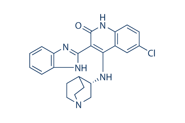
Chemical Structure
Molecular Weight: 419.91
Quality Control
Cell Culture, Treatment & Working Concentration
| Cell Lines | Assay Type | Concentration | Incubation Time | Formulation | Activity Description | PMID |
|---|---|---|---|---|---|---|
| human MDA-MB-435 cells | Cytotoxic assay | Cytotoxicity against human MDA-MB-435 cells, EC50=0.08 μM | ||||
| human MDA-MB-435 cells | Cytotoxic assay | Cytotoxicity against human MDA-MB-435 cells in presence of camptothecin | ||||
| Click to View More Cell Line Experimental Data | ||||||
Solubility
|
In vitro |
DMSO
: Insoluble
Water : Insoluble Ethanol : Insoluble |
Molarity Calculator
|
In vivo |
|||||
In vivo Formulation Calculator (Clear solution)
Step 1: Enter information below (Recommended: An additional animal making an allowance for loss during the experiment)
Step 2: Enter the in vivo formulation (This is only the calculator, not formulation. Please contact us first if there is no in vivo formulation at the solubility Section.)
Calculation results:
Working concentration: mg/ml;
Method for preparing DMSO master liquid: mg drug pre-dissolved in μL DMSO ( Master liquid concentration mg/mL, Please contact us first if the concentration exceeds the DMSO solubility of the batch of drug. )
Method for preparing in vivo formulation: Take μL DMSO master liquid, next addμL PEG300, mix and clarify, next addμL Tween 80, mix and clarify, next add μL ddH2O, mix and clarify.
Method for preparing in vivo formulation: Take μL DMSO master liquid, next add μL Corn oil, mix and clarify.
Note: 1. Please make sure the liquid is clear before adding the next solvent.
2. Be sure to add the solvent(s) in order. You must ensure that the solution obtained, in the previous addition, is a clear solution before proceeding to add the next solvent. Physical methods such
as vortex, ultrasound or hot water bath can be used to aid dissolving.
Chemical Information, Storage & Stability
| Molecular Weight | 419.91 | Formula | C23H22ClN5O |
Storage (From the date of receipt) | |
|---|---|---|---|---|---|
| CAS No. | 405168-58-3 | Download SDF | Storage of Stock Solutions |
|
|
| Synonyms | N/A | Smiles | C1CN2CCC1C(C2)NC3=C(C(=O)NC4=C3C=C(C=C4)Cl)C5=NC6=CC=CC=C6N5 | ||
Mechanism of Action
| Targets/IC50/Ki |
Chk1
(Cell-free assay) 0.3 nM
FLT3
(Cell-free assay) 5.8 nM
PDGFR
(Cell-free assay) 6.6 nM
GSK-3
(Cell-free assay) 23.3 nM
|
|---|---|
| In vitro |
CHIR-124 is a quinolone-based small molecule that is structurally unrelated to other known inhibitors of Chk1. This compound interacts synergistically with topoisomerase poisons (e.g., Camptothecin or SN-38) in causing growth inhibition in a variety of cancer cell lines, including breast carcinoma (MDA-MB-231 and MDA-MB-435) and colon carcinoma (SW-620 and Colo205), all of which contains the mutant p53 gene. It abrogates the SN-38-induced S and G2-M checkpoints and potentiates apoptosis in MDA-MD-435 breast cancer cells. The abrogation of the G2-Mcheckpoint and induction of apoptosis by this chemical are enhanced by the loss of p53. This compound also potently targets other kinases such as PDGFR and Flt3 with IC50 of 6.6 nM and 5.8 nM, respectively. |
| Kinase Assay |
Chk1 Assay
|
|
For the Chk1 assay, the kinase domain is expressed in Sf9 insect cells, and a biotinylated cdc25c peptide containing the consensus Chk1/Chk2 phosphorylation site (*)(biotin-[AHX]SGSGS*GLYRSPSMP-ENLNRPR[CONH2]) is used as the substrate. A dilution series of CHIR-124 is mixed with a kinasereaction buffer containing a final concentration of 30 mM Tris-HCl(pH 7.5), 10 mM MgCl2, 2 mM DTT, 4 mM EDTA, 25 mMβ-glycerophosphate, 5 mM MnCl2, 0.01% bovine serum albumin, 1.35 nM CHK1 kinase domain, 0.5 μM peptide substrate, and 1 AM unlabeled ATP, plus 5 nM 33Pγ-labeled ATP (specific activity = 2,000 Ci/mmol). Reactions and detection of the phosphate transfer are carried out by a radioactive method. Reactions are incubated at room temperature for 1 to 4 hours and the phosphorylated peptide captured on streptavidin-coated microtiter plates containing stop reaction buffer (25 mM EDTA [ethylenediaminetetraacetic acid], 50 mMHEPES, pH 7.5). Phosphorylated peptide is measured with the DELFIA TRF system using a Europium-labeled anti-phosphotyrosine antibody PT66. The concentration of this compound for IC50 is calculated using nonlinear regression with XL-Fit data analysis software.
|
|
| In vivo |
CHIR-124 potentiates the growth inhibitory effects by abrogating the G2-M checkpoint and increasing tumor apoptosis in an orthotopic breast cancer xenograft model. |
References |
|
Tech Support
Tel: +1-832-582-8158 Ext:3
If you have any other enquiries, please leave a message.
Signaling Pathway Map
Products are for research use only. Not for human use. We do not sell to patients.
©Copyright 2013 Selleck Chemicals. All Rights Reserved.