-
 Australia
-
 Austria
-
 Belgium
-
 Brazil
-
 Canada
-
 China
China
-
 Czech Republic
-
 Denmark
-
 Finland
-
 France
-
 Germany
-
 Greece
-
 Hong Kong
-
 Hungary
-
 Iceland
-
 India
India
-
 Ireland
-
 Israel
-
 Italy
Italy
-
 Japan
Japan
-
 Korea
Korea
-
 Luxembourg
-
 Malaysia
-
 Netherlands
-
 New Zealand
-
 Norway
-
 Poland
-
 Qatar
Qatar
-
 Romania
-
 Saudi Arabia
-
 Singapore
-
 Spain
Spain
-
 Sweden
-
 Switzerland
-
 Taiwan
-
 Turkey
-
 United Kingdom
-
 United States
research use only
IU1 DUB inhibitor
Cat.No.S7134
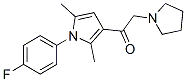
Chemical Structure
Molecular Weight: 300.37
Quality Control
| Related Targets | HDAC Caspase Proteasome Secretase MMP HCV Protease Cysteine Protease Tyrosinase HIV Protease DPP |
|---|---|
| Other DUB Inhibitors | PR-619 P5091 (P005091) b-AP15 P22077 ML323 LDN-57444 TCID VLX1570 EOAI3402143 ML364 |
Cell Culture, Treatment & Working Concentration
| Cell Lines | Assay Type | Concentration | Incubation Time | Formulation | Activity Description | PMID |
|---|---|---|---|---|---|---|
| HEK293T | Function assay | 30 uM | 2 hrs | Inhibition of deubiquitinase in HEK293T cells expressing N-terminal HA-tagged ubiquitin assessed as stabilization of HMW-Ub proteins at 30 uM after 2 hrs by Western blot analysis | 30528168 | |
| Click to View More Cell Line Experimental Data | ||||||
Solubility
|
In vitro |
DMSO
: 60 mg/mL
(199.75 mM)
Ethanol : 60 mg/mL Water : Insoluble |
Molarity Calculator
|
In vivo |
|||||
In vivo Formulation Calculator (Clear solution)
Step 1: Enter information below (Recommended: An additional animal making an allowance for loss during the experiment)
Step 2: Enter the in vivo formulation (This is only the calculator, not formulation. Please contact us first if there is no in vivo formulation at the solubility Section.)
Calculation results:
Working concentration: mg/ml;
Method for preparing DMSO master liquid: mg drug pre-dissolved in μL DMSO ( Master liquid concentration mg/mL, Please contact us first if the concentration exceeds the DMSO solubility of the batch of drug. )
Method for preparing in vivo formulation: Take μL DMSO master liquid, next addμL PEG300, mix and clarify, next addμL Tween 80, mix and clarify, next add μL ddH2O, mix and clarify.
Method for preparing in vivo formulation: Take μL DMSO master liquid, next add μL Corn oil, mix and clarify.
Note: 1. Please make sure the liquid is clear before adding the next solvent.
2. Be sure to add the solvent(s) in order. You must ensure that the solution obtained, in the previous addition, is a clear solution before proceeding to add the next solvent. Physical methods such
as vortex, ultrasound or hot water bath can be used to aid dissolving.
Chemical Information, Storage & Stability
| Molecular Weight | 300.37 | Formula | C18H21FN2O |
Storage (From the date of receipt) | |
|---|---|---|---|---|---|
| CAS No. | 314245-33-5 | Download SDF | Storage of Stock Solutions |
|
|
| Synonyms | N/A | Smiles | CC1=CC(=C(N1C2=CC=C(C=C2)F)C)C(=O)CN3CCCC3 | ||
Mechanism of Action
| Targets/IC50/Ki |
USP14
(cell-free assay) 4.7 μM
|
|---|---|
| In vitro |
IU1 binds specifically to the activated form of USP14. This compound can potentially inhibit USP14 by preventing its docking on the proteasome, exhibiting little or no activity toward 8 other DUBs, IsoT, UCH37, BAP1, UCH-L1, UCH-L3, USP15, USP2, USP7. USP14 inhibition is rapidly established upon addition of this chemical and rapidly reversed upon its removal. It inhibits USP14 induced chain trimming and decreases electrophoretic mobility of Ub-CCNB species. This compound enhances proteasomal degradation of Ub-CCNB in the presence of USP14. It promots degradation of tau and depletes TDP-43, ATXN3, and glial fibrillary acidic protein (GFAP) in proteotoxic mechanisms.
|
| Kinase Assay |
High-throughput screening
|
|
Screening is conducted at the ICCB-Longwood screening facility. 10 μL of recombinant USP14 protein are dispensed into each well of the 384-well low volume plate in duplicate, using a Wellmate plate dispenser. 33.3 nL of compound from the library are pin-transferred into the wells using a Seiko pin transfer robotic system, followed by pre-incubation for about 30 min. The last two columns of each plate are used for positive and negative controls for the assay. To initiate the enzyme reaction, 10 μL of VS-proteasome plus Ub-AMC mixture are added to each well, using a Wellmate dispenser. Samples are then incubated for another 45 min. Ub-AMC hydrolysis is measured at Ex355/Em460 using an Envision plate reader. The final concentrations of USP14, VS-proteasome and Ub-AMC are 15 nM, 1 nM and 0.8 μM, respectively. The final concentration of test compound is approximately 17 μM. Enzymes and substrates are prepared in Ub-AMC assay buffer (50 mM Tris-HCl (pH 7.5), 1 mM EDTA, 1 mM ATP, 5 mM MgCl2, 1 mM DTT, and 1 mg/Ml ovalbumin).
|
References |
Tech Support
Tel: +1-832-582-8158 Ext:3
If you have any other enquiries, please leave a message.
Signaling Pathway Map
Products are for research use only. Not for human use. We do not sell to patients.
©Copyright 2013 Selleck Chemicals. All Rights Reserved.