-
 Australia
-
 Austria
-
 Belgium
-
 Brazil
-
 Canada
-
 China
China
-
 Czech Republic
-
 Denmark
-
 Finland
-
 France
-
 Germany
-
 Greece
-
 Hong Kong
-
 Hungary
-
 Iceland
-
 India
India
-
 Ireland
-
 Israel
-
 Italy
Italy
-
 Japan
Japan
-
 Korea
Korea
-
 Luxembourg
-
 Malaysia
-
 Netherlands
-
 New Zealand
-
 Norway
-
 Poland
-
 Qatar
Qatar
-
 Romania
-
 Saudi Arabia
-
 Singapore
-
 Spain
Spain
-
 Sweden
-
 Switzerland
-
 Taiwan
-
 Turkey
-
 United Kingdom
-
 United States
research use only
CPI-613 (Devimistat) Mitochondrial Metabolism Inhibitor
Cat.No.S2776
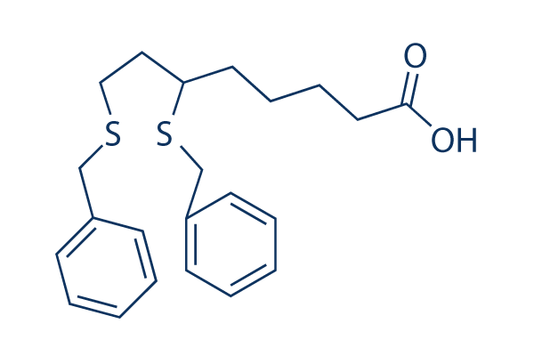
Chemical Structure
Molecular Weight: 388.59
Quality Control
Cell Culture, Treatment & Working Concentration
| Cell Lines | Assay Type | Concentration | Incubation Time | Formulation | Activity Description | PMID |
|---|---|---|---|---|---|---|
| DAOY | qHTS assay | qHTS of pediatric cancer cell lines to identify multiple opportunities for drug repurposing: Primary screen for DAOY cells | 29435139 | |||
| SK-N-MC | qHTS assay | qHTS of pediatric cancer cell lines to identify multiple opportunities for drug repurposing: Primary screen for SK-N-MC cells | 29435139 | |||
| NB1643 | qHTS assay | qHTS of pediatric cancer cell lines to identify multiple opportunities for drug repurposing: Primary screen for NB1643 cells | 29435139 | |||
| RD | qHTS assay | qHTS of pediatric cancer cell lines to identify multiple opportunities for drug repurposing: Primary screen for RD cells | 29435139 | |||
| Click to View More Cell Line Experimental Data | ||||||
Solubility
|
In vitro |
DMSO
: 77 mg/mL
(198.15 mM)
Ethanol : 77 mg/mL Water : Insoluble |
Molarity Calculator
|
In vivo |
|||||
In vivo Formulation Calculator (Clear solution)
Step 1: Enter information below (Recommended: An additional animal making an allowance for loss during the experiment)
Step 2: Enter the in vivo formulation (This is only the calculator, not formulation. Please contact us first if there is no in vivo formulation at the solubility Section.)
Calculation results:
Working concentration: mg/ml;
Method for preparing DMSO master liquid: mg drug pre-dissolved in μL DMSO ( Master liquid concentration mg/mL, Please contact us first if the concentration exceeds the DMSO solubility of the batch of drug. )
Method for preparing in vivo formulation: Take μL DMSO master liquid, next addμL PEG300, mix and clarify, next addμL Tween 80, mix and clarify, next add μL ddH2O, mix and clarify.
Method for preparing in vivo formulation: Take μL DMSO master liquid, next add μL Corn oil, mix and clarify.
Note: 1. Please make sure the liquid is clear before adding the next solvent.
2. Be sure to add the solvent(s) in order. You must ensure that the solution obtained, in the previous addition, is a clear solution before proceeding to add the next solvent. Physical methods such
as vortex, ultrasound or hot water bath can be used to aid dissolving.
Chemical Information, Storage & Stability
| Molecular Weight | 388.59 | Formula | C22H28O2S2 |
Storage (From the date of receipt) | |
|---|---|---|---|---|---|
| CAS No. | 95809-78-2 | Download SDF | Storage of Stock Solutions |
|
|
| Synonyms | N/A | Smiles | C1=CC=C(C=C1)CSCCC(CCCCC(=O)O)SCC2=CC=CC=C2 | ||
Mechanism of Action
| Targets/IC50/Ki |
PDH
(NCI-H460 cells) α-ketoglutarate dehydrogenase
(NCI-H460 cells) |
|---|---|
| In vitro |
Devimistat (CPI-613) produces selective toxicity in vitro against several tumor cell lines, including H460 human lung cancer cells and Saos-2 human sarcoma cells, with EC50 values of 120 μM and 120 μM, respectively. It disrupts H460 cancer cell mitochondrial metabolism, including inhibition of PDH complex activity and loss of mitochondrial membrane potential in a time- and drug dose-dependent fashion. In addition, this compound (240 μM) also induces both apoptotic and non-apoptotic cell death in H460 human lung cancer and Saos-2 human sarcoma cells. |
| Kinase Assay |
E1α phosphorylation
|
|
Cells are seeded and grown overnight. Five dishes per test group are treated with Devimistat (CPI-613) or solvent. Cells are lysed in situ with 150 μL lysis buffer A [455 μL Zoom 2D protein solubilizer 1, 2.5μL 1M Tris base, 5μL 100X protease inhibitor cocktail; 5μL 2M DTT] and lysates from all five dishes are pooled in a 1.5 mL microfuge tube and sonicated on ice for 15 passes at 50% power. After a 10-minute incubation at room temperature, 2.5μL of dimethylacrylamide (DMA) are added and lysates are incubated for an additional 10 minutes. 5μL of 2 M DTT are added to neutralize excess DMA. Lysates are centrifuged for 15 minutes and the supernatant is recovered. Then, 40μL of lysate are mixed with 0.8μL pH 3-10 ampholytes, 0.75 μL 2 M DTT and brought up to 150μL with Zoom 2D protein solubilizer 1. 150μL of sample is loaded into IPG runner, and pH 3-10NL IPG strips are added. Strips are soaked overnight at room temperature. A step protocol is used for isoelectric focusing (250V, 20min.; 450V 15min; 750 V 15 min 2000V 30 minutes). For the second dimension, strips are treated for 15 minutes in 1X loading buffer, followed by 15 minutes in 1X loading buffer plus 160 mM iodoacetatic acid. Strips are electrophoresed on NuPAGE 4-12% Bis Tris ZOOM gels. Proteins are blotted onto PVDF 4.5μm membranes. Serines 232, 293 and 300 are targets of the three well-characterized PDK phosphorylation events controlling E1 activity. Equal amounts of protein from treated and mock-treated cells are loaded on gels and western transfers are probed with anti-E1α Ab as an internal matching control or one of the three anti-phospho-E1α Abs.
|
|
| In vivo |
Devimistat (CPI-613) has potent anticancer activity in a human tumor xenograft model of a pancreatic tumor cell (BxPC-3) at 25 mg/kg. Similarly, this compound (10 mg/kg) also produces significant tumor growth inhibition of H460 human non-small cell lung carcinoma in a mouse model. Besides, it produces little or no side-effect toxicity in expected therapeutic dose ranges in large animal models and has the maximum tolerated dose of 100 mg/kg in mice. |
References |
|
Applications
| Methods | Biomarkers | Images | PMID |
|---|---|---|---|
| Western blot | pE1α p-AMPK / AMPK |
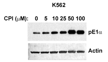
|
25165100 |
Clinical Trial Information
(data from https://clinicaltrials.gov, updated on 2024-05-22)
| NCT Number | Recruitment | Conditions | Sponsor/Collaborators | Start Date | Phases |
|---|---|---|---|---|---|
| NCT05926206 | Withdrawn | Metastatic Pancreatic Adenocarcinoma |
University of Michigan Rogel Cancer Center |
July 2023 | Phase 1|Phase 2 |
| NCT03793140 | Active not recruiting | Lymphoma|Leukemia |
Memorial Sloan Kettering Cancer Center|City of Hope Medical Center|Massachusetts General Hospital|M.D. Anderson Cancer Center|George Washington University |
December 31 2018 | Phase 2 |
Tech Support
Tel: +1-832-582-8158 Ext:3
If you have any other enquiries, please leave a message.
Frequently Asked Questions
Question 1:
How to dissolve it for in vivo applications?
Answer:
It is a suspension in 1% DMSO+30% polyethylene glycol+1% Tween 80 at 30mg/ml, and is for oral gavage.
Signaling Pathway Map
Products are for research use only. Not for human use. We do not sell to patients.
©Copyright 2013 Selleck Chemicals. All Rights Reserved.