-
 Australia
-
 Austria
-
 Belgium
-
 Brazil
-
 Canada
-
 China
China
-
 Czech Republic
-
 Denmark
-
 Finland
-
 France
-
 Germany
-
 Greece
-
 Hong Kong
-
 Hungary
-
 Iceland
-
 India
India
-
 Ireland
-
 Israel
-
 Italy
Italy
-
 Japan
Japan
-
 Korea
Korea
-
 Luxembourg
-
 Malaysia
-
 Netherlands
-
 New Zealand
-
 Norway
-
 Poland
-
 Qatar
Qatar
-
 Romania
-
 Saudi Arabia
-
 Singapore
-
 Spain
Spain
-
 Sweden
-
 Switzerland
-
 Taiwan
-
 Turkey
-
 United Kingdom
-
 United States
research use only
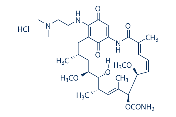
Alvespimycin (17-DMAG) Hydrochloride HSP inhibitor
Cat.No.S1142
Chemical Structure
Molecular Weight: 653.21
Quality Control
Cell Culture, Treatment & Working Concentration
| Cell Lines | Assay Type | Concentration | Incubation Time | Formulation | Activity Description | PMID |
|---|---|---|---|---|---|---|
| MDA-MB-231 | Function assay | Inhibition of Hsp90 in human MDA-MB-231 cells assessed as her2 degradation, IC50=0.0045μM. | 18929486 | |||
| A2058 | Cytotoxicity assay | Cytotoxicity against human A2058 cells by MTT assay, IC50=0.0021μM. | 18929486 | |||
| AGS | Cytotoxicity assay | Cytotoxicity against human AGS cells by MTT assay, IC50=16μM. | 18359631 | |||
| HeLa | Cytotoxicity assay | Cytotoxicity against human HeLa cells by MTT assay, IC50=2.06μM. | 18359631 | |||
| HeLa | Function assay | Inhibition of TNF-alpha-induced NF-kappaB activation in human HeLa cells, IC50=0.15μM. | 18359631 | |||
| AGS | Function assay | Inhibition of hypoxia-induced HIF1 activation in human AGS cells by reporter gene assay, IC50=0.0036μM. | 18359631 | |||
| NCI-H526 | Function assay | 1 uM | 96 hrs | Inhibition of HSP90-mediated proliferation of human NCI-H526 cells at 1 uM after 96 hrs by sulforhodamine B assay | 17603540 | |
| NCI-H526 | Function assay | 1 uM | 24 hrs | Binding affinity to HSP90 in human NCI-H526 cells at 1 uM after 24 hrs by fluorescence polarization assay | 17603540 | |
| AGS | Function assay | 24 hrs | Viability of human AGS cells under normoxic conditions after 24 hrs by MTT assay, IC50=16μM. | 17583950 | ||
| Hep3B | Function assay | 16 hrs | Inhibition of HIF1 activation in human Hep3B cells assessed as inhibition of hypoxia-induced luciferase expression after 16 hrs by reporter assay, IC50=0.061μM. | 17583950 | ||
| AGS | Function assay | 16 hrs | Inhibition of HIF1 activation in human AGS cells assessed as inhibition of hypoxia-induced luciferase expression after 16 hrs by reporter assay, IC50=0.036μM. | 17583950 | ||
| SKOV3 | Function assay | Degradation of Her2 in SKOV3 cells, EC50=0.046μM. | 16854066 | |||
| SKOV3 | Function assay | Upregulation of Hsp70 in SKOV3 cells, EC50=0.014μM. | 16854066 | |||
| SKBR3 | Function assay | Degradation of Her2 in SKBR3 cells, EC50=0.008μM. | 16854066 | |||
| SKBR3 | Function assay | Upregulation of Hsp70 in SKBR3 cells, EC50=0.004μM. | 16854066 | |||
| SKBr3 | Cytotoxicity assay | Cytotoxicity against SKBr3 cells, IC50=0.024μM. | 16165354 | |||
| MDA-MB-231 | Cytotoxicity assay | Cytotoxicity against human MDA-MB-231 cells by MTT assay, IC50=0.0058μM. | 18929486 | |||
| A2058 | Function assay | Inhibition of Hsp90 in human A2058 cells, EC50=0.0079μM. | 18929486 | |||
| MDA-MB-231 | Function assay | Inhibition of Hsp90 in human MDA-MB-231 cells assessed as Akt degradation, IC50=0.0176μM. | 18929486 | |||
| A2058 | Function assay | Inhibition of Hsp90 in human A2058 cells assessed as Akt degradation, IC50=0.0243μM. | 18929486 | |||
| HuH7 | Antiviral assay | 3 days | Antiviral activity against Hepatitis C virus genotype 1b Con1 infected in human HuH7 cells assessed as GAPDH RNA or 18S rRNA level after 3 days by qRT-PCR analysis, EC50=0.0012μM. | 18936191 | ||
| HuH7 | Antiviral assay | 3 days | Antiviral activity against Hepatitis C virus genotype 1b Con1 infected in human HuH7 cells assessed as GAPDH RNA or 18S rRNA level after 3 days selected with 40 nM HCV-796 and 800 nM boceprevir by qRT-PCR analysis, EC50=0.0031μM. | 18936191 | ||
| SKBR3 | Function assay | Binding affinity to Hsp90 in human SKBR3 cells, IC50=0.024μM. | 19017562 | |||
| Hep3B | Function assay | 30 mins | Inhibition of hypoxia-induced HIF1alpha protein accumulation in human Hep3B cells treated for 30 mins measured after 12 hrs by Western blot analysis, IC50=0.0572μM. | 19072214 | ||
| Hep3B | Function assay | 16 hrs | Inhibition of hypoxia-induced VEGF protein secretion in human Hep3B cells after 16 hrs by ELISA, IC50=0.0795μM. | 19072214 | ||
| HCT116 | Cytotoxicity assay | 72 hrs | Cytotoxicity against human HCT116 cells after 72 hrs, IC50=0.057μM. | 19231864 | ||
| SKBR3 | Cytotoxicity assay | 72 hrs | Cytotoxicity against human SKBR3 cells after 72 hrs, IC50=0.058μM. | 19231864 | ||
| MCF7 | Cytotoxicity assay | 72 hrs | Cytotoxicity against human MCF7 cells after 72 hrs, IC50=0.071μM. | 19231864 | ||
| SKOV3 | Cytotoxicity assay | 72 hrs | Cytotoxicity against human SKOV3 cells after 72 hrs, IC50=0.122μM. | 19231864 | ||
| SKBR3 | Cytotoxicity assay | 72 hrs | Cytotoxicity against human SKBR3 cells after 72 hrs in presence of NQO1 inhibitor dicoumarol, IC50=0.23μM. | 19231864 | ||
| MCF7 | Cytotoxicity assay | 72 hrs | Cytotoxicity against human MCF7 cells after 72 hrs in presence of NQO1 inhibitor dicoumarol, IC50=0.862μM. | 19231864 | ||
| NCI-H596 | Cytotoxicity assay | 72 hrs | Cytotoxicity against NQ01-deficient human NCI-H596 cells after 72 hrs, IC50=1.1μM. | 19231864 | ||
| MDA468 | Cytotoxicity assay | 72 hrs | Cytotoxicity against NQ01-deficient human MDA468 cells after 72 hrs, IC50=1.6μM. | 19231864 | ||
| SKBR3 | Cytotoxicity assay | 72 hrs | Cytotoxicity against human SKBR3 cells after 72 hrs by celltiter-glo assay, IC50=0.024μM. | 19405528 | ||
| A549 | Cytotoxicity assay | 72 hrs | Cytotoxicity against human A549 cells after 72 hrs by celltiter-glo assay, IC50=0.068μM. | 19405528 | ||
| SKOV3 | Cytotoxicity assay | 72 hrs | Cytotoxicity against human SKOV3 cells after 72 hrs by celltiter-glo assay, IC50=0.22μM. | 19405528 | ||
| MCF7 | Cytotoxicity assay | 72 hrs | Cytotoxicity against human MCF7 cells after 72 hrs by celltiter-glo assay, IC50=0.23μM. | 19405528 | ||
| CCRF-CEM | Cytotoxicity assay | 72 hrs | Cytotoxicity against human CCRF-CEM cells after 72 hrs by celltiter-96 aqueous one solution assay, IC50=0.54μM. | 19405528 | ||
| CCRF-CEM | Cytotoxicity assay | 72 hrs | Cytotoxicity against human paclitaxel-resistant CCRF-CEM cells after 72 hrs by celltiter-96 aqueous one solution assay, IC50=2.5μM. | 19405528 | ||
| Hep3B | Function assay | 30 mins | Inhibition of hypoxia-induced HIF1alpha protein accumulation in human Hep3B cells treated for 30 mins measured after 12 hrs by Western blot analysis, IC50=0.057μM. | 20469887 | ||
| Hep3B | Function assay | 16 hrs | Inhibition of hypoxia-induced VEGF protein secretion in human Hep3B cells after 16 hrs by ELISA, IC50=0.079μM. | 20469887 | ||
| HCT116 | Cytotoxicity assay | Cytotoxicity against human HCT116 cells by Alamar blue assay, IC50=0.05μM. | 20662534 | |||
| NCI-H1299 | Function assay | 24 hrs | Inhibition of human HSP90 in human NCI-H1299 cells assessed as Akt degradation after 24 hrs by luminex assay, IC50=0.1μM. | 21438541 | ||
| LN229-Lux | Function assay | 2.5 to 10 uM | 1 hr | Inhibition of luciferase activity in human LN229-Lux cells at 2.5 to 10 uM incubated for 1 hr under normoxia followed by 24 hrs under hypoxia by reporter gene assay | 22746274 | |
| MCF7 | Antiproliferative assay | 48 hrs | Antiproliferative activity against human MCF7 cells assessed as inhibition of cell viability after 48 hrs by MTT assay, IC50=0.39μM. | 24582477 | ||
| HCT116 | Antiproliferative assay | 48 hrs | Antiproliferative activity against human HCT116 cells assessed as inhibition of cell viability after 48 hrs by MTT assay, IC50=0.78μM. | 24582477 | ||
| SKBR3 | Antiproliferative assay | 48 hrs | Antiproliferative activity against human SKBR3 cells assessed as inhibition of cell viability after 48 hrs by MTT assay, IC50=1.34μM. | 24582477 | ||
| A231 | Antiproliferative assay | 48 hrs | Antiproliferative activity against human A231 cells after 48 hrs by MTT assay, IC50=0.17μM. | 24763261 | ||
| MCF7 | Antiproliferative assay | 48 hrs | Antiproliferative activity against human MCF7 cells after 48 hrs by MTT assay, IC50=0.8μM. | 24763261 | ||
| HCT116 | Antiproliferative assay | 48 hrs | Antiproliferative activity against human HCT116 cells after 48 hrs by MTT assay, IC50=1.21μM. | 24763261 | ||
| SKBR3 | Antiproliferative assay | 48 hrs | Antiproliferative activity against human SKBR3 cells after 48 hrs by MTT assay, IC50=3.11μM. | 24763261 | ||
| NCI-H1299 | Function assay | 12 hrs | Reduction in oxygen consumption rate in human NCI-H1299 cells incubated for 12 hrs | 25383915 | ||
| PC9 | Cytotoxicity assay | 72 hrs | Cytotoxicity against HGF-induced erlotinib-resistant human PC9 cells assessed as inhibition of cell growth after 72 hrs by MTT assay, IC50=0.01μM. | 26844689 | ||
| Ma1 | Cytotoxicity assay | 72 hrs | Cytotoxicity against HGF-induced erlotinib-resistant human Ma1 cells assessed as inhibition of cell growth after 72 hrs by MTT assay, IC50=0.01μM. | 26844689 | ||
| SKBR3 | Function assay | Inhibition of Hsp90 in human SKBR3 cells, IC50=0.024μM. | 26844689 | |||
| HeLa | Function assay | 10 uM | 6 hrs | Inhibition of HSP90 in human HeLa cells assessed as induction of chk1 degradation at 10 uM after 6 hrs by Western blot method | 28816449 | |
| HeLa | Function assay | 10 uM | 6 hrs | Inhibition of HSP90 in human HeLa cells assessed as induction of Akt degradation at 10 uM after 6 hrs by Western blot method | 28816449 | |
| HeLa | Function assay | 10 uM | 6 hrs | Inhibition of HSP90 in human HeLa cells assessed as induction of HSP70 protein expression at 10 uM after 6 hrs by Western blot method | 28816449 | |
| PC3 | Function assay | 10 uM | 6 hrs | Inhibition of HSP90 in human PC3 cells assessed as induction of chk1 degradation at 10 uM after 6 hrs by Western blot method | 28816449 | |
| PC3 | Function assay | 10 uM | 6 hrs | Inhibition of HSP90 in human PC3 cells assessed as induction of Akt degradation at 10 uM after 6 hrs by Western blot method | 28816449 | |
| PC3 | Function assay | 10 uM | 6 hrs | Inhibition of HSP90 in human PC3 cells assessed as induction of HSP70 protein expression at 10 uM after 6 hrs by Western blot method | 28816449 | |
| Click to View More Cell Line Experimental Data | ||||||
Solubility
|
In vitro |
DMSO
: 100 mg/mL
(153.09 mM)
Water : Insoluble Ethanol : Insoluble |
Molarity Calculator
|
In vivo |
|||||
In vivo Formulation Calculator (Clear solution)
Step 1: Enter information below (Recommended: An additional animal making an allowance for loss during the experiment)
Step 2: Enter the in vivo formulation (This is only the calculator, not formulation. Please contact us first if there is no in vivo formulation at the solubility Section.)
Calculation results:
Working concentration: mg/ml;
Method for preparing DMSO master liquid: mg drug pre-dissolved in μL DMSO ( Master liquid concentration mg/mL, Please contact us first if the concentration exceeds the DMSO solubility of the batch of drug. )
Method for preparing in vivo formulation: Take μL DMSO master liquid, next addμL PEG300, mix and clarify, next addμL Tween 80, mix and clarify, next add μL ddH2O, mix and clarify.
Method for preparing in vivo formulation: Take μL DMSO master liquid, next add μL Corn oil, mix and clarify.
Note: 1. Please make sure the liquid is clear before adding the next solvent.
2. Be sure to add the solvent(s) in order. You must ensure that the solution obtained, in the previous addition, is a clear solution before proceeding to add the next solvent. Physical methods such
as vortex, ultrasound or hot water bath can be used to aid dissolving.
Chemical Information, Storage & Stability
| Molecular Weight | 653.21 | Formula | C32H48N4O8•HCl |
Storage (From the date of receipt) | |
|---|---|---|---|---|---|
| CAS No. | 467214-21-7 | Download SDF | Storage of Stock Solutions |
|
|
| Synonyms | NSC 707545,BMS 826476 HCl,KOS 1022 | Smiles | CC1CC(C(C(C=C(C(C(C=CC=C(C(=O)NC2=CC(=O)C(=C(C1)C2=O)NCCN(C)C)C)OC)OC(=O)N)C)C)O)OC.Cl | ||
Mechanism of Action
| Features |
A synthetic derivative Geldanamycin, with lower hepatotoxicity than parent antibiotic & higher potency and bioavailability than the similar derivative 17-AAG.
|
|---|---|
| Targets/IC50/Ki |
HSP90
(Cell-free assay) 62 nM
|
| In vitro |
17-DMAG displays ~2 times potency against human Hsp90 than 17-AAG, with IC50 of 62 nM versus 119 nM. In SKBR3 and SKOV3 cells which over-express Hsp90 client protein Her2, 17-DMAG causes down-regulation of Her2 with EC50 of 8 nM and 46 nM, respectively, as well as induction of Hsp70 with EC50 of 4 nM and 14 nM, respectively, leading to significant cytotoxicity with GI50 of 29 nM and 32 nM, respectively, consistent with Hsp90 inhibition. 17-DMAG in combination with vorinostat synergistically induces apoptosis of the cultured MCL cells as well as primary MCL cells, more potently than either agent alone, by markedly attenuating the levels of cyclin D1 and CDK4, as well as of c-Myc, c-RAF and Akt. In contrast to 17-AAG which is only active for IKKβ in chronic lymphocytic leukemia (CLL) cells, 17-DMAG treatment effectively leads to depletion of the Hsp90 client protein, resulting in diminished NF-κB p50/p65 DNA binding, decreased NF-κB target gene transcription, and caspase-dependent apoptosis. By targeting the NF-κB family, 17-DMAG selectively mediates dose- and time-dependent cytotoxicity against CLL cells, but not normal T cells or NK cells important for immune surveillance. |
| Kinase Assay |
Fluorescence polarization (FP)-based competition binding assay
|
|
This assay utilizes a boron difluoride dipyrromethene (BODIPY) labeled geldanamycin analogue (BODIPY-AG) as a probe and measured fluorescence polarization upon binding of the probe to a protein. Native human Hsp90 protein (α + β isoforms) is isolated from HeLa cells. BODIPY-AG solution is freshly prepared in FP assay buffer (20 mM HEPES-KOH, pH 7.3, 1.0 mM EDTA, 100 mM KCl, 5.0 mM MgCl2, 0.01% NP-40, 0.1 mg/mL fresh bovine γ-globulin (BGG), 1.0 mM fresh DTT, and protease inhibitor from stock solution in DMSO. Competition curves are obtained by mixing 10 μL each of a solution containing BODIPY-AG and Hsp90, and a serial dilution of 17-DMAG freshly prepared in FP assay buffer from stock solution in DMSO. Final concentrations are 10 nM BODIPY-AG, 40 or 60 nM Hsp90, varying concentration of 17-DMAG (0.10 nM-10 μM), and ≤0.25% DMSO in a 384-well microplate. After 3 hours incubation at 30 °C, fluorescence anisotropy (γEx = 485 nm, γEm = 535 nm) is measured on an EnVision 2100 multilabel plate reader. IC50 value of 17-DMAG is obtained from the competition curves.
|
|
| In vivo |
17-DMAG treatment at 5 mg/kg or 25 mg/kg thrice per week significantly reduces tumor growth of TMK-1 xenografts, by significantly reducing vessel area and numbers of proliferating tumor cells in sections. Consistent the inhibition of FAK signaling in vivo, 17-DMAG treatment at 25 mg/kg three times a week significantly suppresses tumor growth, and metastasis of ME180 and SiHa xenografts in mice. Administration of 17-DMAG at 10 mg/kg for 16 days significantly decreases the white blood cell count and prolongs the survival in a TCL1-SCID transplant mouse model. |
References |
|
Applications
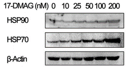
| Methods | Biomarkers | Images | PMID |
|---|---|---|---|
| Western blot | HSP90 / HSP70 p-Akt / Survivin / MMP2 PARP / Cleaved caspase-3 / Cleaved caspase-8 / Cleaved caspase-9 / PUMA p-ALK / ALK / p-Akt / Akt / p-ERK / ERK α-Tax / α-IKKα / α-IKKβ/ α-NEMO / α-TBK1 / α-p65 / α-p50 |
|
28915605 |
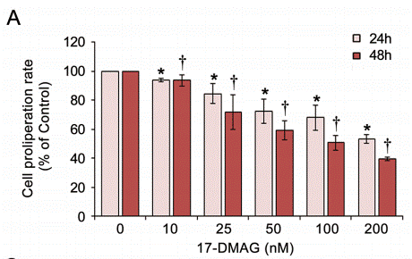
| Growth inhibition assay | Cell proliferation |
|
28915605 |
Clinical Trial Information
(data from https://clinicaltrials.gov, updated on 2024-05-22)
| NCT Number | Recruitment | Conditions | Sponsor/Collaborators | Start Date | Phases |
|---|---|---|---|---|---|
| NCT00780000 | Terminated | Breast Cancer |
Bristol-Myers Squibb |
April 2008 | Phase 2 |
| NCT00248521 | Unknown status | Unspecified Adult Solid Tumor Protocol Specific |
Institute of Cancer Research United Kingdom|National Cancer Institute (NCI) |
October 2005 | Phase 1 |
Tech Support
Tel: +1-832-582-8158 Ext:3
If you have any other enquiries, please leave a message.
Signaling Pathway Map
Products are for research use only. Not for human use. We do not sell to patients.
©Copyright 2013 Selleck Chemicals. All Rights Reserved.