
- Bioactive Compounds
- By Signaling Pathways
- PI3K/Akt/mTOR
- Epigenetics
- Methylation
- Immunology & Inflammation
- Protein Tyrosine Kinase
- Angiogenesis
- Apoptosis
- Autophagy
- ER stress & UPR
- JAK/STAT
- MAPK
- Cytoskeletal Signaling
- Cell Cycle
- TGF-beta/Smad
- DNA Damage/DNA Repair
- Compound Libraries
- Antibodies
- Bioreagents
- qPCR
- 2x SYBR Green qPCR Master Mix
- 2x SYBR Green qPCR Master Mix(Low ROX)
- 2x SYBR Green qPCR Master Mix(High ROX)
- Protein Assay
- Protein A/G Magnetic Beads for IP
- Anti-Flag magnetic beads
- Anti-Flag Affinity Gel
- Anti-Myc magnetic beads
- Anti-HA magnetic beads
- Magnetic Separator
- Poly DYKDDDDK Tag Peptide lyophilized powder
- Protease Inhibitor Cocktail
- Protease Inhibitor Cocktail (EDTA-Free, 100X in DMSO)
- Phosphatase Inhibitor Cocktail (2 Tubes, 100X)
- Cell Biology
- Cell Counting Kit-8 (CCK-8)
- Animal Experiment
- Mouse Direct PCR Kit (For Genotyping)
- New Products
- Contact Us
-
 Australia
-
 Austria
-
 Belgium
-
 Canada
-
 China
China
-
 Czech Republic
-
 Denmark
-
 Finland
-
 France
-
 Germany
-
 Greece
-
 Hong Kong
-
 Hungary
-
 Iceland
-
 India
India
-
 Ireland
-
 Israel
-
 Italy
Italy
-
 Japan
Japan
-
 Korea
Korea
-
 Luxembourg
-
 Malaysia
-
 Netherlands
-
 New Zealand
-
 Norway
-
 Poland
-
 Qatar
Qatar
-
 Romania
-
 Saudi Arabia
-
 Singapore
-
 Spain
Spain
-
 Sweden
-
 Switzerland
-
 Taiwan
-
 Turkey
-
 United Kingdom
-
 United States
-
 Other Countries
PD173074
PD173074 is a potent FGFR1 inhibitor with IC50 of ~25 nM and also inhibits VEGFR2 with IC50 of 100-200 nM in cell-free assays, ~1000-fold selective for FGFR1 than PDGFR and c-Src. PD173074 reduces proliferation and promotes apoptosis in gastric cancer cells.

PD173074 Chemical Structure
CAS No. 219580-11-7
Purity & Quality Control
Batch:
Purity:
99.95%
99.95
PD173074 Related Products
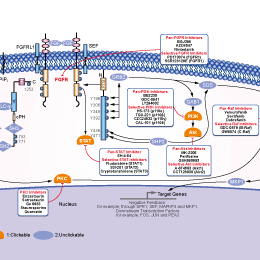
| Related Targets | FGFR1 FGFR2 FGFR3 FGFR4 | Click to Expand |
|---|---|---|
| Related Products | Fexagratinib (AZD4547) BLU9931 LY2874455 Zoligratinib (Debio-1347) Futibatinib (TAS-120) PD-166866 SSR128129E H3B-6527 Fisogatinib (BLU-554) Derazantinib FIIN-2 Ferulic Acid ASP5878 Roblitinib (FGF401) Alofanib (RPT835) NSC12 PRN1371 | Click to Expand |
| Related Compound Libraries | Tyrosine Kinase Inhibitor Library PI3K/Akt Inhibitor Library Angiogenesis Related compound Library HIF-1 Signaling Pathway Compound Library FDA-approved Anticancer Drug Library | Click to Expand |
Signaling Pathway
Cell Data
| Cell Lines | Assay Type | Concentration | Incubation Time | Formulation | Activity Description | PMID |
|---|---|---|---|---|---|---|
| NCI-H1581 | Growth Inhibition Assay | IC50=0.01225 μM | SANGER | |||
| KG-1 | Growth Inhibition Assay | IC50=0.05129 μM | SANGER | |||
| MFM-223 | Growth Inhibition Assay | IC50=0.21576 μM | SANGER | |||
| EoL-1-cell | Growth Inhibition Assay | IC50=0.32984 μM | SANGER | |||
| ECC10 | Growth Inhibition Assay | IC50=0.33898 μM | SANGER | |||
| H-EMC-SS | Growth Inhibition Assay | IC50=0.34715 μM | SANGER | |||
| AN3-CA | Growth Inhibition Assay | IC50=0.40133 μM | SANGER | |||
| HuO-3N1 | Growth Inhibition Assay | IC50=0.54653 μM | SANGER | |||
| RT-112 | Growth Inhibition Assay | IC50=0.54701 μM | SANGER | |||
| NEC8 | Growth Inhibition Assay | IC50=0.56289 μM | SANGER | |||
| D-263MG | Growth Inhibition Assay | IC50=0.71159 μM | SANGER | |||
| SW962 | Growth Inhibition Assay | IC50=0.78988 μM | SANGER | |||
| BV-173 | Growth Inhibition Assay | IC50=0.84623 μM | SANGER | |||
| MFE-280 | Growth Inhibition Assay | IC50=0.85872 μM | SANGER | |||
| HuH-7 | Growth Inhibition Assay | IC50=1.24464 μM | SANGER | |||
| RS4-11 | Growth Inhibition Assay | IC50=1.33886 μM | SANGER | |||
| DMS-114 | Growth Inhibition Assay | IC50=1.36737 μM | SANGER | |||
| MSTO-211H | Growth Inhibition Assay | IC50=1.47378 μM | SANGER | |||
| DU-145 | Growth Inhibition Assay | IC50=1.58217 μM | SANGER | |||
| A172 | Growth Inhibition Assay | IC50=1.70355 μM | SANGER | |||
| SBC-1 | Growth Inhibition Assay | IC50=2.094 μM | SANGER | |||
| H9 | Growth Inhibition Assay | IC50=2.14306 μM | SANGER | |||
| NCI-SNU-1 | Growth Inhibition Assay | IC50=2.18394 μM | SANGER | |||
| NCI-H720 | Growth Inhibition Assay | IC50=2.21283 μM | SANGER | |||
| HCC2218 | Growth Inhibition Assay | IC50=2.37939 μM | SANGER | |||
| G-401 | Growth Inhibition Assay | IC50=2.47189 μM | SANGER | |||
| MPP-89 | Growth Inhibition Assay | IC50=2.48364 μM | SANGER | |||
| 697 | Growth Inhibition Assay | IC50=2.65331 μM | SANGER | |||
| KARPAS-45 | Growth Inhibition Assay | IC50=2.70747 μM | SANGER | |||
| MG-63 | Growth Inhibition Assay | IC50=2.94262 μM | SANGER | |||
| NTERA-S-cl-D1 | Growth Inhibition Assay | IC50=3.03472 μM | SANGER | |||
| G-402 | Growth Inhibition Assay | IC50=3.12727 μM | SANGER | |||
| NKM-1 | Growth Inhibition Assay | IC50=3.13564 μM | SANGER | |||
| RH-18 | Growth Inhibition Assay | IC50=3.19598 μM | SANGER | |||
| NCI-H1092 | Growth Inhibition Assay | IC50=3.1969 μM | SANGER | |||
| RPMI-8226 | Growth Inhibition Assay | IC50=3.23447 μM | SANGER | |||
| GAMG | Growth Inhibition Assay | IC50=3.46576 μM | SANGER | |||
| HH | Growth Inhibition Assay | IC50=3.47676 μM | SANGER | |||
| RO82-W-1 | Growth Inhibition Assay | IC50=3.49855 μM | SANGER | |||
| CCRF-CEM | Growth Inhibition Assay | IC50=3.50488 μM | SANGER | |||
| NBsusSR | Growth Inhibition Assay | IC50=3.63969 μM | SANGER | |||
| CHL-1 | Growth Inhibition Assay | IC50=3.65799 μM | SANGER | |||
| LK-2 | Growth Inhibition Assay | IC50=3.67133 μM | SANGER | |||
| Hs-578-T | Growth Inhibition Assay | IC50=3.67873 μM | SANGER | |||
| CTB-1 | Growth Inhibition Assay | IC50=3.80051 μM | SANGER | |||
| ES5 | Growth Inhibition Assay | IC50=3.83637 μM | SANGER | |||
| A204 | Growth Inhibition Assay | IC50=3.92075 μM | SANGER | |||
| SW780 | Growth Inhibition Assay | IC50=3.92245 μM | SANGER | |||
| EW-3 | Growth Inhibition Assay | IC50=3.98923 μM | SANGER | |||
| A704 | Growth Inhibition Assay | IC50=4.28723 μM | SANGER | |||
| LU-139 | Growth Inhibition Assay | IC50=4.31534 μM | SANGER | |||
| CAL-72 | Growth Inhibition Assay | IC50=4.41746 μM | SANGER | |||
| D-336MG | Growth Inhibition Assay | IC50=4.46817 μM | SANGER | |||
| LAMA-84 | Growth Inhibition Assay | IC50=4.5331 μM | SANGER | |||
| GI-ME-N | Growth Inhibition Assay | IC50=4.5481 μM | SANGER | |||
| KM-H2 | Growth Inhibition Assay | IC50=4.55222 μM | SANGER | |||
| NCI-H209 | Growth Inhibition Assay | IC50=4.58283 μM | SANGER | |||
| IGROV-1 | Growth Inhibition Assay | IC50=4.87168 μM | SANGER | |||
| L-363 | Growth Inhibition Assay | IC50=4.96665 μM | SANGER | |||
| SK-MEL-3 | Growth Inhibition Assay | IC50=5.2406 μM | SANGER | |||
| HuO9 | Growth Inhibition Assay | IC50=5.38843 μM | SANGER | |||
| NOS-1 | Growth Inhibition Assay | IC50=5.72927 μM | SANGER | |||
| NCI-H1770 | Growth Inhibition Assay | IC50=5.95032 μM | SANGER | |||
| SF126 | Growth Inhibition Assay | IC50=6.21406 μM | SANGER | |||
| ML-2 | Growth Inhibition Assay | IC50=6.24977 μM | SANGER | |||
| CHP-134 | Growth Inhibition Assay | IC50=6.25182 μM | SANGER | |||
| NCI-H1355 | Growth Inhibition Assay | IC50=6.41733 μM | SANGER | |||
| TE-12 | Growth Inhibition Assay | IC50=6.72671 μM | SANGER | |||
| A4-Fuk | Growth Inhibition Assay | IC50=6.73142 μM | SANGER | |||
| MV-4-11 | Growth Inhibition Assay | IC50=6.76626 μM | SANGER | |||
| SK-UT-1 | Growth Inhibition Assay | IC50=6.91784 μM | SANGER | |||
| J-RT3-T3-5 | Growth Inhibition Assay | IC50=7.07764 μM | SANGER | |||
| ME-180 | Growth Inhibition Assay | IC50=7.10404 μM | SANGER | |||
| SK-MEL-28 | Growth Inhibition Assay | IC50=7.37819 μM | SANGER | |||
| HAL-01 | Growth Inhibition Assay | IC50=7.48341 μM | SANGER | |||
| ES8 | Growth Inhibition Assay | IC50=7.69626 μM | SANGER | |||
| DB | Growth Inhibition Assay | IC50=8.11504 μM | SANGER | |||
| SK-NEP-1 | Growth Inhibition Assay | IC50=8.48149 μM | SANGER | |||
| COR-L88 | Growth Inhibition Assay | IC50=8.50981 μM | SANGER | |||
| LB1047-RCC | Growth Inhibition Assay | IC50=8.52212 μM | SANGER | |||
| NCI-H520 | Growth Inhibition Assay | IC50=8.62157 μM | SANGER | |||
| SW954 | Growth Inhibition Assay | IC50=8.69786 μM | SANGER | |||
| TE-6 | Growth Inhibition Assay | IC50=8.75143 μM | SANGER | |||
| D-283MED | Growth Inhibition Assay | IC50=9.06534 μM | SANGER | |||
| DBTRG-05MG | Growth Inhibition Assay | IC50=9.09607 μM | SANGER | |||
| NCI-H446 | Growth Inhibition Assay | IC50=9.29526 μM | SANGER | |||
| HOS | Growth Inhibition Assay | IC50=9.35134 μM | SANGER | |||
| ES4 | Growth Inhibition Assay | IC50=9.50595 μM | SANGER | |||
| EW-13 | Growth Inhibition Assay | IC50=9.89055 μM | SANGER | |||
| IST-MES1 | Growth Inhibition Assay | IC50=9.94534 μM | SANGER | |||
| CAS-1 | Growth Inhibition Assay | IC50=9.97659 μM | SANGER | |||
| EM-2 | Growth Inhibition Assay | IC50=10.1393 μM | SANGER | |||
| SW948 | Growth Inhibition Assay | IC50=10.1882 μM | SANGER | |||
| OAW-42 | Growth Inhibition Assay | IC50=10.5267 μM | SANGER | |||
| BE-13 | Growth Inhibition Assay | IC50=10.6576 μM | SANGER | |||
| KU812 | Growth Inhibition Assay | IC50=10.7391 μM | SANGER | |||
| SK-MEL-30 | Growth Inhibition Assay | IC50=10.8901 μM | SANGER | |||
| A2780 | Growth Inhibition Assay | IC50=11.0308 μM | SANGER | |||
| TGBC24TKB | Growth Inhibition Assay | IC50=11.0737 μM | SANGER | |||
| GOTO | Growth Inhibition Assay | IC50=11.2084 μM | SANGER | |||
| NCI-H526 | Growth Inhibition Assay | IC50=11.3837 μM | SANGER | |||
| BHT-101 | Growth Inhibition Assay | IC50=11.4456 μM | SANGER | |||
| NCI-H1155 | Growth Inhibition Assay | IC50=11.4947 μM | SANGER | |||
| MCF7 | Growth Inhibition Assay | IC50=11.6167 μM | SANGER | |||
| MKN45 | Growth Inhibition Assay | IC50=11.7993 μM | SANGER | |||
| MOLT-16 | Growth Inhibition Assay | IC50=11.9692 μM | SANGER | |||
| YH-13 | Growth Inhibition Assay | IC50=12.0346 μM | SANGER | |||
| P12-ICHIKAWA | Growth Inhibition Assay | IC50=12.3845 μM | SANGER | |||
| GR-ST | Growth Inhibition Assay | IC50=12.5295 μM | SANGER | |||
| CAKI-1 | Growth Inhibition Assay | IC50=12.791 μM | SANGER | |||
| LXF-289 | Growth Inhibition Assay | IC50=13.0835 μM | SANGER | |||
| MHH-PREB-1 | Growth Inhibition Assay | IC50=13.2704 μM | SANGER | |||
| EW-16 | Growth Inhibition Assay | IC50=13.3187 μM | SANGER | |||
| NCI-H82 | Growth Inhibition Assay | IC50=13.4195 μM | SANGER | |||
| MMAC-SF | Growth Inhibition Assay | IC50=13.4547 μM | SANGER | |||
| COLO-684 | Growth Inhibition Assay | IC50=13.5318 μM | SANGER | |||
| QIMR-WIL | Growth Inhibition Assay | IC50=13.8109 μM | SANGER | |||
| NB69 | Growth Inhibition Assay | IC50=13.9868 μM | SANGER | |||
| NCI-H2291 | Growth Inhibition Assay | IC50=14.4453 μM | SANGER | |||
| MKN7 | Growth Inhibition Assay | IC50=14.6676 μM | SANGER | |||
| HDLM-2 | Growth Inhibition Assay | IC50=15.1286 μM | SANGER | |||
| A253 | Growth Inhibition Assay | IC50=15.3869 μM | SANGER | |||
| SK-LU-1 | Growth Inhibition Assay | IC50=15.9094 μM | SANGER | |||
| MEG-01 | Growth Inhibition Assay | IC50=15.9107 μM | SANGER | |||
| SK-N-DZ | Growth Inhibition Assay | IC50=15.9376 μM | SANGER | |||
| H4 | Growth Inhibition Assay | IC50=16.088 μM | SANGER | |||
| LU-65 | Growth Inhibition Assay | IC50=16.3384 μM | SANGER | |||
| NCI-H1048 | Growth Inhibition Assay | IC50=16.5165 μM | SANGER | |||
| LCLC-97TM1 | Growth Inhibition Assay | IC50=16.5889 μM | SANGER | |||
| CAL-120 | Growth Inhibition Assay | IC50=16.9879 μM | SANGER | |||
| LU-134-A | Growth Inhibition Assay | IC50=17.3391 μM | SANGER | |||
| SK-MEL-1 | Growth Inhibition Assay | IC50=17.7127 μM | SANGER | |||
| NCI-H69 | Growth Inhibition Assay | IC50=17.9307 μM | SANGER | |||
| MC116 | Growth Inhibition Assay | IC50=17.975 μM | SANGER | |||
| UMC-11 | Growth Inhibition Assay | IC50=18.1788 μM | SANGER | |||
| HCC1395 | Growth Inhibition Assay | IC50=18.4301 μM | SANGER | |||
| no-10 | Growth Inhibition Assay | IC50=18.6388 μM | SANGER | |||
| NY | Growth Inhibition Assay | IC50=19.0809 μM | SANGER | |||
| OS-RC-2 | Growth Inhibition Assay | IC50=19.1252 μM | SANGER | |||
| D-423MG | Growth Inhibition Assay | IC50=19.3952 μM | SANGER | |||
| LC-2-ad | Growth Inhibition Assay | IC50=19.7612 μM | SANGER | |||
| DU-4475 | Growth Inhibition Assay | IC50=19.8852 μM | SANGER | |||
| YKG-1 | Growth Inhibition Assay | IC50=19.962 μM | SANGER | |||
| HCC1569 | Growth Inhibition Assay | IC50=20.2624 μM | SANGER | |||
| TYK-nu | Growth Inhibition Assay | IC50=20.2847 μM | SANGER | |||
| DEL | Growth Inhibition Assay | IC50=20.9808 μM | SANGER | |||
| MHH-ES-1 | Growth Inhibition Assay | IC50=21.3597 μM | SANGER | |||
| KARPAS-299 | Growth Inhibition Assay | IC50=21.529 μM | SANGER | |||
| CTV-1 | Growth Inhibition Assay | IC50=21.6172 μM | SANGER | |||
| NCI-H2452 | Growth Inhibition Assay | IC50=22.6677 μM | SANGER | |||
| D-566MG | Growth Inhibition Assay | IC50=22.7601 μM | SANGER | |||
| EFO-27 | Growth Inhibition Assay | IC50=23.0651 μM | SANGER | |||
| NCI-H596 | Growth Inhibition Assay | IC50=23.8527 μM | SANGER | |||
| KS-1 | Growth Inhibition Assay | IC50=24.2759 μM | SANGER | |||
| 8305C | Growth Inhibition Assay | IC50=24.4045 μM | SANGER | |||
| A427 | Growth Inhibition Assay | IC50=25.0323 μM | SANGER | |||
| COLO-800 | Growth Inhibition Assay | IC50=25.1061 μM | SANGER | |||
| SJRH30 | Growth Inhibition Assay | IC50=25.3908 μM | SANGER | |||
| MEL-HO | Growth Inhibition Assay | IC50=25.4319 μM | SANGER | |||
| FTC-133 | Growth Inhibition Assay | IC50=25.8186 μM | SANGER | |||
| SF295 | Growth Inhibition Assay | IC50=26.296 μM | SANGER | |||
| SW1710 | Growth Inhibition Assay | IC50=26.4123 μM | SANGER | |||
| EFM-19 | Growth Inhibition Assay | IC50=26.8145 μM | SANGER | |||
| NB10 | Growth Inhibition Assay | IC50=28.2297 μM | SANGER | |||
| TK10 | Growth Inhibition Assay | IC50=28.2399 μM | SANGER | |||
| D-502MG | Growth Inhibition Assay | IC50=28.4 μM | SANGER | |||
| EW-18 | Growth Inhibition Assay | IC50=28.4386 μM | SANGER | |||
| VMRC-RCZ | Growth Inhibition Assay | IC50=28.941 μM | SANGER | |||
| Ca9-22 | Growth Inhibition Assay | IC50=29.4557 μM | SANGER | |||
| KYSE-70 | Growth Inhibition Assay | IC50=29.5786 μM | SANGER | |||
| A101D | Growth Inhibition Assay | IC50=29.6472 μM | SANGER | |||
| WM-115 | Growth Inhibition Assay | IC50=29.7607 μM | SANGER | |||
| HCC2157 | Growth Inhibition Assay | IC50=29.8807 μM | SANGER | |||
| TE-9 | Growth Inhibition Assay | IC50=29.8865 μM | SANGER | |||
| K-562 | Growth Inhibition Assay | IC50=30.0933 μM | SANGER | |||
| SN12C | Growth Inhibition Assay | IC50=30.1426 μM | SANGER | |||
| ESS-1 | Growth Inhibition Assay | IC50=30.4759 μM | SANGER | |||
| K5 | Growth Inhibition Assay | IC50=30.764 μM | SANGER | |||
| J82 | Growth Inhibition Assay | IC50=31.0897 μM | SANGER | |||
| HOP-92 | Growth Inhibition Assay | IC50=31.1111 μM | SANGER | |||
| NCI-H2228 | Growth Inhibition Assay | IC50=31.3296 μM | SANGER | |||
| OCI-AML2 | Growth Inhibition Assay | IC50=31.361 μM | SANGER | |||
| NCI-SNU-5 | Growth Inhibition Assay | IC50=31.8137 μM | SANGER | |||
| A3-KAW | Growth Inhibition Assay | IC50=31.9243 μM | SANGER | |||
| LCLC-103H | Growth Inhibition Assay | IC50=32.0171 μM | SANGER | |||
| KY821 | Growth Inhibition Assay | IC50=32.6884 μM | SANGER | |||
| JVM-2 | Growth Inhibition Assay | IC50=32.9079 μM | SANGER | |||
| Mo-T | Growth Inhibition Assay | IC50=33.1005 μM | SANGER | |||
| IA-LM | Growth Inhibition Assay | IC50=33.275 μM | SANGER | |||
| C8166 | Growth Inhibition Assay | IC50=33.3192 μM | SANGER | |||
| TCCSUP | Growth Inhibition Assay | IC50=33.4307 μM | SANGER | |||
| JEG-3 | Growth Inhibition Assay | IC50=33.4768 μM | SANGER | |||
| MS-1 | Growth Inhibition Assay | IC50=33.5551 μM | SANGER | |||
| NCI-H1304 | Growth Inhibition Assay | IC50=33.5725 μM | SANGER | |||
| Ramos-2G6-4C10 | Growth Inhibition Assay | IC50=34.0335 μM | SANGER | |||
| MDA-MB-453 | Growth Inhibition Assay | IC50=34.6395 μM | SANGER | |||
| KYSE-520 | Growth Inhibition Assay | IC50=34.7181 μM | SANGER | |||
| SW900 | Growth Inhibition Assay | IC50=34.8115 μM | SANGER | |||
| HCC2998 | Growth Inhibition Assay | IC50=35.1529 μM | SANGER | |||
| A2058 | Growth Inhibition Assay | IC50=35.6061 μM | SANGER | |||
| OVCAR-3 | Growth Inhibition Assay | IC50=36.2045 μM | SANGER | |||
| MOLT-4 | Growth Inhibition Assay | IC50=36.2294 μM | SANGER | |||
| CAPAN-1 | Growth Inhibition Assay | IC50=36.4699 μM | SANGER | |||
| SCC-9 | Growth Inhibition Assay | IC50=37.4027 μM | SANGER | |||
| SF268 | Growth Inhibition Assay | IC50=38.3433 μM | SANGER | |||
| HGC-27 | Growth Inhibition Assay | IC50=38.3711 μM | SANGER | |||
| DOHH-2 | Growth Inhibition Assay | IC50=38.7158 μM | SANGER | |||
| KE-37 | Growth Inhibition Assay | IC50=38.9828 μM | SANGER | |||
| MOLT-13 | Growth Inhibition Assay | IC50=39.2502 μM | SANGER | |||
| ES1 | Growth Inhibition Assay | IC50=39.385 μM | SANGER | |||
| SK-OV-3 | Growth Inhibition Assay | IC50=39.9643 μM | SANGER | |||
| SNU-449 | Growth Inhibition Assay | IC50=40.0764 μM | SANGER | |||
| KYSE-510 | Growth Inhibition Assay | IC50=40.1295 μM | SANGER | |||
| HL-60 | Growth Inhibition Assay | IC50=40.9783 μM | SANGER | |||
| DJM-1 | Growth Inhibition Assay | IC50=40.9799 μM | SANGER | |||
| TGBC11TKB | Growth Inhibition Assay | IC50=41.0926 μM | SANGER | |||
| U-2-OS | Growth Inhibition Assay | IC50=42.2641 μM | SANGER | |||
| NCI-H2030 | Growth Inhibition Assay | IC50=42.4368 μM | SANGER | |||
| LU-135 | Growth Inhibition Assay | IC50=42.5447 μM | SANGER | |||
| ZR-75-30 | Growth Inhibition Assay | IC50=43.0493 μM | SANGER | |||
| GT3TKB | Growth Inhibition Assay | IC50=43.2679 μM | SANGER | |||
| RPMI-2650 | Growth Inhibition Assay | IC50=43.7816 μM | SANGER | |||
| SAS | Growth Inhibition Assay | IC50=43.9534 μM | SANGER | |||
| MDA-MB-231 | Growth Inhibition Assay | IC50=43.9609 μM | SANGER | |||
| JVM-3 | Growth Inhibition Assay | IC50=44.0533 μM | SANGER | |||
| COLO-320-HSR | Growth Inhibition Assay | IC50=44.5633 μM | SANGER | |||
| SNB75 | Growth Inhibition Assay | IC50=44.6105 μM | SANGER | |||
| NCI-H441 | Growth Inhibition Assay | IC50=44.9328 μM | SANGER | |||
| HCT-116 | Growth Inhibition Assay | IC50=44.9868 μM | SANGER | |||
| NCI-H226 | Growth Inhibition Assay | IC50=45.6368 μM | SANGER | |||
| CAL-33 | Growth Inhibition Assay | IC50=45.9217 μM | SANGER | |||
| NCI-H1437 | Growth Inhibition Assay | IC50=46.321 μM | SANGER | |||
| HCC1187 | Growth Inhibition Assay | IC50=46.4255 μM | SANGER | |||
| NUGC-3 | Growth Inhibition Assay | IC50=46.5709 μM | SANGER | |||
| T98G | Growth Inhibition Assay | IC50=47.547 μM | SANGER | |||
| OVCAR-8 | Growth Inhibition Assay | IC50=47.683 μM | SANGER | |||
| LB2241-RCC | Growth Inhibition Assay | IC50=47.727 μM | SANGER | |||
| NCI-H358 | Growth Inhibition Assay | IC50=48.1152 μM | SANGER | |||
| PANC-08-13 | Growth Inhibition Assay | IC50=48.1853 μM | SANGER | |||
| KP-N-YN | Growth Inhibition Assay | IC50=48.2102 μM | SANGER | |||
| NCI-H1755 | Growth Inhibition Assay | IC50=48.2726 μM | SANGER | |||
| NCI-N87 | Growth Inhibition Assay | IC50=48.2991 μM | SANGER | |||
| SUM52 | Antiproliferative assay | 5 days | Antiproliferative activity against human FGFR2-amplified SUM52 cells after 5 days by SRB assay, IC50 = 0.0123 μM. | 28521156 | ||
| RT112 | Antiproliferative assay | 72 hrs | Antiproliferative activity against human RT112 cells after 72 hrs by MTT assay, GI50 = 0.015 μM. | 27599742 | ||
| insect cells | Function assay | 10 mins | Inhibition of recombinant full length human FGFR1 expressed in baculovirus infected insect cells in presence of [gamma-32P]ATP after 10 mins by scintillation counting based radioactive filter binding assay, IC50 = 0.0215 μM. | 27914362 | ||
| SW780 | Antiproliferative assay | 5 days | Antiproliferative activity against human FGFR3-amplified SW780 cells after 5 days by SRB assay, IC50 = 0.0843 μM. | 28521156 | ||
| NIH/ 3T3 | Function assay | 5 mins | Inhibition of VEGFR2 (unknown origin) overexpressed in mouse NIH/ 3T3 cells incubated for 5 mins followed by stimulation with VEGF for 5 mins by immunoblotting analysis, IC50 = 0.1 μM. | 27326339 | ||
| NCI-H520 | Antiproliferative assay | 7 days | Antiproliferative activity against human FGFR1-amplified NCI-H520 cells after 7 days by SRB assay, IC50 = 0.281 μM. | 28521156 | ||
| HUVEC | Function assay | 10 nM | 14 hrs | Inhibition of FGFR-mediated angiogenesis in human HUVEC cells assessed as decrease in capillary tube sprouting at 10 nM after 14 hrs | 16474387 | |
| HUVEC | Function assay | 100 nM | 14 hrs | Inhibition of FGFR-mediated angiogenesis in human HUVEC cells assessed as decrease in capillary tube sprouting at 100 nM after 14 hrs | 16474387 | |
| SJ-GBM2 | qHTS assay | qHTS of pediatric cancer cell lines to identify multiple opportunities for drug repurposing: Primary screen for SJ-GBM2 cells | 29435139 | |||
| A673 | qHTS assay | qHTS of pediatric cancer cell lines to identify multiple opportunities for drug repurposing: Primary screen for A673 cells | 29435139 | |||
| SK-N-MC | qHTS assay | qHTS of pediatric cancer cell lines to identify multiple opportunities for drug repurposing: Primary screen for SK-N-MC cells | 29435139 | |||
| BT-37 | qHTS assay | qHTS of pediatric cancer cell lines to identify multiple opportunities for drug repurposing: Primary screen for BT-37 cells | 29435139 | |||
| SK-N-SH | qHTS assay | qHTS of pediatric cancer cell lines to identify multiple opportunities for drug repurposing: Primary screen for SK-N-SH cells | 29435139 | |||
| LAN-5 | qHTS assay | qHTS of pediatric cancer cell lines to identify multiple opportunities for drug repurposing: Primary screen for LAN-5 cells | 29435139 | |||
| BT-12 | qHTS assay | qHTS of pediatric cancer cell lines to identify multiple opportunities for drug repurposing: Primary screen for BT-12 cells | 29435139 | |||
| Rh18 | qHTS assay | qHTS of pediatric cancer cell lines to identify multiple opportunities for drug repurposing: Primary screen for Rh18 cells | 29435139 | |||
| OHS-50 | qHTS assay | qHTS of pediatric cancer cell lines to identify multiple opportunities for drug repurposing: Primary screen for OHS-50 cells | 29435139 | |||
| RD | qHTS assay | qHTS of pediatric cancer cell lines to identify multiple opportunities for drug repurposing: Primary screen for RD cells | 29435139 | |||
| fibroblast cells | qHTS assay | qHTS of pediatric cancer cell lines to identify multiple opportunities for drug repurposing: Primary screen for control Hh wild type fibroblast cells | 29435139 | |||
| Rh41 | qHTS assay | qHTS of pediatric cancer cell lines to identify multiple opportunities for drug repurposing: Primary screen for Rh41 cells | 29435139 | |||
| Click to View More Cell Line Experimental Data | ||||||
Biological Activity
| Description | PD173074 is a potent FGFR1 inhibitor with IC50 of ~25 nM and also inhibits VEGFR2 with IC50 of 100-200 nM in cell-free assays, ~1000-fold selective for FGFR1 than PDGFR and c-Src. PD173074 reduces proliferation and promotes apoptosis in gastric cancer cells. | ||||
|---|---|---|---|---|---|
| Targets |
|
| In vitro | ||||
| In vitro | PD173074 is an ATP-competitive inhibitor of FGFR1 with Ki of ~40 nM. PD173074 is also an effective inhibitor of VEGFR2. Compared to FGFR1, PD173074 weakly inhibits the activities of Src, InsR, EGFR, PDGFR, MEK, and PKC with 1000-fold or greater IC50 values. PD173074 inhibits autophosphorylation of FGFR1 and VEGFR2 in a dose-dependent manner with IC50 of 1-5 nM and 100-200 nM, respectively. [1] PD173074 inhibits FGF-2 promotion of granule neuron survival in a dose-dependent manner with IC50 of 12 nM, exhibiting 1,000-fold greater potency than that of SU 5402. [2] PD173074 specifically inhibits FGF-2-mediated effects on proliferation, differentiation, and MAPK activation in oligodendrocyte (OL) lineage cells. [3] PD173074 is active against the WT receptor and FGFR3 mutations in multiple myeloma (MM) cell lines. PD173074 also potently inhibits autophosphorylation of FGFR3 in a dose-dependent manner with IC50 of ~5 nM. PD173074 treatment potently reduces viability of FGFR3-expressing KMS11 and KMS18 cells with IC50 of <20 nM. Inhibition of aFGF-stimulated MM cell growth by PD173074 is highly correlated with the expression of FGFR3. PD173074 treatment completely abolishes NIH 3T3 transformation mediated by Y373C FGFR3 but not by Ras V12, demonstrating that PD173074 specifically targets FGFR3-mediated cell transformation and lacks nonspecific cytotoxic effect. PD173074 also induces functional maturation of KMS11 and KMS18 cells. [4] | |||
|---|---|---|---|---|
| Kinase Assay | In vitro kinase inhibition assays | |||
| Assays using the full-length FGFR-1 kinase are performed in a total volume of 100 μL containing 25 mM HEPES buffer (pH 7.4), 150 mM NaCl, 10 mM MnCl2, 0.2 mM sodium orthovanadate, 750 μg/mL concentration of a random copolymer of glutamic acid and tyrosine (4:1), various concentrations of PD173074 and 60 to 75 ng of enzyme. The reaction is initiated by the addition of [γ-32P]ATP (5 μM ATP containing 0.4 μCi of [γ-32P]ATP per incubation), and samples are incubated at 25°C for 10 minutes. The reaction is terminated by the addition of 30% trichloroacetic acid and the precipitation of material onto glass-fiber filter mats. Filters are washed three times with 15% trichloroacetic acid, and the incorporation of [32P] into the glutamate tyrosine polymer substrate is determined by counting the radioactivity retained on the filters in a Wallac 1250 betaplate reader. Nonspecific activity is defined as radioactivity retained on the filters following incubation of samples without enzyme. Specific activity is determined as total activity (enzyme plus buffer) minus nonspecific activity. The concentration of PD173074 that inhibits FGFR-1 enzymatic activity by 50% (IC50) is determined graphically. | ||||
| Cell Research | Cell lines | KMS11 and KMS18 | ||
| Concentrations | Dissolved in DMSO, final concentrations ~100 nM | |||
| Incubation Time | 48 hours | |||
| Method | Cells are incubated with increasing concentrations of PD173074 in the presence of aFGF/heparin for 48 hours. The percentage of viable cells is determined by MTT. | |||
| Experimental Result Images | Methods | Biomarkers | Images | PMID |
| Western blot | pFGFR2 / FGFR2 p-S6RP / p-PRAS40 / p-p105 NFKB / P105 NFKB / P50 NFKB / p-AMPK / p-CRK II / p-PDK1 |
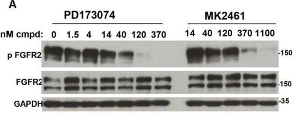
|
24968263 | |
| Growth inhibition assay | Cell viability |
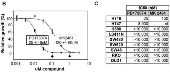
|
24968263 | |
| In Vivo | ||
| In vivo | Administration of PD173074 at 1 mg/kg/day or 2 mg/ka/day in mice can effectively block angiogenesis induced by either FGF or VEGF in a dose-dependent manner with no apparent toxicity. [1] PD173074 inhibits in vivo growth of mutant FGFR3-transfected NIH 3T3 cells in nude mice. Inhibition of FGFR3 by PD173074 delays tumor growth and increases survival of mice in a KMS11 xenograft myeloma model. [4] In the H-510 xenograft, oral aministration of PD173074 blocks tumor growth similar to that seen with single-agent cisplatin administration, increasing median survival compared with control sham-treated animals. In H-69 xenografts, PD173074 induces complete responses lasting >6 months in 50% of mice. These effects are correlated with increased apoptosis in excised tumors, but not a consequence of disrupted tumor vasculature. [5] | |
|---|---|---|
| Animal Research | Animal Models | Swiss Webster mice with induced corneal angiogenesis |
| Dosages | ~2 mg/kg/day | |
| Administration | Administered intraperitoneally | |
|
Chemical Information & Solubility
| Molecular Weight | 523.67 | Formula | C28H41N7O3 |
| CAS No. | 219580-11-7 | SDF | Download PD173074 SDF |
| Smiles | CCN(CC)CCCCNC1=NC2=NC(=C(C=C2C=N1)C3=CC(=CC(=C3)OC)OC)NC(=O)NC(C)(C)C | ||
| Storage (From the date of receipt) | |||
|
In vitro |
DMSO : 105 mg/mL ( (200.5 mM) Moisture-absorbing DMSO reduces solubility. Please use fresh DMSO.) Ethanol : 105 mg/mL Water : Insoluble |
Molecular Weight Calculator |
|
In vivo Add solvents to the product individually and in order. |
In vivo Formulation Calculator |
|||||
Preparing Stock Solutions
Molarity Calculator
In vivo Formulation Calculator (Clear solution)
Step 1: Enter information below (Recommended: An additional animal making an allowance for loss during the experiment)
mg/kg
g
μL
Step 2: Enter the in vivo formulation (This is only the calculator, not formulation. Please contact us first if there is no in vivo formulation at the solubility Section.)
% DMSO
%
% Tween 80
% ddH2O
%DMSO
%
Calculation results:
Working concentration: mg/ml;
Method for preparing DMSO master liquid: mg drug pre-dissolved in μL DMSO ( Master liquid concentration mg/mL, Please contact us first if the concentration exceeds the DMSO solubility of the batch of drug. )
Method for preparing in vivo formulation: Take μL DMSO master liquid, next addμL PEG300, mix and clarify, next addμL Tween 80, mix and clarify, next add μL ddH2O, mix and clarify.
Method for preparing in vivo formulation: Take μL DMSO master liquid, next add μL Corn oil, mix and clarify.
Note: 1. Please make sure the liquid is clear before adding the next solvent.
2. Be sure to add the solvent(s) in order. You must ensure that the solution obtained, in the previous addition, is a clear solution before proceeding to add the next solvent. Physical methods such
as vortex, ultrasound or hot water bath can be used to aid dissolving.
Tech Support
Answers to questions you may have can be found in the inhibitor handling instructions. Topics include how to prepare stock solutions, how to store inhibitors, and issues that need special attention for cell-based assays and animal experiments.
Tel: +1-832-582-8158 Ext:3
If you have any other enquiries, please leave a message.
* Indicates a Required Field
Frequently Asked Questions
Question 1:
What is the half-life of PD173074(S1264) in vivo?
Answer:
According to literature research, PD173074 is given twice daily because it has a short half-life in vivo, please refer to the following link for detailed pharmacokinetic information (Supplementary Figure 8B): http://www.ncbi.nlm.nih.gov/pmc/articles/PMC3990281/#!po=50.0000.
Tags: buy PD173074 | PD173074 supplier | purchase PD173074 | PD173074 cost | PD173074 manufacturer | order PD173074 | PD173074 distributor
Products are for research use only. Not for human use. We do not sell to patients.
©Copyright 2013 Selleck Chemicals. All Rights Reserved.