-
 Australia
-
 Austria
-
 Belgium
-
 Brazil
-
 Canada
-
 China
China
-
 Czech Republic
-
 Denmark
-
 Finland
-
 France
-
 Germany
-
 Greece
-
 Hong Kong
-
 Hungary
-
 Iceland
-
 India
India
-
 Ireland
-
 Israel
-
 Italy
Italy
-
 Japan
Japan
-
 Korea
Korea
-
 Luxembourg
-
 Malaysia
-
 Netherlands
-
 New Zealand
-
 Norway
-
 Poland
-
 Qatar
Qatar
-
 Romania
-
 Saudi Arabia
-
 Singapore
-
 Spain
Spain
-
 Sweden
-
 Switzerland
-
 Taiwan
-
 Turkey
-
 United Kingdom
-
 United States
research use only
Sepantronium Bromide (YM155) Survivin inhibitor
Cat.No.S1130
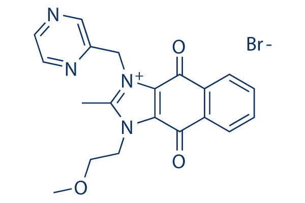
Chemical Structure
Molecular Weight: 443.29
Quality Control
| Related Targets | Bcl-2 Caspase PD-1/PD-L1 Ferroptosis p53 Apoptosis related Synthetic Lethality STAT TNF-alpha Ras |
|---|---|
| Other Survivin Inhibitors | LQZ-7I FL118 |
Cell Culture, Treatment & Working Concentration
| Cell Lines | Assay Type | Concentration | Incubation Time | Formulation | Activity Description | PMID |
|---|---|---|---|---|---|---|
| Kasumi-1 | Growth Inhibition Assay | 72 h | DMSO | IC50=0.009 ± 0.0009 μM | 25659731 | |
| M-07e | Growth Inhibition Assay | 72 h | DMSO | IC50=0.040 ± 0.013 μM | 25659731 | |
| THP-1 | Growth Inhibition Assay | 72 h | DMSO | IC50=0.051 ± 0.013 μM | 25659731 | |
| CMK | Growth Inhibition Assay | 72 h | DMSO | IC50=0.053 ± 0.009 μM | 25659731 | |
| MV4-11 | Growth Inhibition Assay | 72 h | DMSO | IC50=0.055 ± 0.028 μM | 25659731 | |
| AML-193 | Growth Inhibition Assay | 72 h | DMSO | IC50=0.462 ± 0.060 μM | 25659731 | |
| HL-60 | Growth Inhibition Assay | 72 h | DMSO | IC50=0.001 ± 0.0002 μM | 25659731 | |
| ML-2 | Growth Inhibition Assay | 72 h | DMSO | IC50=0.009 ± 0.002 μM | 25659731 | |
| OCI/AML3 | Growth Inhibition Assay | 72 h | DMSO | IC50=0.011 ± 0.002 μM | 25659731 | |
| HEL | Growth Inhibition Assay | 72 h | DMSO | IC50=0.559 ± 0.038 μM | 25659731 | |
| ME-1 | Growth Inhibition Assay | 72 h | DMSO | IC50=0.684 ± 0.179 μM | 25659731 | |
| THP-1 | Apoptosis Assay | 1 μM | 72 h | DMSO | induces apoptosis | 25659731 |
| M-07e | Function Assay | 0–1 μM | 72 h | DMSO | induces downregulation of Survivin | 25659731 |
| THP-1 | Function Assay | 0–1 μM | 72 h | DMSO | induces downregulation of Survivin | 25659731 |
| CMK | Function Assay | 0–1 μM | 72 h | DMSO | induces downregulation of Survivin | 25659731 |
| AML-193 | Function Assay | 0–1 μM | 72 h | DMSO | induces downregulation of Survivin | 25659731 |
| Kasumi-1 | Function Assay | 0–1 μM | 72 h | DMSO | induces downregulation of Survivin | 25659731 |
| MV4-11 | Function Assay | 0–1 μM | 72 h | DMSO | induces downregulation of Survivin | 25659731 |
| MUG-Chor | Growth Inhibition Assay | 0-5 μM | 24/48 h | IC50=7.05 nM for 48h | 25640185 | |
| U-CH1 | Growth Inhibition Assay | 0-5 μM | 24/48 h | IC50=9.03 nM for 48h | 25640185 | |
| KATOIII | Growth Inhibition Assay | 10/20 nM | 48 h | inhibits cell growth in a dose-dependent manner | 25635055 | |
| AGS | Growth Inhibition Assay | 10/20 nM | 48 h | inhibits cell growth in a dose-dependent manner | 25635055 | |
| SACC-83 | Function Assay | 5 nM | 48 h | decreases nuclear expression of HIF-1α | 25485635 | |
| INA-6 | Growth Inhibition Assay | 0-500 nM | 48 h | inhibits cell growth in a dose-dependent manner | 25296978 | |
| U-266 | Growth Inhibition Assay | 0-500 nM | 48 h | inhibits cell growth in a dose-dependent manner | 25296978 | |
| MOLP-8 | Growth Inhibition Assay | 0-500 nM | 48 h | inhibits cell growth in a dose-dependent manner | 25296978 | |
| HG-1 | Growth Inhibition Assay | 0-500 nM | 48 h | inhibits cell growth in a dose-dependent manner | 25296978 | |
| NCI-H929 | Growth Inhibition Assay | 0-500 nM | 48 h | inhibits cell growth in a dose-dependent manner | 25296978 | |
| OPM-2 | Growth Inhibition Assay | 0-500 nM | 48 h | inhibits cell growth in a dose-dependent manner | 25296978 | |
| L-363 | Growth Inhibition Assay | 0-500 nM | 48 h | inhibits cell growth in a dose-dependent manner | 25296978 | |
| MOLP-2 | Growth Inhibition Assay | 0-500 nM | 48 h | inhibits cell growth in a dose-dependent manner | 25296978 | |
| KMS-12-BM | Growth Inhibition Assay | 0-500 nM | 48 h | inhibits cell growth in a dose-dependent manner | 25296978 | |
| SK-MM-2 | Growth Inhibition Assay | 0-500 nM | 48 h | inhibits cell growth in a dose-dependent manner | 25296978 | |
| U-266 | Apoptosis Assay | 0-50 nM | 24 h | induces apoptosis | 25296978 | |
| INA-6 | Apoptosis Assay | 0-50 nM | 24 h | induces apoptosis | 25296978 | |
| MCF7 | Growth Inhibition Assay | 72 h | IC50=13 ± 6 nM | 25220225 | ||
| MCF7-TamR6 | Growth Inhibition Assay | 72 h | IC50=8 ± 6 nM | 25220225 | ||
| MCF7-TamR7 | Growth Inhibition Assay | 72 h | IC50=8 ± 3 nM | 25220225 | ||
| MCF7-TamR8 | Growth Inhibition Assay | 72 h | IC50=15 ± 6 nM | 25220225 | ||
| MCF7-TamC3 | Growth Inhibition Assay | 72 h | IC50=6 ± 3 nM | 25220225 | ||
| MCF7-TamC6 | Growth Inhibition Assay | 72 h | IC50=6 ± 0.1 nM | 25220225 | ||
| MDA-MB-231 | Growth Inhibition Assay | 72 h | IC50=5 ± 1 nM | 25220225 | ||
| SK-BR-3 | Growth Inhibition Assay | 72 h | IC50=7 ± 0.3 nM | 25220225 | ||
| Eca109 | Function Assay | 1-50 nM | 48 h | DMSO | suppresses survivin expression in a dose dependent manner | 25139395 |
| TE13 | Function Assay | 1-50 nM | 48 h | DMSO | suppresses survivin expression in a dose dependent manner | 25139395 |
| Eca109 | Growth Inhibition Assay | 0-100 nM | 24/48 h | DMSO | decreases cell viability in a dose-dependent manner | 25139395 |
| TE13 | Growth Inhibition Assay | 0-100 nM | 24/48 h | DMSO | decreases cell viability in a dose-dependent manner | 25139395 |
| MT-3 | Kinase Assay | 72 h | DMSO | IC50=2.86 ± 0.54 nM for DR4 expression | 24866585 | |
| MDA-MB-468 | Kinase Assay | 72 h | DMSO | IC50=0.11 ± 0.01 nM for DR4 expression | 24866585 | |
| SUM-159 | Kinase Assay | 72 h | DMSO | IC50=1.72 ± 0.33 nM for DR4 expression | 24866585 | |
| MT-3 | Kinase Assay | 72 h | DMSO | IC50=54.11 ± 4.32 nM for DR5 expression | 24866585 | |
| MDA-MB-468 | Kinase Assay | 72 h | DMSO | IC50=0.07 ± 0.02 nM for DR5 expression | 24866585 | |
| SUM-159 | Kinase Assay | 72 h | DMSO | IC50=69.4 ± 4.23 nM for DR5 expression | 24866585 | |
| MT-3 + NAC | Kinase Assay | 72 h | DMSO | IC50=56.2 ± 2.07 nM for DR5 expression | 24866585 | |
| MT-3 + SB203580 | Kinase Assay | 72 h | DMSO | IC50=38.41 ± 5.02 nM for DR5 expression | 24866585 | |
| DB | Growth Inhibition Assay | 10 nM | 24 h | DMSO | inhibits cell proliferation | 24486595 |
| SU-DHL-8 | Growth Inhibition Assay | 10 nM | 24 h | DMSO | inhibits cell proliferation | 24486595 |
| WSU-DLCL2 | Growth Inhibition Assay | 10 nM | 24 h | DMSO | inhibits cell proliferation | 24486595 |
| ACC-2 | Growth Inhibition Assay | 0-100 nM | 24 h | inhibits cell growth in a dose-dependent manner | 24370995 | |
| ACC-2 | Apoptosis Assay | 0-20 nM | 24 h | induces apoptosis in a dose-dependent manner | 24370995 | |
| ACC-2 | Function Assay | 0-20 nM | 24 h | increases the conversion of LC3I to LC3II | 24370995 | |
| BFTC905 | Growth Inhibition Assay | 0-1000 nM | 48 h | DMSO | IC50=20 nM, inhibits cell growth in a dose-dependent manner | 24297644 |
| T24 | Growth Inhibition Assay | 0-1000 nM | 48 h | DMSO | IC50=20 nM, inhibits cell growth in a dose-dependent manner | 24297644 |
| TSGH8301 | Growth Inhibition Assay | 0-1000 nM | 48 h | DMSO | IC50=20 nM, inhibits cell growth in a dose-dependent manner | 24297644 |
| BFTC909 | Growth Inhibition Assay | 0-1000 nM | 48 h | DMSO | IC50=20 nM, inhibits cell growth in a dose-dependent manner | 24297644 |
| BFTC905 | Apoptosis Assay | 20 nM | 48 h | DMSO | induces apoptosis | 24297644 |
| BFTC905 | Function Assay | 20 nM | 48 h | DMSO | decreases the expression of LC3B-II | 24297644 |
| A2780p | Function Assay | 0-100 nM | 24 h | DMSO | induces Survivin downregulation | 24262875 |
| A2780cis | Function Assay | 0-100 nM | 24 h | DMSO | induces Survivin downregulation | 24262875 |
| A2780p | Apoptosis Assay | 5-100 nM | 24/48 h | DMSO | increases apoptosis in a concentration-dependent manner | 24262875 |
| A2780cis | Apoptosis Assay | 5-100 nM | 24/48 h | DMSO | increases apoptosis in a concentration-dependent manner | 24262875 |
| SH-SY5Y | Apoptosis Assay | 1/10/100 µM | 72 h | DMSO | induces apoptosis in a dose-dependent manner | 24254560 |
| HL-60 | Growth Inhibition Assay | 72 h | DMSO | IC50=0.3 nM | 23618862 | |
| U937 | Growth Inhibition Assay | 72 h | DMSO | IC50=0.8 nM | 23618862 | |
| HL-60 | Function Assay | 1 μM | 6/12/24 h | DMSO | inhibits the expression of survivin | 23618862 |
| U937 | Function Assay | 1 μM | 6/12/24 h | DMSO | inhibits the expression of survivin | 23618862 |
| HL-60 | Apoptosis Assay | 0.1/1 μM | 8 h | DMSO | induces apoptosis | 23618862 |
| Sk-NEP-1 | Growth Inhibition Assay | 1-10000 nM | 24 h | DMSO | IC50=100 nM | 23267699 |
| SK-NEP-1 | Apoptosis Assay | 50/100 nM | 12/24 h | DMSO | induces apoptosis | 23267699 |
| TC-32 | Growth Inhibition Assay | 0.1-1000 nM | EC50=3.0 nM, inhibits cell growth in a dose-dependent manner | 22961763 | ||
| TC-71 | Growth Inhibition Assay | 0.1-1000 nM | EC50=5.7 nM, inhibits cell growth in a dose-dependent manner | 22961763 | ||
| SK-ES-1 | Growth Inhibition Assay | 0.1-1000 nM | EC50=2.8 nM, inhibits cell growth in a dose-dependent manner | 22961763 | ||
| RD-ES | Growth Inhibition Assay | 0.1-1000 nM | EC50=6.2 nM, inhibits cell growth in a dose-dependent manner | 22961763 | ||
| HEK293 | Growth Inhibition Assay | 0.1-1000 nM | EC50=23.0 nM, inhibits cell growth in a dose-dependent manner | 22961763 | ||
| M059J | Growth Inhibition Assay | 0-50 nM | 48 h | inhibits cell growth in a dose-dependent manner | 22770110 | |
| M059K | Growth Inhibition Assay | 0-50 nM | 48 h | inhibits cell growth in a dose-dependent manner | 22770110 | |
| M059J | Apoptosis Assay | 30 nM | 24 h | induces apoptosis | 22770110 | |
| M059K | Apoptosis Assay | 30 nM | 24 h | induces apoptosis | 22770110 | |
| PANC-1 | Growth Inhibition Assay | 0.01-1000 nM | 48 h | IC50=3.69 nM | 22723871 | |
| MIAPaCa-2 | Growth Inhibition Assay | 0.01-1000 nM | 48 h | IC50=29.36 nM | 22723871 | |
| BxPC-3 | Growth Inhibition Assay | 0.01-1000 nM | 48 h | IC50=30.26 nM | 22723871 | |
| PANC-1 | Function Assay | 0-1000 nM | 24 h | induces downregulation of XIAP and survivin expression | 22723871 | |
| MIAPaCa-2 | Function Assay | 0-1000 nM | 24 h | induces downregulation of XIAP and survivin expression | 22723871 | |
| BxPC-3 | Function Assay | 0-1000 nM | 24 h | induces downregulation of XIAP and survivin expression | 22723871 | |
| RPMI-7951 | Growth Inhibition Assay | GI50=3.2 nM | 21737502 | |||
| SK-MEL-5 | Growth Inhibition Assay | GI50=4.2 nM | 21737502 | |||
| A375 | Growth Inhibition Assay | GI50=6.3 nM | 21737502 | |||
| SK-MEL-28 | Growth Inhibition Assay | GI50=7.6 nM | 21737502 | |||
| SK-MEL-2 | Growth Inhibition Assay | GI50=11 nM | 21737502 | |||
| DB | Growth Inhibition Assay | 48 h | GI50=3.5 nM | 21237508 | ||
| Pfeiffer | Growth Inhibition Assay | 48 h | GI50=3.9 nM | 21237508 | ||
| SU-DHL-5 | Growth Inhibition Assay | 48 h | GI50=0.23 nM | 21237508 | ||
| SU-DHL-8 | Growth Inhibition Assay | 48 h | GI50=1.4 nM | 21237508 | ||
| WSU-DLCL-2 | Growth Inhibition Assay | 48 h | GI50=1.4 nM | 21237508 | ||
| A549 | Growth inhibition assay | Growth inhibition of human A549 cells, IC50=0.0134μM | 28814374 | |||
| DU145 | Cytotoxicity assay | Cytotoxicity against human DU145 cells by MTT assay, EC50=0.0138μM | 28774426 | |||
| PC3 | Cytotoxicity assay | Cytotoxicity against human PC3 cells by MTT assay, EC50=0.092μM | 28774426 | |||
| A549 | Function assay | Inhibition of PAK1 in human A549 cells, IC50=0.5μM | 28814374 | |||
| Click to View More Cell Line Experimental Data | ||||||
Solubility
|
In vitro |
Water : 89 mg/mL
DMSO
: 55 mg/mL
(124.07 mM)
Ethanol : 6 mg/mL |
Molarity Calculator
|
In vivo |
|||||
In vivo Formulation Calculator (Clear solution)
Step 1: Enter information below (Recommended: An additional animal making an allowance for loss during the experiment)
Step 2: Enter the in vivo formulation (This is only the calculator, not formulation. Please contact us first if there is no in vivo formulation at the solubility Section.)
Calculation results:
Working concentration: mg/ml;
Method for preparing DMSO master liquid: mg drug pre-dissolved in μL DMSO ( Master liquid concentration mg/mL, Please contact us first if the concentration exceeds the DMSO solubility of the batch of drug. )
Method for preparing in vivo formulation: Take μL DMSO master liquid, next addμL PEG300, mix and clarify, next addμL Tween 80, mix and clarify, next add μL ddH2O, mix and clarify.
Method for preparing in vivo formulation: Take μL DMSO master liquid, next add μL Corn oil, mix and clarify.
Note: 1. Please make sure the liquid is clear before adding the next solvent.
2. Be sure to add the solvent(s) in order. You must ensure that the solution obtained, in the previous addition, is a clear solution before proceeding to add the next solvent. Physical methods such
as vortex, ultrasound or hot water bath can be used to aid dissolving.
Chemical Information, Storage & Stability
| Molecular Weight | 443.29 | Formula | C20H19BrN4O3 |
Storage (From the date of receipt) | |
|---|---|---|---|---|---|
| CAS No. | 781661-94-7 | Download SDF | Storage of Stock Solutions |
|
|
Mechanism of Action
| Targets/IC50/Ki |
Survivin
(HeLa-SURP-luc, CHO-SV40-luc cells) 0.54 nM
|
|---|---|
| In vitro |
Sepantronium Bromide (YM155) is not sensitive to survivn gene promoter-driven luciferase reporter activity even at 30 μM. It significantly inhibits endogenous survivin expression in PC-3 and PPC-1 human HRPC cells with deficient p53 through transcriptional inhibition of the survivin gene promoter. On the contrary this compound shows no sufficient effect on protein expression of c-IAP2, XIAP, Bcl-2, Bcl-xL, Bad, α-actin, and β-tubulin at 100 nM. It indicates great apoptosis in human cancer cell lines including PC-3 and PPC-1 with a concomitant increase in caspase-3 activity. YM155 potently inhibits human cancer cell lines (mutated or truncated p53) including PC-3, PPC-1, DU145, TSU-Pr1, 22Rv1, SK-MEL-5 and A375 with IC50 from 2.3 to 11 nM, respectively. It increases the sensitivity of NSCLC cells to γ-radiation. The combination of YM155 and γ-radiation increases both the number of apoptotic cells and the activity of caspase-3. This compound delays the repair of radiation-induced double-strand breaks in nuclear DNA. |
| Kinase Assay |
Promoter-luciferase reporter assay
|
|
A 2,767-bp sequence of human survivin gene promoter is isolated from human genomic DNA by PCR using Pyrobest polymerase and the following primers: 5
|
|
| In vivo |
Sepantronium Bromide (YM155) completely inhibits the tumor growth of PC-3 s.c. xenografted prostate tumors at doses of 3 and 10 mg/kg, without body weight loss and blood cell count decrease. Pharmacokinetic analysis shows that it is highly distributed to tumor tissue. Moreover, this compound shows 80% TGI at a dose of 5 mg/kg in PC-3 orthotopic xenografts. The combination therapy with YM155 and γ-radiation shows great antitumor activity against H460 or Calu6 xenografts in nude mice. |
References |
|
Applications
| Methods | Biomarkers | Images | PMID |
|---|---|---|---|
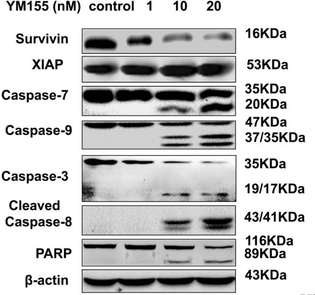
| Western blot | Survivin / XIAP / Caspase-7 / Caspase-9 / Caspase-3 / Cleaved Caspase-8 / PARP β-catenin / c-Myc / Cyclin D1 / CD44 p-STAT3 / STAT3 |
|
26771139 |
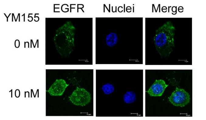
| Immunofluorescence | EGFR PARP-1 / PAR |
|
22723871 |
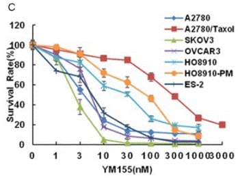
| Growth inhibition assay | Cell viability |
|
29636860 |
Clinical Trial Information
(data from https://clinicaltrials.gov, updated on 2024-05-22)
| NCT Number | Recruitment | Conditions | Sponsor/Collaborators | Start Date | Phases |
|---|---|---|---|---|---|
| NCT05263583 | Recruiting | High-grade B-cell Lymphoma|Burkitt Lymphoma|Lymphoma B-Cell|Lymphoma Large B-Cell Diffuse|Lymphatic Diseases|Lymphoma High-Grade|C-MYC/BCL2 Double-Hit High-Grade B-Cell Lymphoma|C-MYC/BCL6 Double-Hit High-Grade B-Cell Lymphoma|C-Myc Gene Rearrangement |
Cothera Bioscience Inc |
December 9 2022 | Phase 2 |
| NCT01023386 | Completed | Cancer |
Astellas Pharma Inc |
November 2009 | Phase 1 |
Tech Support
Tel: +1-832-582-8158 Ext:3
If you have any other enquiries, please leave a message.
Signaling Pathway Map
Products are for research use only. Not for human use. We do not sell to patients.
©Copyright 2013 Selleck Chemicals. All Rights Reserved.