-
 Australia
-
 Austria
-
 Belgium
-
 Brazil
-
 Canada
-
 China
China
-
 Czech Republic
-
 Denmark
-
 Finland
-
 France
-
 Germany
-
 Greece
-
 Hong Kong
-
 Hungary
-
 Iceland
-
 India
India
-
 Ireland
-
 Israel
-
 Italy
Italy
-
 Japan
Japan
-
 Korea
Korea
-
 Luxembourg
-
 Malaysia
-
 Netherlands
-
 New Zealand
-
 Norway
-
 Poland
-
 Qatar
Qatar
-
 Romania
-
 Saudi Arabia
-
 Singapore
-
 Spain
Spain
-
 Sweden
-
 Switzerland
-
 Taiwan
-
 Turkey
-
 United Kingdom
-
 United States
research use only
Dovitinib (TKI-258) FLT3 inhibitor
Cat.No.S1018
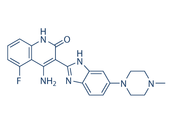
Chemical Structure
Molecular Weight: 392.43
Quality Control
| Related Targets | EGFR VEGFR JAK PDGFR FGFR Src HIF FLT HER2 Bcr-Abl |
|---|---|
| Other FLT3 Inhibitors | UNC2025 Crenolanib (CP-868596) Dovitinib (TKI258) Lactate monohydrate Tandutinib (MLN518) KW-2449 ENMD-2076 AST-487 (NVP-AST487) TCS 359 FF-10101 G-749 |
Cell Culture, Treatment & Working Concentration
| Cell Lines | Assay Type | Concentration | Incubation Time | Formulation | Activity Description | PMID |
|---|---|---|---|---|---|---|
| SupB15 | Growth Inhibition Assay | IC50=0.449 μM | 25202073 | |||
| SupB15-R | Growth Inhibition Assay | IC50=0.558 μM | 25202073 | |||
| BaF3-pSRα | Growth Inhibition Assay | IC50=0.668 μM | 25202073 | |||
| BaF3-p210Bcr-Abl | Growth Inhibition Assay | IC50=0.692 μM | 25202073 | |||
| BaF3-p210Bcr-Abl-T315I | Growth Inhibition Assay | IC50=2.626 μM | 25202073 | |||
| CCRF-CEM | Growth Inhibition Assay | IC50=0.398 μM | 25202072 | |||
| CEM/C2 | Growth Inhibition Assay | IC50=1.125 μM | 25202072 | |||
| Nalm-6 | Growth Inhibition Assay | IC50=0.382 μM | 25202072 | |||
| SEM-K2 | Growth Inhibition Assay | IC50=0.022 μM | 25202072 | |||
| HB-1119 | Growth Inhibition Assay | IC50=0.028 μM | 25202072 | |||
| RS4:11 | Growth Inhibition Assay | IC50=2.81 μM | 25202072 | |||
| Nalm-6 | Apoptosis Assay | 2 μM | 24/48 h | induces apoptosis resulting in about 72% of cell death after 24 h treatment and 81% after 48 h | 25202072 | |
| SEM-K2 | Apoptosis Assay | 0.1/1 μM | 24 h | induces early apoptosis of SEM-K2 cells at 0.1 μM after 24 h | 25202072 | |
| HCT-116 | Growth Inhibition Assay | IC50=3.050.58 μM | 24495750 | |||
| HT-29 | Growth Inhibition Assay | IC50=5.21.93 μM | 24495750 | |||
| SW-480 | Growth Inhibition Assay | IC50=4.330.47 μM | 24495750 | |||
| CaCO2 | Growth Inhibition Assay | IC50=3.230.64 μM | 24495750 | |||
| LS174T | Growth Inhibition Assay | IC50=4.330.47 μM | 24495750 | |||
| HEC-1A | Function Assay | 0.05/0.1/0.5 μM | 72 h | causes a decrease in STAT3, ERK, and AKT phosphorylation | 24495750 | |
| AN3CA | Function Assay | 0.05/0.1/0.5 μM | 72 h | causes a decrease in STAT3, ERK, and AKT phosphorylation | 24495750 | |
| MFE-296 | Function Assay | 0.05/0.1/0.5 μM | 72 h | causes a decrease in STAT3, ERK, and AKT phosphorylation | 24495750 | |
| UMC3 | Cell Viability Assay | 1-10 μM | 72 h | inhibits cell growth in a dose dependent manner | 24325461 | |
| 5637 | Cell Viability Assay | 1-10 μM | 72 h | inhibits cell growth in a dose dependent manner | 24325461 | |
| HU456 | Cell Viability Assay | 1-10 μM | 72 h | inhibits cell growth in a dose dependent manner | 24325461 | |
| MGHU4 | Cell Viability Assay | 1-10 μM | 72 h | inhibits cell growth in a dose dependent manner | 24325461 | |
| HT1376 | Cell Viability Assay | 1-10 μM | 72 h | inhibits cell growth in a dose dependent manner | 24325461 | |
| RT112 | Cell Viability Assay | 1-10 μM | 72 h | inhibits cell growth in a dose dependent manner | 24325461 | |
| T24 | Cell Viability Assay | 1-10 μM | 72 h | inhibits cell growth in a dose dependent manner | 24325461 | |
| BFTC905 | Cell Viability Assay | 1-10 μM | 72 h | inhibits cell growth in a dose dependent manner | 24325461 | |
| TCC-SUP | Cell Viability Assay | 1-10 μM | 72 h | inhibits cell growth in a dose dependent manner | 24325461 | |
| RT4 | Cell Viability Assay | 1-10 μM | 72 h | inhibits cell growth in a dose dependent manner | 24325461 | |
| HONE1 | Growth Inhibition Assay | 0.1-10 μM | 48 h | induces G2/M delay in a concentration-dependent manner | 24238094 | |
| HNE1 | Growth Inhibition Assay | 0.1-10 μM | 48 h | induces G2/M delay in a concentration-dependent manner | 24238094 | |
| CNE2 | Growth Inhibition Assay | 0.1-10 μM | 48 h | induces G2/M delay in a concentration-dependent manner | 24238094 | |
| C666-1 | Growth Inhibition Assay | 0.1-10 μM | 48 h | induces G2/M delay in a concentration-dependent manner | 24238094 | |
| HeLa | Growth Inhibition Assay | 0.1-10 μM | 24 h | induces G2/M arrest in a concentration-dependent manner | 24238094 | |
| Hep3B | Growth Inhibition Assay | 0.1-10 μM | 24 h | induces G2 arrest | 24238094 | |
| HepG2 | Growth Inhibition Assay | 48 h | IC50=2.727 ± 0.429 μM | 23546591 | ||
| Hep3B | Growth Inhibition Assay | 48 h | IC50=4.223 ± 0.839 μM | 23546591 | ||
| PLC/PRF5 | Growth Inhibition Assay | 48 h | IC50=16.120 ± 4.001 μM | 23546591 | ||
| Huh7 | Growth Inhibition Assay | 48 h | IC50=15.007 ± 7.334 μM | 23546591 | ||
| HepG2 | Growth Inhibition Assay | 72 h | IC50=1.200 ± 0.226 μM | 23546591 | ||
| Hep3B | Growth Inhibition Assay | 72 h | IC50=0.892 ± 0.044 μM | 23546591 | ||
| PLC/PRF5 | Growth Inhibition Assay | 72 h | IC50=3.110 ± 0.337 μM | 23546591 | ||
| Huh7 | Growth Inhibition Assay | 72 h | IC50=3.980 ± 0.803 μM | 23546591 | ||
| MFE280 | Growth Inhibition Assay | IC50=0.42 ± 0.06 μM | 23443805 | |||
| AN3CA | Growth Inhibition Assay | IC50=0.50 ± 0.10 μM | 23443805 | |||
| HEC155 | Growth Inhibition Assay | IC50=0.66 ± 0.09 μM | 23443805 | |||
| MFE296 | Growth Inhibition Assay | IC50=0.66 ± 0.19 μM | 23443805 | |||
| SPAC1S | Growth Inhibition Assay | IC50=0.77 ± 0.08 μM | 23443805 | |||
| RL952 | Growth Inhibition Assay | IC50=0.93 ± 0.01 μM | 23443805 | |||
| EN1 | Growth Inhibition Assay | IC50=1.02 ± 0.25 μM | 23443805 | |||
| SNGII | Growth Inhibition Assay | IC50=1.24 ± 0.28 μM | 23443805 | |||
| ISHIKAWA | Growth Inhibition Assay | IC50=1.30 ± 0.11 μM | 23443805 | |||
| HEC1A | Growth Inhibition Assay | IC50=1.34 ± 0.30 μM | 23443805 | |||
| KLE | Growth Inhibition Assay | IC50=1.37 ± 0.02 μM | 23443805 | |||
| SNGM | Growth Inhibition Assay | IC50=1.42 ± 0.13 μM | 23443805 | |||
| USPC2 | Growth Inhibition Assay | IC50=1.62 ± 0.01 μM | 23443805 | |||
| EN | Growth Inhibition Assay | IC50=1.66 ± 0.01 μM | 23443805 | |||
| MFE319 | Growth Inhibition Assay | IC50=1.87 ± 0.45 μM | 23443805 | |||
| EFE184 | Growth Inhibition Assay | IC50=2.04 ± 0.13 μM | 23443805 | |||
| ECC1 | Growth Inhibition Assay | IC50=2.07 ± 0.01 μM | 23443805 | |||
| HEC1B | Growth Inhibition Assay | IC50=2.57 ± 0.23 μM | 23443805 | |||
| USPC1 | Growth Inhibition Assay | IC50=2.60 ± 0.13 μM | 23443805 | |||
| SPAC1L | Growth Inhibition Assay | IC50=3.06 ± 1.14 μM | 23443805 | |||
| HUVEC | Cell Viability Assay | 0-25 μM | 72 h | DMSO | inhibits cell growth in a dose dependent manner | 23228017 |
| HMVEC | Cell Viability Assay | 0-25 μM | 72 h | DMSO | inhibits cell growth in a dose dependent manner | 23228017 |
| MHCC-97H | Cell Viability Assay | 0-25 μM | 72 h | DMSO | inhibits cell growth in a dose dependent manner | 23228017 |
| SMMC7721 | Cell Viability Assay | 0-25 μM | 72 h | DMSO | inhibits cell growth in a dose dependent manner | 23228017 |
| Huh-7 | Apoptosis Assay | 0-12.5 μM | 24 h | DMSO | sensitizes HCC cells to TRAIL- and tigatuzumab-induced apoptosis in a dose-dependent manner | 22230479 |
| Sk-Hep1 | Apoptosis Assay | 0-12.5 μM | 24 h | DMSO | sensitizes HCC cells to TRAIL- and tigatuzumab-induced apoptosis in a dose-dependent manner | 22230479 |
| Hep3B | Apoptosis Assay | 0-12.5 μM | 24 h | DMSO | sensitizes HCC cells to TRAIL- and tigatuzumab-induced apoptosis in a dose-dependent manner | 22230479 |
| PLC5 | Apoptosis Assay | 0-12.5 μM | 24 h | DMSO | sensitizes HCC cells to TRAIL- and tigatuzumab-induced apoptosis in a dose-dependent manner | 22230479 |
| PLC5 | Cell Viability Assay | 0-15 μM | 72 h | reduces cell viability in a dose-dependent manner | 22180308 | |
| Hep3B | Cell Viability Assay | 0-15 μM | 72 h | reduces cell viability in a dose-dependent manner | 22180308 | |
| Sk-Hep1 | Cell Viability Assay | 0-15 μM | 72 h | reduces cell viability in a dose-dependent manner | 22180308 | |
| Huh-7 | Cell Viability Assay | 0-15 μM | 72 h | reduces cell viability in a dose-dependent manner | 22180308 | |
| PLC5 | Apoptosis Assay | 0-15 μM | 24 h | increases apoptotic cell death in a dose-dependent manner | 22180308 | |
| Hep3B | Apoptosis Assay | 0-15 μM | 24 h | increases apoptotic cell death in a dose-dependent manner | 22180308 | |
| Sk-Hep1 | Apoptosis Assay | 0-15 μM | 24 h | increases apoptotic cell death in a dose-dependent manner | 22180308 | |
| Huh-7 | Apoptosis Assay | 0-15 μM | 24 h | increases apoptotic cell death in a dose-dependent manner | 22180308 | |
| PLC5 | Function Assay | 0-10 μM | 24 h | causes dose-dependent DNA fragmentation | 22180308 | |
| Hep3B | Function Assay | 0-10 μM | 24 h | causes dose-dependent DNA fragmentation | 22180308 | |
| Sk-Hep1 | Function Assay | 0-10 μM | 24 h | causes dose-dependent DNA fragmentation | 22180308 | |
| Huh-7 | Function Assay | 0-10 μM | 24 h | causes dose-dependent DNA fragmentation | 22180308 | |
| SW780 | Growth Inhibition Assay | 5 d | IC50=50 nM | 21119661 | ||
| RT112 | Growth Inhibition Assay | 5 d | IC50=15 nM | 21119661 | ||
| RT4 | Growth Inhibition Assay | 5 d | IC50=5 nM | 21119661 | ||
| JMSU1 | Growth Inhibition Assay | 5 d | IC50=50 nM | 21119661 | ||
| J82 | Growth Inhibition Assay | 5 d | IC50=1400 nM | 21119661 | ||
| 97-7 | Growth Inhibition Assay | 5 d | IC50=1000 nM | 21119661 | ||
| RT112 | Function Assay | 500 nM | 24 h | increases the proportion of cells in G1 accompanied by a decrease in S and G2/M phases | 21119661 | |
| RT4 | Function Assay | 500 nM | 24 h | increases the proportion of cells in G1 accompanied by a decrease in S and G2/M phases | 21119661 | |
| MGH-U3 | Function Assay | 500 nM | 24 h | increases the proportion of cells in G1 accompanied by a decrease in S and G2/M phases | 21119661 | |
| SW780 | Function Assay | 500 nM | 24 h | increases the proportion of cells in G1 accompanied by a decrease in S and G2/M phases | 21119661 | |
| 97-7 | Function Assay | 500 nM | 24 h | increases the proportion of cells in G1 accompanied by a decrease in S and G2/M phases | 21119661 | |
| J807C | Cell Viability Assay | 0-400 nM | 48 h | inhibits cell growth in a dose dependent manner | 15598814 | |
| Y373C | Cell Viability Assay | 0-400 nM | 48 h | inhibits cell growth in a dose dependent manner | 15598814 | |
| K650E | Cell Viability Assay | 0-400 nM | 48 h | inhibits cell growth in a dose dependent manner | 15598814 | |
| G384D | Cell Viability Assay | 0-400 nM | 48 h | inhibits cell growth in a dose dependent manner | 15598814 | |
| F384L | Cell Viability Assay | 0-400 nM | 48 h | inhibits cell growth in a dose dependent manner | 15598814 | |
| KMS11 | Growth Inhibition Assay | 72 h | IC50=90 nM | 15598814 | ||
| KMS18 | Growth Inhibition Assay | 72 h | IC50=550 nM | 15598814 | ||
| OPM2 | Growth Inhibition Assay | 72 h | IC50=90 nM | 15598814 | ||
| H929 | Growth Inhibition Assay | 72 h | IC50> 2500 nM | 15598814 | ||
| 8226 | Growth Inhibition Assay | 72 h | IC50> 2500 nM | 15598814 | ||
| U266 | Growth Inhibition Assay | 72 h | IC50> 2500 nM | 15598814 | ||
| KM12L4A | Function assay | Inhibition of VEGFR2 phosphorylation expressed in human KM12L4A cells by Western blot analysis, EC50=0.046μM | 19113866 | |||
| KM12L4A | Function assay | Inhibition of PDGFRbeta phosphorylation expressed in human KM12L4A cells Western blot analysis, EC50=0.051μM | 19113866 | |||
| KM12L4A | Function assay | Inhibition of FGFR1 phosphorylation expressed in human KM12L4A cells by Western blot analysis, EC50=0.166μM | 19113866 | |||
| insect cells | Function assay | 1 to 4 hrs | Inhibition of recombinant PDGFRbeta (unknown origin) expressed in baculovirus infected insect cells using biotinylated peptide substrate GGLFDDPSYVNVQNL in presence of ATP incubated for 1 to 4 hrs by time resolved fluorescence assay, IC50=0.001μM | 27914362 | ||
| Sf9 | Function assay | 1 to 4 hrs | Inhibition of recombinant human N-terminal GST/His6-tagged c-KIT (544 to 976 residues) expressed in baculovirus infected sf9 cells using biotinylated peptide substrate GGLFDDPSYVNVQNL in presence of ATP incubated for 1 to 4 hrs by time resolved fluorescen, IC50=0.001μM | 27914362 | ||
| Sf9 | Function assay | 1 to 4 hrs | Inhibition of recombinant human N-terminal GST/His6-tagged FLT3 (571 to 993 residues) expressed in baculovirus infected sf9 cells using biotinylated peptide substrate GGLFDDPSYVNVQNL in presence of ATP incubated for 1 to 4 hrs by time resolved fluorescenc, IC50=0.001μM | 27914362 | ||
| insect cells | Function assay | 1 to 4 hrs | Inhibition of recombinant FGFR1 (unknown origin) expressed in baculovirus infected insect cells using biotinylated peptide substrate GGGGQDGKDYIVLPI in presence of ATP incubated for 1 to 4 hrs by time resolved fluorescence assay, IC50=0.008μM | 27914362 | ||
| insect cells | Function assay | 1 to 4 hrs | Inhibition of recombinant VEGFR3 (unknown origin) expressed in baculovirus infected insect cells using biotinylated peptide substrate GGGGQDGKDYIVLPI in presence of ATP incubated for 1 to 4 hrs by time resolved fluorescence assay, IC50=0.008μM | 27914362 | ||
| insect cells | Function assay | 1 to 4 hrs | Inhibition of recombinant VEGFR1 (unknown origin) expressed in baculovirus infected insect cells using biotinylated peptide substrate GGGGQDGKDYIVLPI in presence of ATP incubated for 1 to 4 hrs by time resolved fluorescence assay, IC50=0.01μM | 27914362 | ||
| insect cells | Function assay | 1 to 4 hrs | Inhibition of recombinant VEGFR2 (unknown origin) expressed in baculovirus infected insect cells using biotinylated peptide substrate GGGGQDGKDYIVLPI in presence of ATP incubated for 1 to 4 hrs by time resolved fluorescence assay, IC50=0.013μM | 27914362 | ||
| TC32 | qHTS assay | qHTS of pediatric cancer cell lines to identify multiple opportunities for drug repurposing: Primary screen for TC32 cells | 29435139 | |||
| SJ-GBM2 | qHTS assay | qHTS of pediatric cancer cell lines to identify multiple opportunities for drug repurposing: Primary screen for SJ-GBM2 cells | 29435139 | |||
| A673 | qHTS assay | qHTS of pediatric cancer cell lines to identify multiple opportunities for drug repurposing: Primary screen for A673 cells | 29435139 | |||
| SK-N-MC | qHTS assay | qHTS of pediatric cancer cell lines to identify multiple opportunities for drug repurposing: Primary screen for SK-N-MC cells | 29435139 | |||
| BT-37 | qHTS assay | qHTS of pediatric cancer cell lines to identify multiple opportunities for drug repurposing: Primary screen for BT-37 cells | 29435139 | |||
| NB-EBc1 | qHTS assay | qHTS of pediatric cancer cell lines to identify multiple opportunities for drug repurposing: Primary screen for NB-EBc1 cells | 29435139 | |||
| U-2 OS | qHTS assay | qHTS of pediatric cancer cell lines to identify multiple opportunities for drug repurposing: Primary screen for U-2 OS cells | 29435139 | |||
| Saos-2 | qHTS assay | qHTS of pediatric cancer cell lines to identify multiple opportunities for drug repurposing: Primary screen for Saos-2 cells | 29435139 | |||
| SK-N-SH | qHTS assay | qHTS of pediatric cancer cell lines to identify multiple opportunities for drug repurposing: Primary screen for SK-N-SH cells | 29435139 | |||
| NB1643 | qHTS assay | qHTS of pediatric cancer cell lines to identify multiple opportunities for drug repurposing: Primary screen for NB1643 cells | 29435139 | |||
| LAN-5 | qHTS assay | qHTS of pediatric cancer cell lines to identify multiple opportunities for drug repurposing: Primary screen for LAN-5 cells | 29435139 | |||
| BT-12 | qHTS assay | qHTS of pediatric cancer cell lines to identify multiple opportunities for drug repurposing: Primary screen for BT-12 cells | 29435139 | |||
| Rh18 | qHTS assay | qHTS of pediatric cancer cell lines to identify multiple opportunities for drug repurposing: Primary screen for Rh18 cells | 29435139 | |||
| OHS-50 | qHTS assay | qHTS of pediatric cancer cell lines to identify multiple opportunities for drug repurposing: Primary screen for OHS-50 cells | 29435139 | |||
| RD | qHTS assay | qHTS of pediatric cancer cell lines to identify multiple opportunities for drug repurposing: Primary screen for RD cells | 29435139 | |||
| insect cells | Function assay | 1 to 4 hrs | Inhibition of recombinant FGFR1 (unknown origin) expressed in baculovirus infected insect cells using GGGGQDGKDYIVLPI as substrate after 1 to 4 hrs by time-resolved fluorescence assay, IC50=0.008μM | 30503938 | ||
| NCI-H1703 | Function assay | 10 uM | 24 hrs | Inhibition of TNIK in human NCI-H1703 cells transfected with lentiviral vector 7TFP assessed as reduction of GSK3 inhibitor X activated TNIK-mediated Wnt/TCF/beta-catenin-dependent transcription at 10 uM after 24 hrs by luciferase reporter assay | ChEMBL | |
| LoVo | Cytotoxicity assay | 10 uM | 72 hrs | Cytotoxicity against Wnt/beta-catenin signalling dependent human LoVo cells assessed as cell viability at 10 uM after 72 hrs by ATPlite assay | ChEMBL | |
| HCT116 | Cytotoxicity assay | 10 uM | 72 hrs | Cytotoxicity against Wnt/beta-catenin signalling dependent human HCT116 cells assessed as cell viability at 10 uM after 72 hrs by ATPlite assay | ChEMBL | |
| Click to View More Cell Line Experimental Data | ||||||
Solubility
|
In vitro |
DMSO
: 30 mg/mL
(76.44 mM)
Water : Insoluble Ethanol : Insoluble |
Molarity Calculator
|
In vivo |
|||||
In vivo Formulation Calculator (Clear solution)
Step 1: Enter information below (Recommended: An additional animal making an allowance for loss during the experiment)
Step 2: Enter the in vivo formulation (This is only the calculator, not formulation. Please contact us first if there is no in vivo formulation at the solubility Section.)
Calculation results:
Working concentration: mg/ml;
Method for preparing DMSO master liquid: mg drug pre-dissolved in μL DMSO ( Master liquid concentration mg/mL, Please contact us first if the concentration exceeds the DMSO solubility of the batch of drug. )
Method for preparing in vivo formulation: Take μL DMSO master liquid, next addμL PEG300, mix and clarify, next addμL Tween 80, mix and clarify, next add μL ddH2O, mix and clarify.
Method for preparing in vivo formulation: Take μL DMSO master liquid, next add μL Corn oil, mix and clarify.
Note: 1. Please make sure the liquid is clear before adding the next solvent.
2. Be sure to add the solvent(s) in order. You must ensure that the solution obtained, in the previous addition, is a clear solution before proceeding to add the next solvent. Physical methods such
as vortex, ultrasound or hot water bath can be used to aid dissolving.
Chemical Information, Storage & Stability
| Molecular Weight | 392.43 | Formula | C21H21FN6O |
Storage (From the date of receipt) | |
|---|---|---|---|---|---|
| CAS No. | 405169-16-6 | Download SDF | Storage of Stock Solutions |
|
|
| Synonyms | CHIR-258 | Smiles | CN1CCN(CC1)C2=CC3=C(C=C2)N=C(N3)C4=C(C5=C(C=CC=C5F)NC4=O)N | ||
Mechanism of Action
| Targets/IC50/Ki |
FLT3
(Cell-free assay) 1 nM
c-Kit
(Cell-free assay) 2 nM
FGFR1
(Cell-free assay) 8 nM
VEGFR3/FLT4
(Cell-free assay) 8 nM
FGFR3
(Cell-free assay) 9 nM
VEGFR1/FLT1
(Cell-free assay) 10 nM
VEGFR2/Flk1
(Cell-free assay) 13 nM
PDGFRβ
(Cell-free assay) 27 nM
CSF-1R/c-Fms
(Cell-free assay) 36 nM
|
|---|---|
| In vitro |
Dovitinib (TKI-258) potently inhibits the FGF-stimulated growth of WT and F384L-FGFR3-expressing B9 cells with IC50 of 25 nM. In addition, it inhibits proliferation of B9 cells expressing each of the various activated mutants of FGFR3. Interestingly, there are minimal observed differences in the sensitivity of the different FGFR3 mutations to this compound, with the IC50 ranging from 70 to 90 nM for each of the various mutations. IL-6-dependent B9 cells containing vector only (B9-MINV cells are resistant to its inhibitory activity at concentrations up to 1 μM. It inhibits cell proliferation of KMS11 (FGFR3-Y373C), OPM2 (FGFR3-K650E), and KMS18 (FGFR3-G384D) cells with IC50 of 90 nM (KMS11 and OPM2) and 550 nM, respectively. The compound inhibits FGF-mediated ERK1/2 phosphorylation and induces cytotoxicity in FGFR3-expressing primary MM cells. BMSCs does confer a modest degree of resistance with 44.6% growth inhibition for cells treated with 500 nM Dovitinib and cultured on stroma compared with 71.6% growth inhibition for cells grown without BMSCs. It inhibits proliferation of M-NFS-60, an M-CSF growth-driven mouse myeloblastic cell line with a median effective concentration (EC50) of 220 nM. Treatment of SK-HEP1 cells with this compound results in a dose-dependent reduction in cell number and G2/M phase arrest with reduction in the G0/G1 and S phases, inhibition of anchorage-independent growth and blockage of bFGF-induced cell motility. The IC50 for it in SK-HEP1 cells is approximately 1.7 μM. It also significantly reduces the basal phosphorylation levels of FGFR-1, FGFR substrate 2α (FRS2-α) and ERK1/2 but not Akt in both SK-HEP1 and 21-0208 cells. In 21-0208 HCC cells, it significantly inhibits bFGF-induced phosphorylation of FGFR-1, FRS2-α, ERK1/2 but not Akt. |
| Kinase Assay |
In vitro kinase assays
|
|
The inhibitory concentration of 50% (IC50) values for the inhibition of RTKs by Dovitinib (TKI-258) are determined in a time-resolved fluorescence (TRF) or radioactive format, measuring its inhibition of phosphate transfer to a substrate by the respective enzyme. The kinase domains of FGFR3, FGFR1, PDGFRβ, and VEGFR1-3 are assayed in 50 mM HEPES (N-2-hydroxyethylpiperazine-N′-2-ethanesulfonic acid), pH 7.0, 2 mM MgCl2, 10 mM MnCl2 1 mM NaF, 1 mM dithiothreitol (DTT), 1 mg/mL bovine serum albumin (BSA), 0.25 μM biotinylated peptide substrate (GGGGQDGKDYIVLPI), and 1 to 30 μM adenosine triphosphate (ATP) depending on the Km for the respective enzyme. ATP concentrations are at or just below Km. For c-KIT and FLT3 reactions the pH is raised to 7.5 with 0.2 to 8 μM ATP in the presence of 0.25 to 1 μM biotinylated peptide substrate (GGLFDDPSYVNVQNL). Reactions are incubated at room temperature for 1 to 4 hours and the phosphorylated peptide captured on streptavidin-coated microtiter plates containing stop reaction buffer (25 mM EDTA [ethylenediaminetetraacetic acid], 50 mM HEPES, pH 7.5). Phosphorylated peptide is measured with the DELFIA TRF system using a Europium-labeled antiphosphotyrosine antibody PT66. The concentration of this compound for IC50 is calculated using nonlinear regression with XL-Fit data analysis software version 4.1 (IDBS). Inhibition of colony-stimulating factor-1 receptor (CSF-1R), PDGFRα, insulin receptor (InsR), and insulin-like growth factor receptor 1 (IGFR1) kinase activity is determined at ATP concentrations close the Km for ATP.
|
|
| In vivo |
Dovitinib (TKI-258) induces both cytostatic and cytotoxic responses in vivo resulting in regression of FGFR3-expressing tumors. It shows a dose- and exposure-dependent inhibition of target receptor tyrosine kinases (RTKs) expressed in tumor xenografts. This compound potently inhibits tumor growth of six HCC lines. Inhibition of angiogenesis correlated with inactivation of FGFR/PDGFRβ/VEGFR2 signaling pathways. In an orthotopic model, it potently inhibits primary tumor growth and lung metastasis and significantly prolonged mouse survival. Administration of Dovitinib results in significant tumor growth inhibition and tumor regressions, including large, established tumors (500-1,000 mm3). |
References |
|
Applications
| Methods | Biomarkers | Images | PMID |
|---|---|---|---|
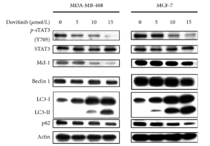
| Western blot | p-STAT3 / STAT3 / Mcl-1 / LC3 / Beclin 1 / p62 p-VEGFR-2 / VEGFR-2 / p-FGFR-1 / FGFR-1 p-PDGFR-β / PDGFR-β / p-ERK / ERK CDK1 / p-CDK1 / p53 / p21 |
|
31485222 |
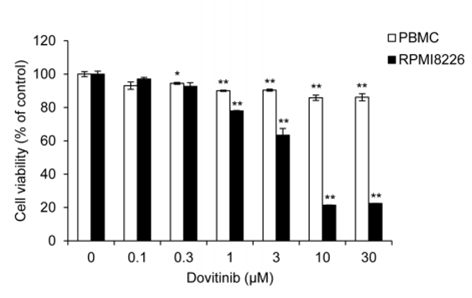
| Growth inhibition assay | Cell viability |
|
28467797 |
Clinical Trial Information
(data from https://clinicaltrials.gov, updated on 2024-05-22)
| NCT Number | Recruitment | Conditions | Sponsor/Collaborators | Start Date | Phases |
|---|---|---|---|---|---|
| NCT05571969 | Recruiting | Advanced Solid Tumors |
Allarity Therapeutics|Amarex Clinical Research |
February 20 2023 | Phase 1 |
| NCT02268435 | Withdrawn | Gastrointestinal Stromal Tumors |
Asan Medical Center |
March 2015 | Phase 1 |
| NCT01700270 | Completed | Advanced Solid Tumors Excluding Breast Cancer |
Novartis Pharmaceuticals|Novartis |
May 2013 | Phase 1 |
| NCT01680796 | Withdrawn | Multiple Myeloma |
University of Florida|Novartis Pharmaceuticals |
February 2013 | Phase 1 |
| NCT01266070 | Terminated | Von Hippel-Lindau Syndrome |
M.D. Anderson Cancer Center|Novartis |
November 2012 | Phase 2 |
Tech Support
Tel: +1-832-582-8158 Ext:3
If you have any other enquiries, please leave a message.
Signaling Pathway Map
Products are for research use only. Not for human use. We do not sell to patients.
©Copyright 2013 Selleck Chemicals. All Rights Reserved.