
- Bioactive Compounds
- By Signaling Pathways
- PI3K/Akt/mTOR
- Epigenetics
- Methylation
- Immunology & Inflammation
- Protein Tyrosine Kinase
- Angiogenesis
- Apoptosis
- Autophagy
- ER stress & UPR
- JAK/STAT
- MAPK
- Cytoskeletal Signaling
- Cell Cycle
- TGF-beta/Smad
- DNA Damage/DNA Repair
- Compound Libraries
- Antibodies
- Bioreagents
- qPCR
- 2x SYBR Green qPCR Master Mix
- 2x SYBR Green qPCR Master Mix(Low ROX)
- 2x SYBR Green qPCR Master Mix(High ROX)
- Protein Assay
- Protein A/G Magnetic Beads for IP
- Anti-Flag magnetic beads
- Anti-Flag Affinity Gel
- Anti-Myc magnetic beads
- Anti-HA magnetic beads
- Magnetic Separator
- Poly DYKDDDDK Tag Peptide lyophilized powder
- Protease Inhibitor Cocktail
- Protease Inhibitor Cocktail (EDTA-Free, 100X in DMSO)
- Phosphatase Inhibitor Cocktail (2 Tubes, 100X)
- Cell Biology
- Cell Counting Kit-8 (CCK-8)
- Animal Experiment
- Mouse Direct PCR Kit (For Genotyping)
- New Products
- Contact Us
-
 Australia
-
 Austria
-
 Belgium
-
 Canada
-
 China
China
-
 Czech Republic
-
 Denmark
-
 Finland
-
 France
-
 Germany
-
 Greece
-
 Hong Kong
-
 Hungary
-
 Iceland
-
 India
India
-
 Ireland
-
 Israel
-
 Italy
Italy
-
 Japan
Japan
-
 Korea
Korea
-
 Luxembourg
-
 Malaysia
-
 Netherlands
-
 New Zealand
-
 Norway
-
 Poland
-
 Qatar
Qatar
-
 Romania
-
 Saudi Arabia
-
 Singapore
-
 Spain
Spain
-
 Sweden
-
 Switzerland
-
 Taiwan
-
 Turkey
-
 United Kingdom
-
 United States
-
 Other Countries
Masitinib
Synonyms: AB1010
Masitinib is a novel inhibitor for Kit (c-Kit) and PDGFRα/β with IC50 of 200 nM and 540 nM/800 nM, weak inhibition to ABL and c-Fms. Phase 3.
Masitinib Chemical Structure
CAS No. 790299-79-5
Purity & Quality Control
Batch:
Purity:
99.99%
99.99
Masitinib Related Products
| Related Products | Amuvatinib (MP-470) OSI-930 Sitravatinib (MGCD516) ISCK03 PDGFR inhibitor 1 | Click to Expand |
|---|---|---|
| Related Compound Libraries | Kinase Inhibitor Library Tyrosine Kinase Inhibitor Library PI3K/Akt Inhibitor Library Cell Cycle compound library Angiogenesis Related compound Library | Click to Expand |
Signaling Pathway
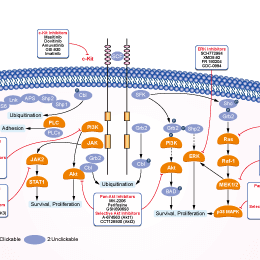
Biological Activity
| Description | Masitinib is a novel inhibitor for Kit (c-Kit) and PDGFRα/β with IC50 of 200 nM and 540 nM/800 nM, weak inhibition to ABL and c-Fms. Phase 3. | |||||||||||
|---|---|---|---|---|---|---|---|---|---|---|---|---|
| Features | Potential low side-effect profile. | |||||||||||
| Targets |
|
| In vitro | ||||
| In vitro | Masitinib is a competitive inhibitor against ATP at concentrations ≤500 nM. Masitinib also potently inhibits recombinant PDGFR and the intracellular kinase Lyn, and to a lesser extent, fibroblast growth factor receptor 3. In contrast, Masitinib demonstrates weak inhibition of Abl and c-Fms. Masitinib more strongly inhibits degranulation, cytokine production, and bone marrow mast cell migration than imatinib. In Ba/F3 cells expressing human wild-type Kit, Masitinib inhibits SCF (stem cell factor)-induced cell proliferation with an IC50 of 150 nM, while the IC50 for inhibition of IL-3-stimulated proliferation is at approximately >10 µM. In Ba/F3 cells expressing PDGFRα, Masitinib inhibits PDGF-BB-stimulated proliferation and PDGFRα tyrosine phosphorylation with IC50 of 300 nM. Masitinib also causes inhibition of SCF-stimulated tyrosine phosphorylation of human Kit in mastocytoma cell-lines and BMMC. Masitinib inhibits Kit gain-of-function mutants, including V559D mutant and Δ27 mouse mutant with IC50 of 3 and 5 nM in Ba/F3 cells. Masitinib inhibits the cell proliferation of mastocytoma cell lines including HMC-1α155 and FMA3 with IC50 of 10 and 30 nM, respectively. [1] Masitinib inhibits cell growth and PDGFR phosphorylation in two novel ISS cell lines, which suggest that Masitinib displays activity against both primary and metastatic ISS cell line and may aid in the clinical management of ISS. [2] | |||
|---|---|---|---|---|
| Kinase Assay | In vitro enzyme-linked immunoassay with recombinant protein kinases | |||
| A 96-well microtitre plateis coated overnight with 0.25 mg/ml poly(Glu,Tyr 4:1), rinsed twice with 250 µL of washing buffer (10 mM phosphate-buffered saline [pH 7.4] and 0.05% Tween 20) and dried for 2 hours at room temperature. Assays are performed at room temperature with a final volume of 50 µL in kinase buffer (10 mM MgCl2, 1 mM MnCl2, 1 mM sodium orthovanadate, 20 mM HEPES, pH 7.8) containing ATP at a concentration of at least twice the Km for each enzyme and an appropriate amount of recombinant enzyme to ensure a linear reaction rate. Reactions are initiated upon introduction of the enzyme and terminated with the addition of one reaction volume (50 μL) of 100 mM EDTA per 5 M urea mix. Plates are washed three times and incubated with 1:30,000 horseradish peroxidase-conjugated anti-phosphotyrosine monoclonal antibody, then washed three times and incubated with tetramethylbenzidine. The final reaction product is quantified by spectrophotometry at 450 nm. | ||||
| Cell Research | Cell lines | Ba/F3 cells expressing wild-type or mutant human Kit, HMC1, HMC-1α155 | ||
| Concentrations | 0.1 nM - 10 μM | |||
| Incubation Time | 48 hours | |||
| Method | For the assay of Ba/F3 cell proliferation, microtitre plates are seeded with a total of 104 cells/well in 100 μL of RPMI 1640 medium with 10% foetal bovine serum at 37 °C. These are supplemented, or not, with either 0.1% conditioned medium from X63-IL-3 cells or 250 ng/mL murine SCF. The murine SCF, which activates Kit, is purified from the conditioned medium of SCF-producing CHO cells. Cells are grown for 48 hours at 37 °C with Masitinib and then incubated with 10 μL/well of WST-1 reagent for 3 hours at 37 °C. The amount of formazan dye formed is quantified by its absorbance at 450 nm using a scanning multiwell spectrophotometer. A blank well without cells is used as a background control for the spectrophotometer. | |||
| In Vivo | ||
| In vivo | Masitinib inhibits tumour growth and increases the median survival time in Δ27-expressing Ba/F3 tumor models at 30 mg/kg, without cardiotoxicity or genotoxicity. [1] Masitinib (12.5 mg/kg/d PO) increases overall TTP (time-to-tumor progression) compared with placebo in dogs. [3] The combination of masitinib/gemcitabine shows synergy in vitro on proliferation of gemcitabine-refractory cell lines Mia Paca2 and Panc1, and to a lesser extent on Mia Paca-2 pancreatic tumours in Nog璖CID mice. [4] | |
|---|---|---|
| Animal Research | Animal Models | Ba/F3 Δ27 tumour model in female MBRI Nu/Nu mice |
| Dosages | 30 mg/kg (intraperitoneal) or 10, 30, or 45 mg/kg (orally). | |
| Administration | Intraperitoneal or orally administered. | |
| NCT Number | Recruitment | Conditions | Sponsor/Collaborators | Start Date | Phases |
|---|---|---|---|---|---|
| NCT00866138 | Completed | Multiple Myeloma |
AB Science |
February 2005 | Phase 2 |
| NCT00831974 | Completed | Mastocytosis |
AB Science |
October 2004 | Phase 2 |
Chemical Information & Solubility
| Molecular Weight | 498.64 | Formula | C28H30N6OS |
| CAS No. | 790299-79-5 | SDF | Download Masitinib SDF |
| Smiles | CC1=C(C=C(C=C1)NC(=O)C2=CC=C(C=C2)CN3CCN(CC3)C)NC4=NC(=CS4)C5=CN=CC=C5 | ||
| Storage (From the date of receipt) | |||
|
In vitro |
DMSO : 99 mg/mL ( (198.54 mM) Moisture-absorbing DMSO reduces solubility. Please use fresh DMSO.) Ethanol : 99 mg/mL Water : Insoluble |
Molecular Weight Calculator |
|
In vivo Add solvents to the product individually and in order. |
In vivo Formulation Calculator |
||||
Preparing Stock Solutions
Molarity Calculator
In vivo Formulation Calculator (Clear solution)
Step 1: Enter information below (Recommended: An additional animal making an allowance for loss during the experiment)
mg/kg
g
μL
Step 2: Enter the in vivo formulation (This is only the calculator, not formulation. Please contact us first if there is no in vivo formulation at the solubility Section.)
% DMSO
%
% Tween 80
% ddH2O
%DMSO
%
Calculation results:
Working concentration: mg/ml;
Method for preparing DMSO master liquid: mg drug pre-dissolved in μL DMSO ( Master liquid concentration mg/mL, Please contact us first if the concentration exceeds the DMSO solubility of the batch of drug. )
Method for preparing in vivo formulation: Take μL DMSO master liquid, next addμL PEG300, mix and clarify, next addμL Tween 80, mix and clarify, next add μL ddH2O, mix and clarify.
Method for preparing in vivo formulation: Take μL DMSO master liquid, next add μL Corn oil, mix and clarify.
Note: 1. Please make sure the liquid is clear before adding the next solvent.
2. Be sure to add the solvent(s) in order. You must ensure that the solution obtained, in the previous addition, is a clear solution before proceeding to add the next solvent. Physical methods such
as vortex, ultrasound or hot water bath can be used to aid dissolving.
Tech Support
Answers to questions you may have can be found in the inhibitor handling instructions. Topics include how to prepare stock solutions, how to store inhibitors, and issues that need special attention for cell-based assays and animal experiments.
Tel: +1-832-582-8158 Ext:3
If you have any other enquiries, please leave a message.
* Indicates a Required Field
Tags: buy Masitinib | Masitinib supplier | purchase Masitinib | Masitinib cost | Masitinib manufacturer | order Masitinib | Masitinib distributor
Products are for research use only. Not for human use. We do not sell to patients.
©Copyright 2013 Selleck Chemicals. All Rights Reserved.