
- Bioactive Compounds
- By Signaling Pathways
- PI3K/Akt/mTOR
- Epigenetics
- Methylation
- Immunology & Inflammation
- Protein Tyrosine Kinase
- Angiogenesis
- Apoptosis
- Autophagy
- ER stress & UPR
- JAK/STAT
- MAPK
- Cytoskeletal Signaling
- Cell Cycle
- TGF-beta/Smad
- Compound Libraries
- Antibodies
- Bioreagents
- qPCR
- 2x SYBR Green qPCR Master Mix
- 2x SYBR Green qPCR Master Mix(Low ROX)
- 2x SYBR Green qPCR Master Mix(High ROX)
- Protein Assay
- Protein A/G Magnetic Beads for IP
- Anti-Flag magnetic beads
- Anti-Flag Affinity Gel
- Anti-Myc magnetic beads
- Anti-HA magnetic beads
- Poly FLAG Peptide lyophilized powder
- Protease Inhibitor Cocktail
- Protease Inhibitor Cocktail (EDTA-Free, 100X in DMSO)
- Phosphatase Inhibitor Cocktail (2 Tubes, 100X)
- Cell Biology
- Cell Counting Kit-8 (CCK-8)
- Animal Experiment
- Mouse Direct PCR Kit (For Genotyping)
- New Products
- Contact Us
-
 Australia
-
 Austria
-
 Belgium
-
 Brazil
-
 Canada
-
 China
China
-
 Czech Republic
-
 Denmark
-
 Finland
-
 France
-
 Germany
-
 Greece
-
 Hong Kong
-
 Hungary
-
 Iceland
-
 India
India
-
 Ireland
-
 Israel
-
 Italy
Italy
-
 Japan
Japan
-
 Korea
Korea
-
 Luxembourg
-
 Malaysia
-
 Netherlands
-
 New Zealand
-
 Norway
-
 Poland
-
 Qatar
Qatar
-
 Romania
-
 Saudi Arabia
-
 Singapore
-
 Spain
Spain
-
 Sweden
-
 Switzerland
-
 Taiwan
-
 Turkey
-
 United Kingdom
-
 United States
-
 Other Countries
Bafetinib
Synonyms: INNO-406, NS-187
Bafetinib is a potent and selective dual Bcr-Abl/Lyn inhibitor with IC50 of 5.8 nM/19 nM in cell-free assays, does not inhibit the phosphorylation of the T315I mutant and is less potent to PDGFR and c-Kit.
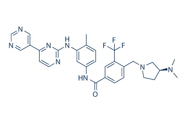
Bafetinib Chemical Structure
CAS: 859212-16-1
Selleck's Bafetinib has been cited by 26 Publications
1 Customer Review
Purity & Quality Control
Batch:
Purity:
99.89%
99.89
Bafetinib Related Products
| Related Targets | Abl c-Abl v-Abl | Click to Expand |
|---|---|---|
| Related Compound Libraries | Tyrosine Kinase Inhibitor Library PI3K/Akt Inhibitor Library Angiogenesis Related compound Library HIF-1 Signaling Pathway Compound Library FDA-approved Anticancer Drug Library | Click to Expand |
Signaling Pathway
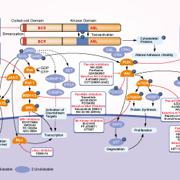
Choose Selective Bcr-Abl Inhibitors
Biological Activity
| Description | Bafetinib is a potent and selective dual Bcr-Abl/Lyn inhibitor with IC50 of 5.8 nM/19 nM in cell-free assays, does not inhibit the phosphorylation of the T315I mutant and is less potent to PDGFR and c-Kit. | ||||
|---|---|---|---|---|---|
| Features | Dual Bcr-Abl/Lyn inhibitor. | ||||
| Targets |
|
| In vitro | ||||
| In vitro | Bafetinib blocks WT Bcr-Abl autophosphorylation and its downstream kinase activity with IC50 of 11 nM and 22 nM in K562 and 293T cells, respectively. Bafetinib suppresses the growth of the Bcr-Abl-positive cell lines including K562, KU812, and BaF3/wt cells potently without effects on the proliferation of the Bcr-Abl-negative U937 cell line. Moreover, Bafetinib exhibits a dose-dependent antiproliferative effect against Bcr-Abl point mutant cell lines, such as BaF3/E255K cells. [1] In Bcr-Abl+ leukemia cell lines, Bafetinib induces both caspase-mediated and caspase-independent cell death by blocking the phosphorylation of Bcr-Abl. [2] |
|||
|---|---|---|---|---|
| Kinase Assay | Kinase assay | |||
| Bcr-Abl kinase assays are performed in 25 μL of reaction mixture containing 250 μM peptide substrate, 740 Bq/μL [γ-33P]ATP, and 20 μM cold adenosine triphosphate (ATP) by using the SignaTECT protein tyrosine kinase assay system. Each Bcr-Abl kinase is used at a concentration of 10 nM. Kinase assays for Abl, Src, and Lyn are carried out with an enzyme-linked immunosorbent assay (ELISA) kit. The inhibitory effects of NS-187 against 79 tyrosine kinases are tested with KinaseProfiler. | ||||
| Cell Research | Cell lines | K562, BaF3/wt, BaF3/E255K, and BaF3/T315I cells | ||
| Concentrations | 0-10 μM | |||
| Incubation Time | 72 hours | |||
| Method | K562, BaF3/wt, BaF3/E255K, and BaF3/T315I cells are plated at 1 × 10³ in 96-well plates, whereas KU812 and U937 cells are plated at 5 × 10³ in 96-well plates. Cells are incubated with serial dilutions of Bafetinib for 3 days. Cell proliferation is measured by MTT (3-(4,5-dimethylthiazol-2-yl)-2,5-diphenyltetrazolium bromide; Nacalai Tesque) assay, and the 50% inhibitory concentration (IC50) values are calculated by fitting the data to a logistic curve. |
|||
| In Vivo | ||
| In vivo |
In Bcr-Abl–positive KU812 mouse model, Bafetinib (0.2 mg/kg/day) significantly inhibits tumor growth, and completely inhibits tumor growth without adverse effects at 20 mg/kg/day. For Balb/c mice, Bafetinib shows maximal tolerated dose of 200 mg/kg/d and bioavailability value (BA) of 32%. [1] In a Central nervous system (CNS) leukemia model bearing Ba/F3/wt bcr-ablGFP, Ba/F3/Q252H, or Ba/F3/M351T cells, combination treatment of Bafetinib (60 mg/kg) and cyclosporine A (CsA) (50 mg/kg) leads to more significant inhibition of leukemia growth in the brain than either Bafetinib or CsA alone. [3] |
|
|---|---|---|
| Animal Research | Animal Models | KU812 xenograft is established by subcutaneous injection of KU812 cells into the right flank of Balb/c-nu/nu female mice. |
| Dosages | ≤20 mg/kg/day | |
| Administration | Administered via p.o. | |
| NCT Number | Recruitment | Conditions | Sponsor/Collaborators | Start Date | Phases |
|---|---|---|---|---|---|
| NCT00352677 | Completed | Chronic Myeloid Leukemia|Acute Lymphocytic Leukemia |
CytRx |
July 2006 | Phase 1 |
Chemical Information & Solubility
| Molecular Weight | 576.62 | Formula | C30H31F3N8O |
| CAS No. | 859212-16-1 | SDF | Download Bafetinib SDF Download Bafetinib SDF |
| Smiles | CC1=C(C=C(C=C1)NC(=O)C2=CC(=C(C=C2)CN3CCC(C3)N(C)C)C(F)(F)F)NC4=NC=CC(=N4)C5=CN=CN=C5 | ||
| Storage (From the date of receipt) | |||
|
In vitro |
DMSO : 100 mg/mL ( (173.42 mM); Moisture-absorbing DMSO reduces solubility. Please use fresh DMSO.) Water : Insoluble Ethanol : Insoluble |
Molecular Weight Calculator |
|
In vivo Add solvents to the product individually and in order. |
In vivo Formulation Calculator |
||||
Preparing Stock Solutions
Molarity Calculator
In vivo Formulation Calculator (Clear solution)
Step 1: Enter information below (Recommended: An additional animal making an allowance for loss during the experiment)
mg/kg
g
μL
Step 2: Enter the in vivo formulation (This is only the calculator, not formulation. Please contact us first if there is no in vivo formulation at the solubility Section.)
% DMSO
%
% Tween 80
% ddH2O
%DMSO
%
Calculation results:
Working concentration: mg/ml;
Method for preparing DMSO master liquid: mg drug pre-dissolved in μL DMSO ( Master liquid concentration mg/mL, Please contact us first if the concentration exceeds the DMSO solubility of the batch of drug. )
Method for preparing in vivo formulation: Take μL DMSO master liquid, next addμL PEG300, mix and clarify, next addμL Tween 80, mix and clarify, next add μL ddH2O, mix and clarify.
Method for preparing in vivo formulation: Take μL DMSO master liquid, next add μL Corn oil, mix and clarify.
Note: 1. Please make sure the liquid is clear before adding the next solvent.
2. Be sure to add the solvent(s) in order. You must ensure that the solution obtained, in the previous addition, is a clear solution before proceeding to add the next solvent. Physical methods such
as vortex, ultrasound or hot water bath can be used to aid dissolving.
Tech Support
Answers to questions you may have can be found in the inhibitor handling instructions. Topics include how to prepare stock solutions, how to store inhibitors, and issues that need special attention for cell-based assays and animal experiments.
Tel: +1-832-582-8158 Ext:3
If you have any other enquiries, please leave a message.
* Indicates a Required Field
Frequently Asked Questions
Question 1:
Can you suggest the route of in vivo administration for S1369?
Answer:
S1369 in 0.5% methylcellulose+0.2% Tween 80 at 30mg/ml is a suspension, and it is fine for oral gavage. If you dissolved the compound in 0.5% methyl cellulose and got a suspension, it is fine. And 0.2% Tween 80 can help the suspension dissolving more homogeneous.
Tags: buy Bafetinib | Bafetinib supplier | purchase Bafetinib | Bafetinib cost | Bafetinib manufacturer | order Bafetinib | Bafetinib distributor
Products are for research use only. Not for human use. We do not sell to patients.
©Copyright 2013 Selleck Chemicals. All Rights Reserved.