- Bioactive Compounds
- By Signaling Pathways
- PI3K/Akt/mTOR
- Epigenetics
- Methylation
- Immunology & Inflammation
- Protein Tyrosine Kinase
- Angiogenesis
- Apoptosis
- Autophagy
- ER stress & UPR
- JAK/STAT
- MAPK
- Cytoskeletal Signaling
- Cell Cycle
- TGF-beta/Smad
- DNA Damage/DNA Repair
- Compound Libraries
- Popular Compound Libraries
- Customize Library
- Clinical and FDA-approved Related
- Bioactive Compound Libraries
- Inhibitor Related
- Natural Product Related
- Metabolism Related
- Cell Death Related
- By Signaling Pathway
- By Disease
- Anti-infection and Antiviral Related
- Neuronal and Immunology Related
- Fragment and Covalent Related
- FDA-approved Drug Library
- FDA-approved & Passed Phase I Drug Library
- Preclinical/Clinical Compound Library
- Bioactive Compound Library-I
- Bioactive Compound Library-Ⅱ
- Kinase Inhibitor Library
- Express-Pick Library
- Natural Product Library
- Human Endogenous Metabolite Compound Library
- Alkaloid Compound LibraryNew
- Angiogenesis Related compound Library
- Anti-Aging Compound Library
- Anti-alzheimer Disease Compound Library
- Antibiotics compound Library
- Anti-cancer Compound Library
- Anti-cancer Compound Library-Ⅱ
- Anti-cancer Metabolism Compound Library
- Anti-Cardiovascular Disease Compound Library
- Anti-diabetic Compound Library
- Anti-infection Compound Library
- Antioxidant Compound Library
- Anti-parasitic Compound Library
- Antiviral Compound Library
- Apoptosis Compound Library
- Autophagy Compound Library
- Calcium Channel Blocker LibraryNew
- Cambridge Cancer Compound Library
- Carbohydrate Metabolism Compound LibraryNew
- Cell Cycle compound library
- CNS-Penetrant Compound Library
- Covalent Inhibitor Library
- Cytokine Inhibitor LibraryNew
- Cytoskeletal Signaling Pathway Compound Library
- DNA Damage/DNA Repair compound Library
- Drug-like Compound Library
- Endoplasmic Reticulum Stress Compound Library
- Epigenetics Compound Library
- Exosome Secretion Related Compound LibraryNew
- FDA-approved Anticancer Drug LibraryNew
- Ferroptosis Compound Library
- Flavonoid Compound Library
- Fragment Library
- Glutamine Metabolism Compound Library
- Glycolysis Compound Library
- GPCR Compound Library
- Gut Microbial Metabolite Library
- HIF-1 Signaling Pathway Compound Library
- Highly Selective Inhibitor Library
- Histone modification compound library
- HTS Library for Drug Discovery
- Human Hormone Related Compound LibraryNew
- Human Transcription Factor Compound LibraryNew
- Immunology/Inflammation Compound Library
- Inhibitor Library
- Ion Channel Ligand Library
- JAK/STAT compound library
- Lipid Metabolism Compound LibraryNew
- Macrocyclic Compound Library
- MAPK Inhibitor Library
- Medicine Food Homology Compound Library
- Metabolism Compound Library
- Methylation Compound Library
- Mouse Metabolite Compound LibraryNew
- Natural Organic Compound Library
- Neuronal Signaling Compound Library
- NF-κB Signaling Compound Library
- Nucleoside Analogue Library
- Obesity Compound Library
- Oxidative Stress Compound LibraryNew
- Plant Extract Library
- Phenotypic Screening Library
- PI3K/Akt Inhibitor Library
- Protease Inhibitor Library
- Protein-protein Interaction Inhibitor Library
- Pyroptosis Compound Library
- Small Molecule Immuno-Oncology Compound Library
- Mitochondria-Targeted Compound LibraryNew
- Stem Cell Differentiation Compound LibraryNew
- Stem Cell Signaling Compound Library
- Natural Phenol Compound LibraryNew
- Natural Terpenoid Compound LibraryNew
- TGF-beta/Smad compound library
- Traditional Chinese Medicine Library
- Tyrosine Kinase Inhibitor Library
- Ubiquitination Compound Library
-
Cherry Picking
You can personalize your library with chemicals from within Selleck's inventory. Build the right library for your research endeavors by choosing from compounds in all of our available libraries.
Please contact us at [email protected] to customize your library.
You could select:
- Antibodies
- Bioreagents
- qPCR
- 2x SYBR Green qPCR Master Mix
- 2x SYBR Green qPCR Master Mix(Low ROX)
- 2x SYBR Green qPCR Master Mix(High ROX)
- Protein Assay
- Protein A/G Magnetic Beads for IP
- Anti-Flag magnetic beads
- Anti-Flag Affinity Gel
- Anti-Myc magnetic beads
- Anti-HA magnetic beads
- Magnetic Separator
- Poly DYKDDDDK Tag Peptide lyophilized powder
- Protease Inhibitor Cocktail
- Protease Inhibitor Cocktail (EDTA-Free, 100X in DMSO)
- Phosphatase Inhibitor Cocktail (2 Tubes, 100X)
- Cell Biology
- Cell Counting Kit-8 (CCK-8)
- Animal Experiment
- Mouse Direct PCR Kit (For Genotyping)
- New Products
- Contact Us
4μ8C
Synonyms: IRE1 Inhibitor III
4μ8C (IRE1 Inhibitor III) is a potent and selective IRE1 Rnase inhibitor with IC50 of 76 nM.

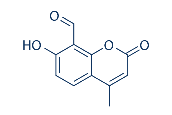
4μ8C Chemical Structure
CAS No. 14003-96-4
Purity & Quality Control
Batch:
Purity:
99.75%
99.75
4μ8C Related Products
| Related Products | STF-083010 kira6 MKC-3946 MKC8866 APY29 Kira8 IXA4 | Click to Expand |
|---|---|---|
| Related Compound Libraries | FDA-approved Drug Library Natural Product Library Bioactive Compound Library-I Bioactive Compound Library-Ⅱ Highly Selective Inhibitor Library | Click to Expand |
Cell Data
| Cell Lines | Assay Type | Concentration | Incubation Time | Formulation | Activity Description | PMID |
|---|---|---|---|---|---|---|
| SF21 cells | Function assay | 30 mins | Inhibition of human recombinant puritin-His-tagged IRE-1 RNase expressed in SF21 cells using XBP-1 RNA stem loop as substrate incubated for 30 mins prior to substrate addition measured after 2 hrs by FRET-suppression assay, IC50=0.206 μM | 24749861 | ||
| human Jeko cells | Function assay | 24 h | Inhibition of XBP-1s expression in human Jeko cells after 24 hrs by immunoblotting analysis, IC50=1.57 μM | 24749861 | ||
| human Mino cells | Function assay | 24 h | Inhibition of XBP-1s expression in human Mino cells after 24 hrs by immunoblotting analysis, IC50=1.62 μM | 24749861 | ||
| Click to View More Cell Line Experimental Data | ||||||
Biological Activity
| Description | 4μ8C (IRE1 Inhibitor III) is a potent and selective IRE1 Rnase inhibitor with IC50 of 76 nM. | ||
|---|---|---|---|
| Features | IRE1 Rnase-selective inhibitor, used as a platform for developing new locally acting drugs. | ||
| Targets |
|
| In vitro | ||||
| In vitro | 4μ8C blocks substrate(RIDD) access to the active site of IRE1 and selectively inactivates both Xbp1 splicing and IRE1-mediated mRNA degradation. IRE1 inhibition subsequently induces ER stress without measureable acute toxicity. [1] 4μ8C, as an IRE1 inhibitor, blocks IL-4, IL-5, and IL-13 production from CD4+ T cells. [2] |
|||
|---|---|---|---|---|
| Kinase Assay | In Vitro IRE1 RNase and RIDD Assays | |||
| Analysis of radiolabeled Xbp1 substrate cleavage is performed as previously except that mammalian IRE1 reaction buffer is used. In vitro RIDD substrates are synthesized by in vitro transcription using the T7-MAXIscript Kit in the presence of 32P ATP or Cy5-UTP on templates isolated by RT-PCR from mouse Min6 cells (Ins2) or PCR from cloned XBP1 cDNA. The resulting products are gel purified to obtain full-length substrate. Reactions are then separated by 15% UREA-PAGE for analysis by phosphorimaging or by near-infrared imaging using the LI-COR Odyssey scanner. | ||||
| Cell Research | Cell lines | Wild-type MEFs | ||
| Concentrations | ~128 μM | |||
| Incubation Time | 24 hours | |||
| Method | Cells are seeded in phenol red-free cell culture medium in 96 or 24 well dishes at a density of 5 × 10³ or 5 × 10⁴ cells per well, respectively. Cultures are incubated for 16 h before treatment with 4μ8C for 24 h. Cultures are then analyzed by the addition of 200 μM WST1 and 10 μM phenazine metho-sulfate. After development of the reagent for 2 h at 37°C, the hydrolyzed dye is detected by absorbance at 450 nm, after subtracting background and absorbance at 595 nm. Alternatively, cell viability is determined by staining of the adherent culture with crystal violet. Quantitation of the dye uptake is analyzed by extensive washing of the stained cells with water and solublization of the crystal violet in methanol followed by absorbance measurements at 595 nm. |
|||
| In Vivo | ||
| In vivo | 4μ8c is an IRE1 Inhibitor III that reduces atherosclerotic lesion and effectively mitigate plaque development in mice. |
|
|---|---|---|
| Animal Research | Animal Models | C57BL/6 mice |
| Dosages | 10 mg/kg | |
| Administration | i.p. | |
Chemical Information & Solubility
| Molecular Weight | 204.18 | Formula | C11H8O4 |
| CAS No. | 14003-96-4 | SDF | Download 4μ8C SDF |
| Smiles | CC1=CC(=O)OC2=C1C=CC(=C2C=O)O | ||
| Storage (From the date of receipt) | |||
|
In vitro |
DMSO : 71 mg/mL ( (347.73 mM) Moisture-absorbing DMSO reduces solubility. Please use fresh DMSO.) Water : Insoluble Ethanol : Insoluble |
Molecular Weight Calculator |
|
In vivo Add solvents to the product individually and in order. |
In vivo Formulation Calculator |
||||
Preparing Stock Solutions
Molarity Calculator
In vivo Formulation Calculator (Clear solution)
Step 1: Enter information below (Recommended: An additional animal making an allowance for loss during the experiment)
mg/kg
g
μL
Step 2: Enter the in vivo formulation (This is only the calculator, not formulation. Please contact us first if there is no in vivo formulation at the solubility Section.)
% DMSO
%
% Tween 80
% ddH2O
%DMSO
%
Calculation results:
Working concentration: mg/ml;
Method for preparing DMSO master liquid: mg drug pre-dissolved in μL DMSO ( Master liquid concentration mg/mL, Please contact us first if the concentration exceeds the DMSO solubility of the batch of drug. )
Method for preparing in vivo formulation: Take μL DMSO master liquid, next addμL PEG300, mix and clarify, next addμL Tween 80, mix and clarify, next add μL ddH2O, mix and clarify.
Method for preparing in vivo formulation: Take μL DMSO master liquid, next add μL Corn oil, mix and clarify.
Note: 1. Please make sure the liquid is clear before adding the next solvent.
2. Be sure to add the solvent(s) in order. You must ensure that the solution obtained, in the previous addition, is a clear solution before proceeding to add the next solvent. Physical methods such
as vortex, ultrasound or hot water bath can be used to aid dissolving.
Tech Support
Answers to questions you may have can be found in the inhibitor handling instructions. Topics include how to prepare stock solutions, how to store inhibitors, and issues that need special attention for cell-based assays and animal experiments.
Tel: +1-832-582-8158 Ext:3
If you have any other enquiries, please leave a message.
* Indicates a Required Field
Tags: buy 4μ8C | 4μ8C supplier | purchase 4μ8C | 4μ8C cost | 4μ8C manufacturer | order 4μ8C | 4μ8C distributor


